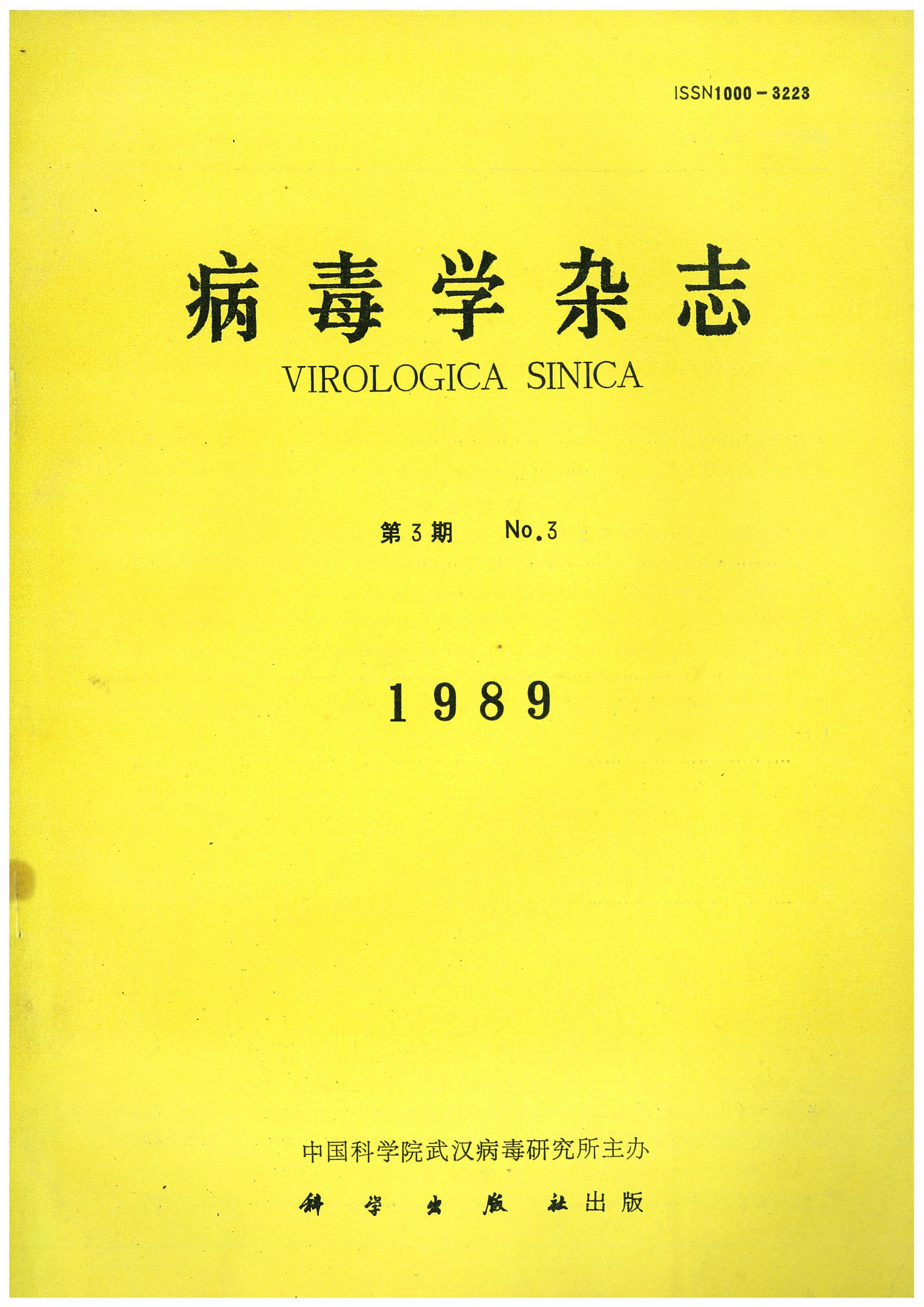
YANG Ying-Zhen, JIN Pei-Ying, GUO Qi, YANG Xue-Yi, PU Shou-Yue, CHEN Hao-Zhu, YANG Jun-Hua, WANG Ke-Jiang, SHI Jian-Ying, GONG Zu-Xun, CHEN Ju-Yang and PENG Bao-Zhen. Experimental Acute Coxsackle B-3 Viral Myocarditis in Mice[J]. Virologica Sinica, 1989, 4(3).
Citation: YANG Ying-Zhen, JIN Pei-Ying, GUO Qi, YANG Xue-Yi, PU Shou-Yue, CHEN Hao-Zhu, YANG Jun-Hua, WANG Ke-Jiang, SHI Jian-Ying, GONG Zu-Xun, CHEN Ju-Yang, PENG Bao-Zhen. Experimental Acute Coxsackle B-3 Viral Myocarditis in Mice .VIROLOGICA SINICA, 1989, 4(3) : 233.

实验性小鼠急性Coxsackie B-3病毒性心肌炎的研究

  • 小鼠感染Coxsackie B-3病毒后7天,各鼠心脏均出现巨检病变,组织学检查心肌细胞示变性、坏死及炎性细胞浸润,心肌切片作图象分析示病灶总面积/所测心肌总面积为11.3%,并有明显心肌超微结构改变。心肌中分离到病毒。对照鼠心脏均无上述改变,亦未分离到病毒。本模型可作为进一步探索病毒性心肌炎药物治疗的基础。

Experimental Acute Coxsackle B-3 Viral Myocarditis in Mice

  • An experimental acute myocarditis caused hy Coxsaekie B-3 virus(CB_3V)was developed in male BALB/c mice 4 weeks of age. The animals were szc-rificed at the 7th day after infection, After gross examination, the heart was cut transversely into 3 pieces for light, electron microscopic examinations andvirus assay. In addition, myocardial lesions were also quantitatively measuredby image analysis. White spots, streaks or patches were seen on the surface of ventrieles ofhearts in the infected group (14 mice) .The...

  • 加载中
  • 加载中

Article Metrics

Article views(4011) PDF downloads(793) Cited by(0)

Related
Proportional views
    通讯作者: 陈斌, bchen63@163.com
    • 1. 

      沈阳化工大学材料科学与工程学院 沈阳 110142

    1. 本站搜索
    2. 百度学术搜索
    3. 万方数据库搜索
    4. CNKI搜索

    Experimental Acute Coxsackle B-3 Viral Myocarditis in Mice

    • 1. Shanghai Institute of Cardiovascular Diseases
    • 2. Zhongshan Hospital
    • 3. shanghai Medical University
    • 4. Shanghai

    Abstract: An experimental acute myocarditis caused hy Coxsaekie B-3 virus(CB_3V)was developed in male BALB/c mice 4 weeks of age. The animals were szc-rificed at the 7th day after infection, After gross examination, the heart was cut transversely into 3 pieces for light, electron microscopic examinations andvirus assay. In addition, myocardial lesions were also quantitatively measuredby image analysis. White spots, streaks or patches were seen on the surface of ventrieles ofhearts in the infected group (14 mice) .The...

    Relative (20)

    目录

    /

    DownLoad:  Full-Size Img  PowerPoint
    Return
    Return