-

-
-
-
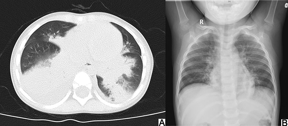
No. Age Gender Main manifestation Physical sign Peak fever (℃) Fever duration (Days) PB discovered (Days) Chest CT and airway reconstruction PB location Bronchoscopy therapy (Times) Oxygen Outcome 1 2 y Female Fever, cough, dyspnea Reduced left side breath sound, wet rales 40.5 17 13 Extensive consolidation of both lungs, bilateral pleural effusions; no significant abnormalities in airway reconstruction Basal segmental bronchus of the right lower lobe 2 CPAP Discharged, recurrent wheezing 2 4 y and 10 m Female Fever, cough Reduced right side breath sound, wet rales 40.0 8 12 Right lung lesions, especially the right middle lobe consolidation, pleural effusion on the right; airway reconstruction showed no obvious abnormalities Right medial and lateral segmental bronchus 1 Unused Discharged 3 15 y and 7 m Female Fever, cough, dyspnea Reduced left side breath sound 38.6 1 5 Left lung consolidation with atelectasis, right middle lobe consolidation atelectasis, right middle lobe bronchiectasis, airway reconstruction shows left main bronchus unclear Left superior lobar bronchus 4 CPAP, mask Discharged 4 5 y and 2 m Female Fever, cough, dyspnea, dispirited Reduced left side breath sound 40.4 8 13 Multiple consolidation and atelectasis in the left lung, with left pleural effusion, partial bronchial occlusion, and a small amount of pericardial effusion; No airway reconstruction Apicoposterior segmental bronchus of the left upper lobe 2 Nasal catheter Discharged 5 1 y and 6 m Male Fever, cough, dyspnea Reduced both sides breath sound, wet rales 41.0 20 17 Large consolidation of both lungs, bilateral pleural effusion; no airway reconstruction Broad secondary bronchus 4 Intubation Dead 6 4 y and 1 m Male Fever, cough Reduced left side breath sound, wet rales and wheezing 40.2 4 7 Left upper lobe atelectasis and consolidation; the upper left lobe bronchial lumen is interrupted Left superior lobar bronchus 2 Nasal catheter Discharged 7 10 m Male Fever, cough, wheezing, dyspnea Reduced left side breath sound, wet rales and wheezing 39.0 2 7 Each segment of the left lower lobe, the apicoposterior and the lingular segment atelectasis; the left main bronchus and the left lower lobe are occluded, the apicoposterior and lingual segments of the left upper bronchus are not clear Left inferior lobar bronchus 2 CPAP Discharged 8 8 m and 15 d Male Fever, cough, wheezing, dyspnea Wet rales and wheezing 40.5 11 8 Extensive lesions of both lungs, exudation and consolidation, especially in the right lung Right middle and inferior lobar bronchus 1 Intubation Dead 9 3 y and 5 m Female Fever, cough, dyspnea Reduced right side breath sound, wet rales 40.5 10 10 Consolidation and atelectasis in the right middle lobe and the left lower lobe, right pleural effusion; no airway reconstruction Right middle lobar bronchus 1 Nasal catheter Discharged 10 10 m and 7 d Female Fever, cough, dyspnea Reduced right side breath sound, wet rales and wheezing 40.0 17 15 Both lung lesions, segmental consolidation and atelectasis, especially in right lung, bilateral pleural thicken; no airway reconstruction Right superior and inferior lobar bronchus 4 Nasal catheter Discharged, recurrent wheezing Table 1. Clinical Data of Ten HAdV PB Patients.
-
No. Sputum HAdV load (copies/mL) BALF HAdV load (copies/mL) BALF nucleated cell count (× 106/L, percentage in total nucleated cell) Co-infected pathogens 1 1.56 × 10⁷ 6.77 × 10⁶ 2459 (MØ 69%, Lymph18%, Nuet 12%) EBV 2 2.44 × 10⁷ 1.27 × 10⁵ Undone Streptococcus pneumoniae 3 9.34 × 10³ Undone 42, 800 (Nuet 87%, MØ 5%, Lymph 3%) Pseudomonas aeruginosa 4 1.62 × 10⁶ 3.16 × 10⁵ 19, 680 (Lymph 52%, MØ 15%, Nuet 14%) Streptococcus pneumoniae, EBV, CVB 5 1.14 × 10⁸ 5.19 × 10⁷ Undone Haemophilus influenzae 6 2.93 × 10⁴ Undone 2000 (Lymph 18%, Nuet 52%, MØ 23%) Moraxella catarrhalis 7 6.98 × 10⁶ Undone 24, 800 (Nuet 37%, Lymph 32%, MØ 29%) Not found 8 1.02 × 10⁷ Undone Undone Escherichia coli 9 Negative (antigen-positive) 1.18 × 10⁶ Undone Streptococcus pneumoniae 10 Undone (antigen-positive) Positive(viral load unknown) Undone Not found MØ, Macrophage; Lymph, Lymphocyte; Nuet, Neutrophile; CVB, Coxsackie group B virus. EBV, Epstein-Barr virus. Table 2. Pathogen detection in ten adenovirus plastic bronchitis patients.
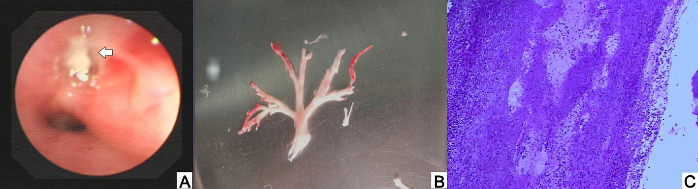
Figure 3 个
Table 2 个