-

-

-
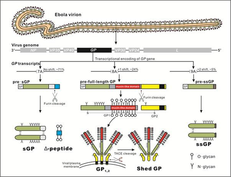
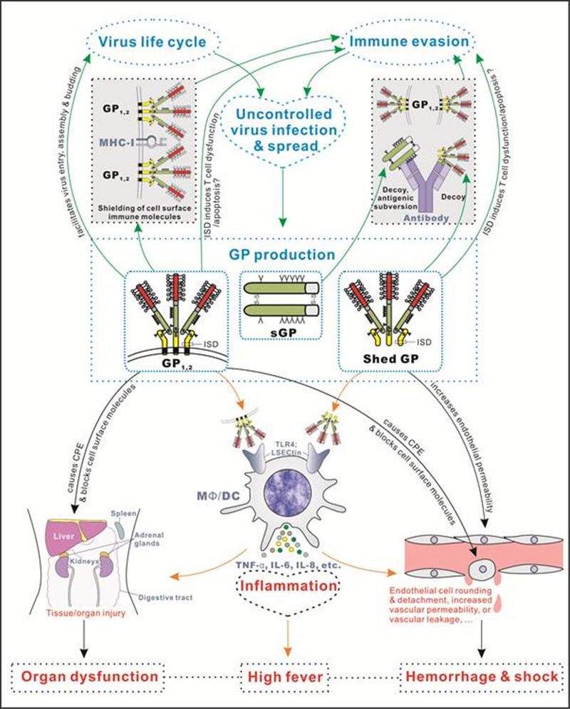
GPs Functions References Full-length GP (GP1, 2) Mediates virus entry as the virion surface spike Lee and Saphire, 2009 Promotes virus budding by antagonizing tetherin Kaletsky et al., 2009 Sterically shields the epitopes and functions of cellular surface proteins via the MLD, causing rounding and detachment of cultured cells, endothelial cell damage, leakage of explanted blood vessels, and loss of cell physiological functions (such as antigen presentation by MHC-Ⅰ) Chan et al., 2000; Takada et al., 2000; Yang et al., 2000; Simmons et al., 2002; Reynard et al., 2009; Francica et al., 2010 Sterically shields the epitopes of the GP1, 2 core via the MLD, blocking recognition by neutralizing antibodies (?) Reynard et al., 2009; Francica et al., 2010 Activates MΦ/DCs and triggers the secretion of inflammatory cytokines by the MLD (?), likely contributing to the excessive inflammation in EVD Wahl-Jensen et al., 2005a; Ye et al., 2006; Martinez et al., 2007 Contains a putative ISD, mediating T cell dysfunction/apoptosis (?) Volchkov et al., 1992; Yaddanapudi et al., 2006 Shed GP Functions as a decoy for anti-GP1, 2 antibodies, contributing to viral immune evasion Dolnik et al., 2004 Activates MΦ/DCs leading to the secretion of inflammatory cytokines; increases the permeability of HUVEC monolayers Escudero-Perez et al., 2014 Its release modulates the abundance of surface GP1, 2, likely orchestrating virus cytotoxicity, infectivity, and spread (?) Dolnik et al., 2015 Contains a putative ISD, mediating T cell dysfunction/apoptosis (?) Volchkov et al., 1992; Yaddanapudi et al., 2006 sGP Functions as a decoy of anti-GP1, 2 antibodies, or mediates “antigenic subversion”, diverting the immune response away from GP1, 2(?) Wilson et al., 2000; Ito et al., 2001; Mohan et al., 2012 Inactivates neutrophils and reverses TNF-α-induced injury of endothelial barriers, playing anti-inflammatory roles (?) Kindzelskii et al., 2000; Sui and Marasco, 2002; Wahl-Jensen et al., 2005b Assembles with GP2 as a substitute for GP1, perhaps as a structural protein (?) Iwasa et al., 2011 Δ-peptide Binds to filovirus-permissive cells and inhibits filovirus GP1, 2-mediated cell entry Radoshitzky et al., 2011 Contains an amphipathic region similar to the cytolytic peptide motif of rotavirus NSP4 and may serve as a membrane-damaging viroporin (?) Gallaher and Garry, 2015 ssGP Unknown; unlike sGP, does not display the anti-inflammatory activity that reverses TNF-α-induced damage of endothelial barriers Mehedi et al., 2011 Notes: GP, glycoprotein; GPs, glycoproteins; sGP, soluble glycoprotein; ssGP, small soluble glycoprotein; MLD, mucin-like domain; MHC-Ⅰ, major histocompatibility complex class I; MΦ, macrophages; DCs, dendritic cells; EVD, Ebola virus disease; ISD, immunosuppressive domain; HUVEC, human umbilical vein endothelial cell; TNF-α, tumor necrosis factor-α; NSP4, nonstructural protein 4; “(?)” indicates putative functions that especially require additional verification. Table 1. Summary of the known or potential functions of ebolavirus GPs
Figure 2 个
Table 1 个