


-
It was my good fortune to meet personally the three invertebrate cell culture pioneers, Richard Goldschmidt, Zan-Yin Gaw, and Thomas D. C. Grace (Fig. 1). In 1951 I met Goldschmidt at a symposium in Cold Spring Harbor, but I only knew that he was a prominent geneticist. I had no idea about his insect cell culture work at Yale University and daily contacts with Ross G. Harrison. In 1959 Zan Yin Gaw in Wuhan successfully cultured monolayers of silkworm cells for more than one year. I reported his breakthrough achievement at the 11th International Congress of Entomology in Vienna in 1960, but his work was completely ignored outside China. In 1982 Gaw invited me to Wuhan where he told me that he studied in the United States in the 1930s, working as postdoctoral scientist at the Rockefeller Institute, where he was daily meeting William Trager, and later at Yale University in the Osborn Laboratory, where he was inspired by Harrison. T. D. C. Grace worked in my laboratory at Rockefeller University during 1957 and 1958, then returned to CSIRO in Canberra, Australia. In 1962 he successfully established a cell line from the ovarian tissues of Antherea eucalypti pupae. The subsequent expansion of invertebrate cell culture involved the Nobel laureate Rita Levi Montalcini. I met her at a Growth Symposium, where I saw Harrison the last time. In 1969 she published the first of a dozen papers on in vitro studies of the embryonic nerve system of Periplaneta americana that led to her milestone discovery of the nerve growth factors. The "3 G-s" insect cell culture pioneers were directly or indirectly influenced and inspired by Harrison. Gaw was the only one who created a large following of insect cell culture researchers, who continued and expanded the work started by him in China.
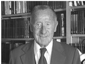
Figure 1. Provided by Karl Maramorosch
HTML
-
In 1947 I worked as a technician at the Brooklyn Botanical Garden and studied for my Ph.D. degree at Columbia University in New York City. In August, my mentor, Dr. Lindsay M. Black, suggested that I accompany him to the symposium of the Society for the Study of Development and Growth, held at Storrs, Connecticut. It was my first opportunity to attend a symposium in the United States but I did not realize that at that meeting I would be the only one without a doctoral degree. The less than 100 invited participants included famous university professors, botanists, zoologists, chemists, and physicians, and my mentor introduced me to several Nobel Prize laureates and others, whose names I knew only from the literature. I was greatly impressed by Ross G. Harrison (Fig. 2), from the Osborn Zoological Laboratory at Yale University, who, at 77 years, was the oldest scientist at this meeting. I learned that Harrison pioneered animal tissue culture in 1904 and that he was twice nominated for the Nobel Prize [12].
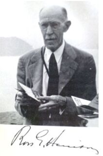
Figure 2. Provided by Karl Maramorosch
At a later Growth Symposium I met Rita Levi Montalcini for the first time, who came from Italy to the United States to work with Victor Hamburger at Washington University in St. Louis, Missouri. I was greatly impressed by her scientific presentation as well as with her charming personality. A few years later Rita devoted her time to the study of tissue culture of Periplaneta americana [14].
The name of Richard Goldschmidt was known to me from my study of genetics at the Agricultural University in Warsaw in 1935. There Prof. Malinowski published his text, in Polish, listing some English terms for the eye color of Drosophila. I learned my first English words, including the word "yellow", from Malinowski's book. When I met Goldschmidt the first time, at a Cold Spring Harbor symposium in June of 1951, I did not know that he pioneered invertebrate tissue culture, or that I would become interested in this subject.
In 1956 I demonstrated that aster yellows phytoplasma could complete its intrinsic incubation in insect tissues in vitro [10]. The entomologist Max. F. Day, who at that time was Australia's scientific attache in Washington, DC, visited my laboratory at Rockefeller University, and suggested that I invite one of his Australian associates, Thomas D. C. Grace, who had just published, in Nature (London), his first attempts to culture invertebrate cells [4]. I obtained a grant from SPH National Institutes of Health in Bethesda, MD, which supported Grace for the following 2 years, 1957-1958. Together, we travelled to visit Harry Eagle at NIH in Bethesda, Maryland. At Rockefeller University we often visited William Trager, who already in 1935 grew the virus of grasserie in silkworm tissue culture [17]. At that time I became interested not only in insect cell culture carried out in different laboratories, but also in the origin of research ideas and interactions between scientists.
-
Goldschmidt was born in Frankfurt-am-Main, Germany to a wealthy old German-Jewish family (Fig. 3). His interest in biology started at 13, and by the time he was 18 he decided to become a biologist. He first enrolled at Heidelberg University as a medical student, two years later went to Munich to study zoology, then returned to Heidelberg where he received his Ph.D. degree. He started compulsory army service in the German army where an officer's commission was withheld because of his Jewish background. Similar experiences throughout his life in Germany had an impact on his personality. He returned to Munich University and remained there till 1914. At the age of 29 he founded the Archiv fur Zellforschung and in 1911 his first textbook on genetics appeared. Working with Lymantria dispar, he crossed the European moth with isolates from Japan and obtained normal males, but abnormal females that were sex integrates. The results were outside the accepted laws of Mendelian genetics and they formed the basis for Goldschmidt's balance theory of sex determination.
Figure 3. Provided by Karl Maramorosch
In 1914 Goldschmidt was appointed as a member of the newly created Kaiser Wilhelm Institute in Berlin and at the same time he obtained a fellowship to Japan to collect races of Lymantria. The outbreak of World War I prevented his return to Germany and he came to the United States, where he began working at the Osborn Zoological Laboratory at Yale University in New Haven, Connecticut. There, under Ross G. Harrison's influence, he decided to attempt the cultivation of insect cells and thus became the pioneer of invertebrate cell culture. He obtained the spermatogenesis of the Cecropia moth in vitro and published his first paper on this subject, entitled "Some experiments on spermatogenesis in vitro " [2]. The following year his German paper was published in Biologisches Zentralblatt [3]. Goldschmidt's work ended abruptly, when an incident occurred on the night of May 1, 1918.
Graduate students who lived in the Osborn Tower were awakened by a Military Intelligence officer who wanted to know why lights were burning in certain rooms. The rooms were those of Goldschmidt's laboratory and the incident gave rise to rumors that Goldschmidt was sending signals to German submarines off the coast of New Haven. Many accepted this version even though the windows were at the rear of the Osborn Laboratory and the lights could not be seen from the harbor. Goldschmidt was interned at Fort Oglethorpe in Georgia. After the war he was released, returned to Germany, and continued his work on genetics. He advanced at the Kaiser Wilhelm Institute and became its president but in 1936 he was removed from his position and forced to leave. He was offered a professorship at the University of California in Berkeley and he described the event as one of the happiest in his life. In 1942 he became a United States citizen and in 1947 he was elected to the U.S. National Academy of Sciences. His theory of evolution and his outdated concepts of genes and alleles were not appreciated by modern geneticists. When I met him the first time in 1951, he was invited to deliver the opening address at the Cold Spring Harbor Symposium on Chromosomes and Genes. At that time I knew nothing about his pioneering work on insect tissue culture. At the symposium I was sitting between Max Delbruck and Joshua Lederberg, listening to the presentation of Barbara McClintock, who, described her "jumping genes" findings. When she finished, only Goldschmidt favorably commented and congratulated her. One of the two future Nobelists next to me, either Delbruck or Lederberg, quietly said: Goldschmidt is already senile. It took more than 30 years, till 1982, before Barbara McClintock received the Nobel Prize for the work that in 1951 only Goldschmidt correctly judged and appreciated.
In September 1953 I saw Goldschmidt the last time, when I attended the 9th International Congress of Genetics in Bellagio, Italy, where Goldschmidt was honored as the President of the congress. Shortly thereafter he suffered a severe heart attack. Despite his illness, he was able to write an extensive treatise, "Theoretical genetics". Two years later he died. I was wondering why Goldschmidt never returned to insect cell culture. He impressed me as a typical European professor, who issues orders to his assistants, but does not himself work at the bench. Did he work by himself at the Osborn Laboratory or did he have an assistant? The following is my hypothesis which could not be confirmed, because the person, who worked at the laboratory at that time and later became a world-famous cancer cell culture expert, never mentioned that she was assisting Goldschmidt. In 1915, when Goldschmidt started insect tissue culture at the Osborn Laboratory, a German parasitologist, Anna Maria Rhoda Erdmann, was also working there [13].
-
The war hysteria was mounting between 1915 & 1918 and anything German was anathema. In 1918 Erdman was arrested and taken incommunicado to Hartford, accused of poisoning the New Haven drinking water and of introducing chicken cholera to the United States. Jailed until February 1919, she was finally released and permitted to leave for Germany. Erdman became Europe 's most prominent cell culture researcher. She organized the first, second, and third European tissue culture congresses and she created the section of Cell Research at the Cancer Institute in Berlin. After the Nazi takeover, when Goldschmidt was forced to leave the Kaiser Wilhelm Institute and left Germany, Erdman was jailed for 2 weeks while the Gestapo searched her institute and her apartment. Her laboratory was permanently closed and she was forced to retire. Erdman was not Jewish, but the Gestapo suspected at first that she, her parents, or her grandparents were. Perhaps they knew something about her years at the Osborn Laboratory or her association with Goldschmidt? Dr. Erdman died in 1935 at the age of 64. In 1992, the German section of the European Tissue Culture Association created a research fund named for Rhoda Erdman. I suspect that Erdmann participated in Goldschmidt's tissue culture work and thus was the first woman pioneer of invertebrate cell culture, although Goldschmidt never mentioned her name.
-
William Trager received his BS degree at Rutgers University in 1930 and his Ph.D.degree at Harvard in 1933 (Fig. 4). He came to the Rockefeller Institute branch in Princeton, NJ in 1933 and worked at Rockefeller University for the remainder of his life. In 1935, drawing on the experience of Rudolf W. Glaser with axenic culture techniques, Trager grew the grasserie virus in silkworm tissue cultures [17]. He observed the multiplication of the polyhedral bodies in cultures containing cells that grew from the lining of the ovariole of the silkworm. Trager's medium and the technique were the basis for all further work on insect tissue culture. The classical work of Trager in 1938 showed that equine encephalomyeliltis virus multiplied in tissue cultures of mosquitoes for 28 days. The titer of virus was 100, 000 times higher than the original suspension [18].
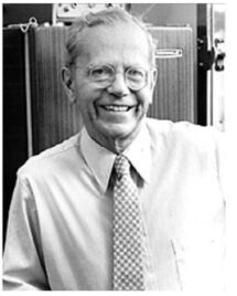
Figure 4. Courtesy of William Trager to Karl Maramorosch
Trager's most important work, carried out in later years, concerned the cultivation of the human malaria parasite Plasmodium falciparum. His influence on invertebrate cell culturists had a lasting effect around the world. In 1956, when I started my first trial to culture leafhopper tissues, I was influenced and advised by Trager. I followed his classical experiments in which he achieved the multiplication of equine encephalitis virus in hanging drop tissue culture of mosquito vectors. I cultured pieces of leafhopper nymphs Macrosteles fascifrons, exposed previously by feeding on plants infected with the aster yellows phytoplasma. After maintaining the cut pieces in the culture medium for 10 days, I was able to recover the phytoplasma from the cultured tissues. Using this method, I demonstrated the multiplication of the phytoplasma in cultured vector tissues for the first time [9].
At that time a conference was arranged in Canada by Paul Weiss, who was secretary of the US National Academy of Sciences. The assembled participants concluded that insect cells would never be grown like cells of vertebrate animals, because, as determined earlier by Trager, the entire growth of the insect larva results from increase in size, not in number of the cells composing the larval tissue. Trager found that some tissues grew by increase of cell size only, but others by cell division. Only the first of Trager's findings was considered by the conference participants. Fortunately, their conclusion did not stop progress in invertebrate cell culture.
-
The first successful cultivation of cells in monolayers was reported in 1959 by Gaw Zan-Yin (Fig. 5), Lien Nien Tsui and Zia Tien Un from the Wuhan Laboratory of Microbiology of the China Academy [1]. Although the paper appeared in English in Acta Virologica 3, Supplement: 55-60, it was ignored, except for my extensive presentation of it at the XI International Congress of Entomology in Vienna in 1960 [10].
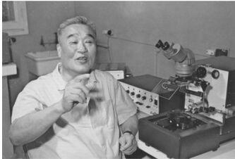
Figure 5. Provided by Prof.Yuanyang Hu
I reported that Gao et al used a medium composed of 90% of Trager's solution (A) and 10% of healthy silkworm serum. Its initial pH was 6.7. This silkworm serum was obtained by bleeding full-grown silkworms aseptically from the leg, centrifuging the blood at 2000 rpm for 10 min and adding penicillin and streptomycin to the final solution. The cells were separated by trypsinization, using bacto-trypsin.
A variety of silkworm tissues, such as male and female gonads, muscle, trachea, silk-gland and intestine were used. For silk gland tissues young third instar larvae were used. These had small glands that contained no silk fluid. In the case of intestine, the larvae were starved for 36 h before use and the tissue was immersed in mercuric chloride solution for 15 min, and then washed 3 times in sterile distilled water. The various tissues were washed, explanted and stored separately in Petri dishes or test tubes throughout the experiments. Tissues were cut and then washed by centrifugation in Trager's solution.
A modification of the hanging drop method, the cover slip method, was applied first. Tissue fragments were fastened to the surface of cover glasses with a droplet of serum and incubated at 36℃ for 10 min. Then a drop of nutrient solution was added to each cover slip. In this way, very small quantities of material were needed for the cultures, and it was easy to observe the growth through a microscope.
In other experiments pieces of tissues were fastened with serum to stationary test tubes, then medium was added and the test tubes stoppered and held in a slightly inclined position to permit the nutrient solution to cover the fragments. The cultures were incubated at 26-27℃. Low power microscopic observations of these cultures were possible.
Two methods were used to prepare monolayer cultures. The first was a cell suspension culture, in which tissues were first cut into small pieces, then centrifuged at 3000 rpm for 20 min with 5 mL of Trager's solution A. After the supernatant had been discarded and the sediment resuspended in Trager's solution by pipetting for 20 min, the material was left to settle for 15 min. During this time larger particles settled at the bottom of the tube, and the supernatant consisted mostly of a single cell suspension. The concentration of the suspension was determined by a haemocytometer. Nutrient solution was added to make 6 x 10, 000 cells per mL and this was placed in Carrel flasks. The second method for the preparation of monolayers involved cells obtained by trypsinization. In these experiments trachea, muscle and silk-gland tissues were successfully used. The tissues were cut into small pieces, washed in Trager's solution, covered with trypsin and held in a water bath at 26 ℃ for 15 min. Clumps were broken up by vigorous pipetting. The cultures were maintained for 7-10 days by changing the medium at intervals of 3-4 days. After removal of the medium the cultures were twice washed in Trager's solution A, and then fresh medium was added. Subcultures succeeded only with ovarian and testicular cell cultures. After culture tubes had been incubated at 26 ℃ for 2-3 days, the cells formed a continuous sheet and the cell population was high enough for subculturing. The cells were trypsinized with 5 mL of a 0.25% trypsin solution in Trager's solution A in a water bath at 26 ℃ for 15 min, until the cell sheet was broken up and the cells became detached from the glass surface. Then clumps were broken up by pipetting 20 to 30 times.
In most cultures new cells began to grow after 24 h of incubation. The maximum amount of growth was attained on the 3rd or 4th day. By changing the nutrient medium, cultures could be maintained for 7-10 days (with 2 changes of the medium). Cells that grew from various tissues differed in shape and size. For instance, in muscle tissue cultures, wandering cells first appeared from the connective tissues under the skin on the first day of incubation. Two days later muscle cells began to grow, while the wandering cells became more numerous. In female gonads wandering cells grew out from the ovary sheath.
Monolayers of insect cells were obtained from male and female gonads, trachea, muscle, intestine and silk-gland tissue cultures. These monolayers were formed after 24 h of incubation. In monolayers of muscle cells the shape of fibroblasts predominated, while in others rectangular epithelial cells were formed. Other types of cells must have been present in these cultures, but they were generally outnumbered by the fast growing epithelial cells or fibroblasts.
Gaw used his cultures for virus work. After the cultures were inoculated with grasserie virus, studies of cytological changes were made. The continuous cell sheet broke up into irregular patches and fragmentation of the cells took place. The female gonad epithelial cells became more or less rounded while the trachea cells maintained their rectangular shape. Virus infection caused a tremendous enlargement of the cell nuclei which consequently moved to an off-center position. The cells became isolated and eventually degenerated. Changes were pronounced in the nuclei, which changed into horseshoe, ring or half-moon shapes and then polyhedra began to appear in them. Generally one to four polyhedra were found in each nucleus, but sometimes many more were formed, filling the nucleus completely. Finally the cell burst, setting the polyhedra free in the culture medium. These polyhedra had the shape of dodecahedra.
The great success of Gaw's work can be seen from the fact that he was able to maintain subcultures of male and female gonad cells for twenty-two generations in one year, and continued to maintain these cultures without encountering difficulties. The cells of subcultures were normal in appearance. In the area of Wuhan where these investigations were made, silkworm could only be raised twice a year in natural conditions, and therefore Gaw's technique permitted continuous studies of the virus disease without raising silkworms.
Why did Gaw succeed to grow insect cells continuously while workers elsewhere had only partial success? I discussed this in 1960 with Trager at Rockefeller University. Trager speculated that perhaps the difference was due to the use of a different strain of silkworm. Trager did not remember that he and Gaw knew each other when Gaw was working in Wendel M. Stanley's group at the Princeton laboratory next door to Trager, and that they were eating lunch in the same room every day.
When I reported Gaw's work at the Entomology Congress in Vienna [11], Prof. Gershenson, from Kharkov, U.S.S.R., called my attention to the work, carried out by his co-worker N. B. Medvedeva. She obtained good results with tissue cultures using cells from ovaries and testes as well as blood cells of insects. In 1959 she published, in Russian, about the multiplication of a polyhedral virus in insect tissue cultures, and on the cultivation of insect tissues in vitro [15, 16]. Her papers, like Gaw's, were completely overlooked and, unfortunately, forgotten. Gershenson's remark stressed my awareness of the importance of attending international congresses and meeting with colleagues from different laboratories and different countries.
I was fortunate to meet Prof. Gaw in Wuhan in 1982. Only then did I find out that he studied in the United States, worked in Stanley's laboratory of the Rockefeller Institute in Princeton, where he met every day both Glaser and Trager, and that he also worked at the Osborn Laboratory at Yale University where he knew well Ross G. Harrison. The influence of Harrison and Trager apparently affected the progress of invertebrate cell culture.
-
In 1956, M. F. Day, an Australian entomologist from CSIRO, Canberra, who worked in Washington as scientific attaché, suggested that I invite Thomas D. C. Grace (Fig. 6) to my laboratory at Rockefeller University, to try to grow insect cells in vitro. In Canberra Grace had already tried to grow the cells of silkworms, mosquitoes, Drosophila, and cockroaches in hanging drop cultures and plasma clots of chick embryo extract and various salt solutions, which have been formulated for use in insect physiology and biochemistry. To formulate his cell culture medium, Grace first used the Australian Emperor gum moth, Antherea eucalypti, believing that the medium must contain hemolymph to support the growth of cells. He also obtained hemolymph from Antherea pernyi from Japan. 15, 000 pupae supplied 30 liters of hemolymph.
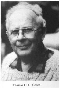
Figure 6. Provided by Karl Maramorosch
When Grace came to my laboratory, he worked first on developing the medium that became the famous medium for insect tissue culture for years to come. He added to the Silver-Wyatt [19] medium cholesterol and 10 members of the vitamin B complex, and reduced the concentration of the blood from 10 to 5%. In this modified medium ovarian tissues of 3 species of Lepidoptera could be maintained alive almost indefinitely [5]. After his return from Rockefeller University to Australia, he continued his work on insect tissue culture, establishing some strains of cells from Antherea eucalypti and the ovarian tissue of the cabbage white butterfly Pieris rapae. In the beginning it seemed no virus could be found in nature in Antherea. Then, during the warm period in the middle of January, some larvae started to die. Grace found that they were killed by a cytoplasmic polyhedrosis virus.
For a very long time it was not possible to maintain cell multiplication of Antherea at a continuously high rate. Contraction of ovariole muscles continued and initially Grace was discarding the cultures after about 3 months. Eventually, however, he decided to keep them in, what he described "benign neglect", changing half of the medium from time to time. After more than a year, a rapid increase in cell number and cell divisions occurred. Grace then subcultured the insect cells every 6 days. What caused the cells, after nearly a year in culture, to suddenly start to divide rapidly? Grace found that the number of chromosomes was much higher than the diploid number of the original cells. Some cells had 128n chromosomes [6]. All of the first 4 lines established at that time were polyploidy, and when transplanted back into their hosts they would form tumors. Over the next 3 years Grace established cell lines from mosquitoes, Aedes aegypti [7] and silkworm [8]. Both were maintained in continuous culture and in liquid nitrogen for many years.
While Grace became well known and admired in Europe, Asia and America, he was hardly appreciated in Australia, where Day, his former chief, became the executive of CSIRO. Frustrated, Grace retired, purchased a farm in Broulee, NSW and with two sons started to grow native Australian flowers. After a few years, the flowers grown by Grace became very popular, often replacing roses and carnations at weddings, and are currently appearing at the flower auction in Aalsmere, Netherlands, flown from Grace's farm to Europe.
-
Rita Levi Montalcini was born in Turin (Fig. 7), Italy on April 22, 1909. She studied medicine in the early 1930s at Turin University, where she was a colleague and friend of Renato Dulbecco and Salvador E. Luria. Years later all three were awarded Nobel Prizes. At 103 years, Montalcini is the longest lived Nobel laureate in history.
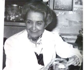
Figure 7. Courtesy of Rita Levi-Montalcini to Karl Maramorosch
I met Rita Montalcini the first time at a symposium of the Society for Growth and Development, to which she came from Victor Hamburger's laboratory at Washington University in St. Louis. Missouri. She told me that in the beginning of her career she was under the spell of Giuseppe Levi, and later under the influence of Victor Hamburger, who channeled her interest toward problems of growth and differentiation of nerve cells. I learned from her that she became a research scientist rather than a practicing physician due to the influence of her anatomy professor at Turin, Giuseppe Levi [14]. He influenced not only Rita, but also Dulbecco and Luria to devote their life to scientific research. Rita's study of nerve cells in vitro followed Levi's 1928 work, and Levi followed the early work of Ross G. Harrison. Years after meeting Rita, I found out that her tissue culture activity was not solely devoted to the study of the nerve growth factor, for which she, together with Stanley Cohen, received the Nobel Prize in 1972. Rita prepared whole mount cultures and dissociated nerve cells of Periplaneta americana [9] Her long term cultures of dissociated nerve cells of the cockroach were discontinued because of the length of time the cultures required, that made it impossible for her to pursue at the same time her studies of the nerve growth factor in vivo and in vitro.
Rita told me how, though Jewish, she survived World War Ⅱ. In 1938 Mussolini issued his anti-Jewish manifest that barred all academic and professional careers for Jews. Rita decided to build a small laboratory in her bedroom where she assembled a microscope, a microtome, and an incubator. Sewing needles, sharpened by hand on a stone, provided her micro-instruments. In 1942, after a heavy bombing of Turin, she moved to a small country home where she rebuilt her laboratory. In 1943 Italy was occupied by Nazi Germany and Italian Jews became the object of mass killings and deportation to the gas chambers at Auschwitz. Rita escaped to Florence where she hid with false identity cards till the end of the war. In 1945 she returned to Turin and a year later, in 1947, she and Renato Dulbecco sailed from Genoa to the United States, where Rita accepted an invitation from Victor Hamburger to work in his laboratory.
-
I have mentioned the most important highlights or milestones in the early study of invertebrate cell culture and shown their significance in the development of insect cell culture. Although Goldschmidt was the first to attempt to culture insect tissues in vitro, he did not return to this work after leaving Yale University. The first important advance began with Trager's studies in the mid-1930s. He carried out a series of studies which were primarily concerned with the multiplication of viruses in cells and not specifically the growth of the cells themselves. One of the problems encountered by Trager was the difficulty of maintaining sterility. There were no antibiotics known at that time. Elaborate precautions had to be taken to ensure sterility and, even then, the majority of cultures would be contaminated within 24 h. Another difficulty was the preparation of the culture media. Today many can be purchased commercially, but the early cell culture workers had to formulate their media before antibiotics and fetal calfserum became available.
The influence of Ross Granville Harrison and personal contacts with him and with Trager have stimulated directly or indirectly the pioneers, who were able to overcome great difficulties and achieve success. Of the three G's, only Gaw, with his brilliant work, created a large following of insect cell culture researchers who established the world's foremost center of in vitro invertebrate cell biology studies in Wuhan, It was my privilege to meet personally the early pioneers, who stimulated my own work and influenced my desire to attend scientific meetings and congresses. I believe that science recognizes no political, religious, ethnic, or geographic borders and we, scientists, speak only one language -the language of science.

 DownLoad: