


-
Dear Editor,
Since April 2010, an outbreak of a new disease has elicited symptoms of high fever, loss of appetite, and reduction in egg production in layer ducks in eastern China; this phenomenon has now spread throughout China (Cao et al., 2011; Su et al., 2011). The causative agent of the disease was identified as Tembusu virus (TMUV), which was classified into the genus Flavivirus, family Flaviviridae. The TMUV virion is enveloped and contains a 10, 990 nt single-stranded, positive-sense RNA genome which contains a long open reading frame. The reading frame encodes a polyprotein that is possessed into three structural (Capsid, prM and E) and seven non-structural (NS1, NS2A, NS2B, NS3, NS4A, NS4B, and NS5) proteins by viral-encoded proteases (Tang et al., 2012; Yun et al., 2012). TMUV isolated from poultry (including chicken, duck and goose), field birds, and mosquitos, can trigger neurological symptoms and even death in mice (Liu et al., 2012; Li et al., 2013a; Liu et al., 2013; Tang et al., 2013; Tang et al., 2015). To date, the elements of TMUV required for viral replication and virulence have not been definitively determined. Reverse genetics is an effective molecular biology technique to determine the function of genomic elements responsible for the replication and virulence in positive-strand RNA viruses. Infectious cDNA clones and recombinant progeny of flaviviruses including Japanese encephalitis virus, West Nile virus, Dengue virus, Yellow fever virus, Tick-borne encephalitis virus are available. There are two major strategies to construct infectious cDNA clones of animal viruses. For example, a duck TMUV infectious cDNA clone that was transcribed into RNA via in vitro transcription has been reported (Li et al., 2013b). However, no infectious cDNA clones based on a DNA-launching strategy have been constructed and produced for progeny virus.
In our study, we report and characterize the first full-length infectious clone of the TMUV genome from which infectious RNA can be transcribed in vivo. The viral proliferation and pathogenicity of progeny virus were examined and compared with those of parental virus.
A TMUV strain ZC-1 isolated from breeder ducks in Shandong province in 2012 was utilized for construction of a full-length TMUV cDNA clone. The genomic sequence of ZC-1 (GenBank accession no. KF557894) has been reported previously (Chen et al., 2013). The virus stock was inoculated into the chorioallantoic cavity of 9-day-old embryonated duck eggs and then propagated in BHK21 cells. Viral RNA was extracted from virus allantoic fluid as described previously. The low-copy number plasmid pWSK29 was modified to replace the multiple cloning sites between Apa Ⅰ and Not Ⅰ restriction sites. The introduced sequence included Apa Ⅰ, Aat Ⅱ, Xho Ⅰ, Nhe Ⅰ, EcoR V, Hind Ⅲ and Not Ⅰ to facilitate the cDNA fragments of TMUV. It was difficult to construct flavivirus infectious clones because the viral genome did not stably replicate in Escherichia coli. Six overlapping fragments of TMUV-ZC-1 cDNA were amplified by RT-PCR using Ex Taq DNA polymerase. The 5′-terminal region was amplified using a forward primer that contained a T7 polymerase promoter sequence (TAATACG ACTCACTATAGG) and an Apa Ⅰ restriction site, and the 3′-UTR terminal region was amplified using a reverse primer-linked T7 terminator and Hepatitis D virus ribozyme (HdvRz) sequence to halt transcription in vivo. All six fragments were cloned into the pMD18-T vector, then transformed into DH5α competent cells, and positive clones were sequenced by Sanger sequencing (Invitrogen, Shanghai, China). The strategy for assembly of a full-length cDNA clone of TMUV was as shown in Figure 1A. Firstly, F1, F3, and F5 were double digested using appropriate restriction endonucleases and inserted into pWSK29, which was digested with the same enzymes to create P1, P3, and P5. Then, F2, F4, and F6 were digested and cloned into P1, P3, and P5, respectively to create P1-2, P3-4 and P5-6. Afterwards, P3-4 was digested with Xho Ⅰ and EcoR Ⅰ. The minor band was gel purified and inserted into P1-2 digested with the same enzymes to create P1-4. Finally, P5-6 was digested with EcoR Ⅰ and Not Ⅰ and cloned into P1-4, which was cut with the same enzymes to obtain the full-length cDNA clone designated as pWSK29-TMUV. All these recombinant plasmids were transformed into E. coli DH5α competent cells, and the bacteria were cultured in LB medium containing 30 ng/mL ampicillin at 25℃ to reduce the replication of mutated recombinant plasmids. To confirm the identity between infectious cDNA sequence and TMUV-ZC-1 viral genomic cDNA sequence, the recombinant plasmid was further sequenced. Replacement of three nucleotides resulted in sense mutations: A to C (nt 2058) resulted in a Q→P change in E protein; and T to C (nt 5385) resulted in an L→S change in the NS3 protein; and T to G (nt 9708) resulted in an F→C change in the NS5 protein. Plasmids were extracted from several passages of E. coli and sequenced, and no nucleotide mutations were detected. These data indicate that the full-length cDNA clone was stable amplified in E. coli. Thus, the nucleotide mutation at nt 5385 (T to C) was used as a genetic marker for the infectious clone.
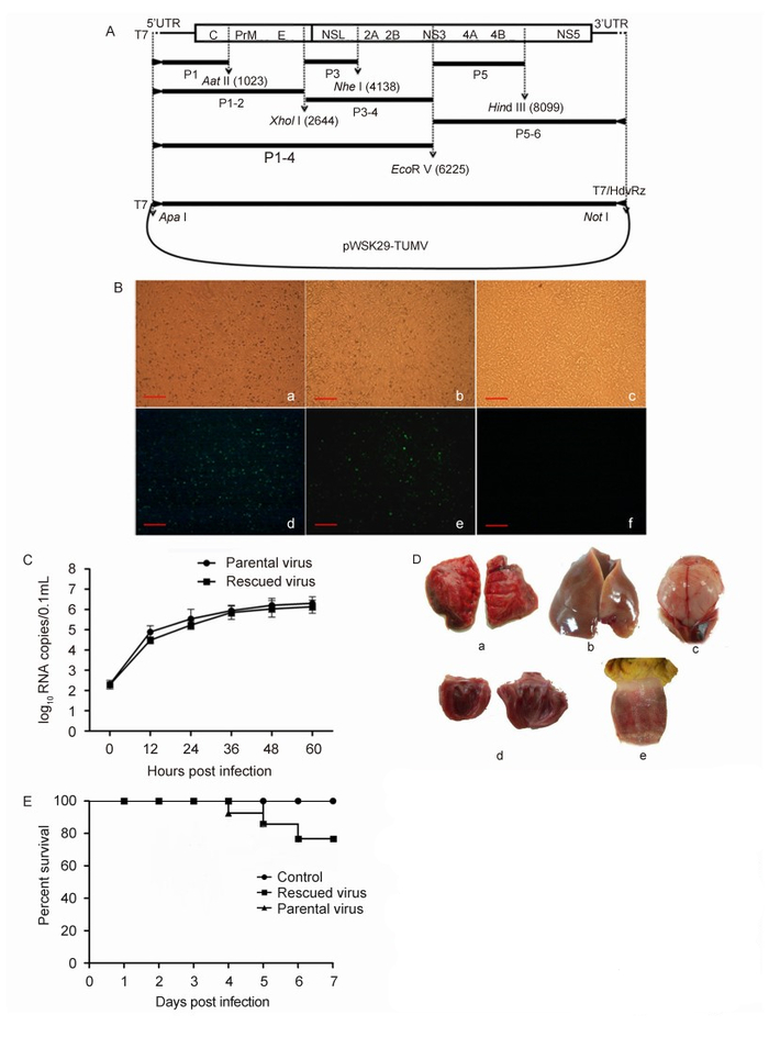
Figure 1. (A) Schematic diagram of the strategy for assembly of the TMUV ZC-1 cDNA clones using the low-copy-number plasmid pWSK29. The T7 RNA polymerase promoter and Apa Ⅰ restriction enzyme site upstream of 5′-terminus of viral cDNA and T7 RNA terminator, Hepatitis D virus ribozyme and Not Ⅰ restriction enzyme site downstream of 3′-terminus of viral cDNA used for plasmid construction are indicated. (B) Micrographs and indirect immunofluorescence assays of infected and control BHK-21 cells: (a, b) parental virus infected cells; (c, d) rescued virus infected cells; (e, f) mock-infected cells (Scale bar: 100 μm). (C) Comparison of the growth of parental and rescued viruses in BHK-21 cells. (D) Pathological changes in ducks infected with rescued Tembusu virus. (E) Survival curves of the inoculated 1-day old ducklings.
The recombinant plasmid was extracted using the E.Z.N.A. Endo-Free Plasmid Mini Kit Ⅱ, and BHK-21 cells stably expressing T7 RNA polymerase were established. BHK-21 cells were subsequently transfected with 0.8 μg pWSK29-TMUV using Lipofectamine 2000 reagent according to the manufacturer's instructions. Five hours later, the supernatant was discarded and Dulbecco's modified Eagle's medium (DMEM) supplied with 2% FBS was added. The cells were incubated at 37℃ and monitored for 5 days. After three additional passages, the cell supernatant was harvested and identified by RT-PCR using a pair of primers based on the NS3 gene sequence. The viral E protein was detected by an indirect immunofluorescence assay using mAb 12B1 (anti-TMUV E protein) as the primary antibody (Chen et al., 2014). The cells were cultured for 48 h and fixed with pre-chilled methanol and acetone (1:1) mixture at -20℃ for 10 min, then washed with PBS and incubated with 12B1 (1:100) at 37℃ for 1 h. Finally, FITC conjugated goat-anti-mouse IgG (1:500) was added and the green fluorescence was visible in both ZC-1 strain and progeny virus-infected cells (Figure 1B, panels b and d).
TMUV infection had relatively little cytopathic effect in BHK21 cells (Figure 1B, panels a and c). A quantitative TaqMan-based real-time PCR assay was established and utilized to evaluate the kinetics of viral replication of the progeny virus in BHK 21 cells. PCR primers (forward primer: 5′-GAGTGAACTCATCATACCTA-3′; reverse primer: 5′-GGAACTCTGAACCTTGTA-3′) and TaqMan probe (5′-FAM-ACCGAAGAGTCATCACAACACGA-TAMRA-3′) were designed based on NS3 genes and the length of amplified fragment was 87 bp. BHK21 cells were cultured in 24-well plates and infected with 10 MOI doses of progeny virus and parental virus. After a 1 h adsorption, cell culture supernatant was discarded and DMEM supplied with 2% FBS was added. Culture supernatant (0.1 mL) was harvested at 0 h, 12 h, 24 h, 36 h, 48 h, and 60 h post infection. RNA extracted from each sample and treated with DNAse I, was reverse-transcribed to cDNA using M-MLV reverse transcriptase. Viral genomic RNA copies were calculated according to the standard curve and the threshold cycles. The results demonstrated that the growth curve of progeny virus was similar to that of parental virus (Figure 1C).
The rescued virus was inoculated into chorioallantoic cavities of 9-day-old duck embryos for three passages. The ELD50 of rescued virus and parental strain were 10-3.8/mL and 10-3.6/mL, respectively, calculated by the Reed-Muench method(Reed and Muench, 1938). To evaluate the pathogenicity of these isolates, forty-five 5-day-old ducklings free of TMUV or antibody were inoculated separately with the rescued virus, the parental ZC-1 strain or PBS buffer and cultured in isolates. The animal procedures were approved by the Animal Care and Use Committee of Shandong Agricultural University and ducklings were euthanized in CO2. All ducklings were monitored for 10 days post-challenge, during which time the ducklings in the PBS group were all healthy. Infected ducklings displayed similar symptoms to those that accompany pathological changes that follow TMUV infection (Figure 1D). The viral RNA was extracted from lungs and brains of dead ducklings and utilized as templates for RT-PCR amplification (Tang et al., 2012). A 406-bp fragment was amplified and sequenced. The results confirmed that the rescued virus and parental strain were detected in ducklings, respectively. No more death occurred until seven days post-challenge (Figure 1E). In addition, the recombinant plasmid was amplified in E. coli for ten passages, and plasmids from the fifth and tenth generations were transformed into BHK21 cells. Finally, virus was detected in both cell cultures.
In conclusion, we have constructed a stable TMUV cDNA clone using a low-copy number plasmid. The infectious RNA was transcribed in vivo and the progeny virus was successfully rescued. The biological and biochemical properties of rescued virus were similar to those of the parental strain. Our TMUV infectious clone provides a more concise tool for investigating the construction and function of viral genome sequence and the function of viral-coded proteins using structure prediction software, as well as offering an advance in mutation technology.
HTML
-
This work was supported by the National Natural Science Foundation of China (no. 31272583, 31472199) and China Agriculture Research System (CARS-43-10). The low-copy number plasmid pWSK29 was provided by Prof. Dabing Zhang (China Agricultural University). All animal experiments were performed in accordance with the "Guidelines for Experimental Animals" of the Ministry of Science and Technology (Beijing, China). All the authors declare that they have no competing interests.

 DownLoad: