-
Dear Editor,
Rift Valley fever (RVF) is an anthropozoonosis caused by Rift Valley fever virus (RVFV). RVFV belongs to the Phlebovirus genus in the family Bunyaviridae, which is circulating among ruminants. Human infection with RVFV is generally asymptomatic, however, minority of patients develop severe RVF diseases like encephalitis or hemorrhagic complications, with a fatality rate up to 50%. RVF mostly distributed in eastern and southern Africa, including Kenya, Zimbabwe, Zambia, Nambia and Somalia (LaBeaud et al., 2011; Shivanna et al., 2016). Infected cases were also reported in Egypt, Saudi Arabia and Yemen (Ikegami and Makino, 2011). Large outbreaks of RVF occurred in Kenya, Madagascar and South Africa with rapid spread (Jean Jose Nepomichene et al., 2015). In recent years, the import and export trade of animals increased remarkably along with the de-velopment of transportation and tourism, which raise the risk of RVFV transmission. The epidemic in Niger caused wide spread concern since the first case in August, 2016. With regards to China, RVFV had not been reported until July 23, 2016, when a Chinese worker returning from Angola was diagnosed with RVFV infection and was confirmed to be the first imported case of RVF in China (World Health Organization, 2016).
Here, we describe the RVFV isolation in serum sample and its complete genome from the first imported case to China. The 45-year-old case returning to China from Angola showed symptoms of fever (38.8 °C), chills, headache, arthralgia, anorexia and enervation on July 13 and returned to China for treatment on July 21 through the port of Beijing Airport. Because of yellow fever outbreak in Angola, differential diagnosis of more than ten pathogens in the lab were carried out, including yellow fever virus, dengue virus, Chikungunya virus and Plasmodium. The real-time fluorescent RT-PCR was performed with specific primers (RVFVf: GAAAATTCCTGAGACAC ATGGCAT; RVFVr: TTCCACTTCCTTGCATCATCTGA) and probe (RVFVp: FAM-CACAA- GTCCACACAGGCCCCTTACATT-BHQ1) to detect RVFV, and the serum sample was determined to be RVFV positive.
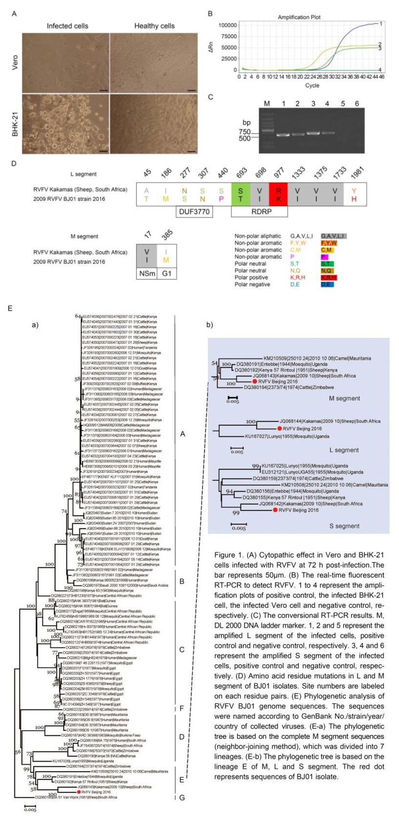
RVFV isolation and culture identification were done in biosafety level 3 lab of Guangdong Inspection and Quarantine Technology Center. Vero and BHK-21 cells were used for virus isolation from serum according to the reference (Barzon et al., 2014). The pre-treated serum sample was inoculated into monolayer Vero and BHK-21 cells in six-well plate. Cells were cultured at 37 °C in 5% CO2 for up to 7 days. Obvious cytopathic effect (CPE) was observed on Vero and BHK-21 cells 72 h post-inoculation. Vero cells showed CPE of swelling, particle increasing, disruption and necrocytosis, with BHK-21 turning round, shrinkage and rupture (Figure 1A). The supernatant of infected Vero and BHK-21 cells was collected 5 days post inoculation and viral RNA was extracted. The real-time fluorescent RT-PCR and RT-PCR were performed to detect RVFV proliferation in cells (Figure 1B, 1C). RT-PCR showed two amplified bands of 564 bp for L segment (RVFL-FP: GGATCACATATCTGAATTTCTCTCT; RVFL-RP: CAAGTCACCAACAAAGTTCCAG) and 635 bp for S segment (RVFS-FP: CTGAACATCTGGGATTGGAG; RVFS-RP: CCCTGTTGTGTCTTTCTCAT) (Figure 1C).

Figure 1. (A) Cytopathic effect in Vero and BHK-21 cells infected with RVFV at 72 h post-infection.The bar represents 50pm. (B) The real-time fluorescent RT-PCR to detect RVFV. 1 to 4 represent the ampli¬fication plots of positive control, the infected BHK-21 cell, the infected Vero cell and negative control, re¬spectively. (C) The conversional RT-PCR results. M, DL 2000 DNA ladder marker. 1, 2 and 5 represent the amplified L segment of the infected cells, positive control and negative control, respectively. 3, 4 and 6 represent the amplified S segment of the infected cells, positive control and negative control, respec¬tively. (D) Amino acid residue mutations in L and M segment of BJ01 isolates. Site numbers are labeled on each residue pairs. (E) Phylogenetic analysis of RVFV BJ01 genome sequences. The sequences were named according to GenBank No./strain/year/country of collected viaises. (E-a) The phylogenetic tree is based on the complete M segment sequences (neighbor-joining method), which was divided into 7 lineages. (E-b) The phylogenetic tree is based on the lineage E of M, L and S segment. The red dot represents sequences of BJ01 isolate
The viral RNA of RVFV extracted from cell culture supernatant was subjected to next generation sequencing according to the in-house SOP of BGI library construction for BGI Seq500 (BGI-Shenzhen, China). About 13.63 Gb short reads were obtained and analyzed using Kraken system (Wood and Salzberg, 2014), most of which are from host cells or unassigned. To obtain the genome of RVFV, all short reads were first mapped to whole genomes of Phlebovirus retrieved from GenBank (until Aug., 2016) with a match rate threshold at 0.9 using TMAP (version 3.4.1, https://github.com/iontorrent/ TMAP). Best whole genomes reference was selected based on the coverage mapped by short reads. Then the consensus genome sequences were called from the dominant bases at each nucleotide site. Full length RVFV genome sequences could be obtained with an average depth at 7.5 × from 160 M SE100 reads, which were submitted to GenBank under accession No. of KX632066, KX632067 and KX632068, corresponding to L, M and S segment respectively. The length of L segment is 6402 bp with the region of 17 nt~6295 nt encoding the RNA polymerase (L protein). M segment contains 3872 bp and three proteins of NSm, Gn (G2) and Gc (G1) are encoded with 21 nt~3614 nt region. S segment is composed of 1686 bp, with 38 nt~775 nt encoding nucleocapsid protein (NP), and 859 nt~1656 nt encoding non-structural protein (NSs).
Alignment of the full genomes sequences of our RVFV isolate (named BJ01 strain) revealed 100% identity to those of serum sample (named BIME-01, GenBank No. KX609031, KX609032 and KX609033 corresponding to L, M and S segment respectively) (Liu et al., 2016). BJ01 strain sequences were 98% homologous to Kakamas isolate from sheep in South Africa in 2009 (GenBank No. JQ068144, JQ068143 and JQ068142). Comparing with RVFV Kakamas, 104, 61, 15 and 9 nucleotide mutation sites occurred in L, M, S1 and S2 fragments respectively, but most of the nucleotide mutations in BJ01 were synonymous. Remarkably, twelve amino acid residue mutations occurred on L segment with 2 mutations (N277S, S307N) located in DUF3770 domain, and 3 mutations (S693T, V698I, R977K) located in RDRP domain (Figure 1D). Interestingly, all four Val mutations became Ile in human with keeping biochemistry property (Figure 1D). Further study is needed to figure out the possible relation of these mutations in RVFV spreading from animal to human. On M segment, six glycosylation sites in NSm, G2 and G1 proteins were completely conserved. One V17I mutation occurred in NSm protein relating to anti-apoptosis and viral pathogenesis with an N terminal exposed to the cytoplasm. The mutation in NSm protein located in the N terminal, whether the mutation affects the function of NSm, remains to be investigated (Terasaki et al., 2013). One I385M mutation occurred in G1 protein. Based on RVFV genome sequences retrieved from the NCBI database using the neighbor-joining method, phylogenetic analysis showed that RVFV was separated into seven distinct genetic lineages A to G (Figure 1E). While representatives from one geographic area do tend to cluster together within each RVFV lineage, strains from geographically distinct areas can be found within each lineage (Bird et al., 2007; Ikegami, 2012). BJ01 strain and Kakamas strain were clustered into the E group, which were in areas of low-level endemicity and localized epizootics and only infected animals such as mosquito, camel, cattle and sheep in the past. Remarkably, BJ01 strain was the first reported RVFV infecting human on E group to date. It demonstrated that BJ01 strain might realize interspecies spread from animals to people because of some important and key amino acid sites mutation.
This imported case of RVF was reported by World Health Organization (WHO) in the news of disease outbreak with a special risk assessment (World Health Organization, 2016). At the same time, the ministry of health of Angola established a specific investigation group under the support of WHO to investigate this case. All of the information mentioned above indicated that the importance of the imported case aroused the attention of the associated organization and country. Thus, it is urgently required to enhance the inspection work at port and cooperate with related section for effective prevention of RVF. More education should be provided for people traveling or working at the epidemic area to remind them of the personal protection. At the meanwhile, the health quarantine department should reinforce the monitoring work towards people and animal from epidemic area to discover and deal with the suspicious cases timely. Moreover, establishing a rapid and efficient lab detection method is in urgent need, to detect the suspected RFV patients and close contacts at the port, which would benefit the other department to take action for effective control and treatment to prevent the local epidemic of RVF in China.
Here, laboratory detection, virus isolation, whole genome sequencing and phylogenetic analysis were performed to characterize the first imported case of Rift Valley fever virus infection returning from Angola. Typical CPE was observed in both Vero and BHK-21 cells, and RVFV was detected in cell culture supernatant by real-time fluorescent RT-PCR and RT-PCR. The whole genome of BJ01 virus was sequenced and submitted to GenBank (No. KX632066, KX632067 and KX632068 for L, M and S segment, respectively). The blast analysis indicated the sequences of BJ01 isolate were closely related to three corresponding segments of RVFV Kakamas strain isolated from sheep in 2009 in South Africa. In L segment, two mutations were found in DUF3770 (N277S and S307N) domain and three in RDRP domain. And for M segment, one non-synonymous substitution occurred in NSm protein (V17I) and one in G1 (I385M). Phylogenetic analysis showed that BJ01 isolate was clustered into lineage E only infecting animals. Further studies are needed to investigate the relationship between some important and key amino acid sites mutation and interspecies spread from animals to people.
HTML
-
We thank General Administration of Quality Supervision, Inspection and Quarantine of the People’s Republic of China, and Beijing Entry-Exit Inspection and Quarantine Bureau for providing the sample. This study was funded by the Science and Technology Planning Project of Guangdong province (Grant No. 2015A020213007) and the Science and Technology Planning Project of General Administration of Quality Supervision, Inspection and Quarantine of China (Grant No. 2015IK066). The authors declare that they have no conflict of interest. The study has been approved by the Ethics Committee of Guangdong Inspection and Quarantine Technology Center. The information and serum sample of patient was obtained under approval. A written consent from the patient was obtained. All authors have read and approved the final manuscript.


 DownLoad: