


HTML
-
Saffold virus (SAFV) is a recently discovered human virus under the Cardiovirus genus of Picornaviridae family. Since first reported by Jones et al. in 2007, there are 11 genotypes of SAFV detected worldwide from clinical samples of young patients (Jones et al. 2007; Naeem et al. 2014). The genome of SAFV is an 8050 nucleotides (nt) long RNA molecule and is divided into three sections: the 5' untranslated regions (5' UTR), one open reading frame (ORF), and the 3' untranslated regions (3' UTR). The single ORF is translated into a polyprotein precursor and cotranslationally cleaved to yield 12 separate functional mature viral proteins: Leader (L), VP4, VP3, VP2, VP1 (1D), 2A, 2B, 2C, 3A, 3B, 3C, and 3D.
Apoptosis is a common defense mechanism employed by the host cells to minimize virus replication and spread during virus infection (Barber 2001; Elmore 2007). Over the last two decades, picornaviruses have been shown to induce apoptotic activity in the host cells, both in cell culture and animal study (Jelachich et al. 1995; Obuchi et al. 1997; Shaw-Jackson and Michiels 1997; Jelachich et al. 1999; Jelachich and Lipton 1999; Ohara et al. 2002). Previous studies in our laboratory have demonstrated that the SAFV infection could induce a number of different types of mammalian cells to undergo apoptosis (Xu et al. 2014), but the viral protein(s) that is responsible for causing the apoptotic activity in cells remains to be identified. Although the essential viral protein of SAFV in apoptotic activity lacks study currently, that of Theiler's murine encephalitis virus (TMEV)–a cardiovirus genetically related to SAFV–has been well studied. In 2009, Fan et al. reported that the induction of apoptotic activity of BHK-21 cells between 24 h and 48 h after the transfection of the TMEV Daniels (DA) L protein, and the apoptotic process was via intrinsic pathway (Fan et al. 2009). Accordingly, based on the similarity between the L protein of TMEV and SAFV, the SAFV L protein is predicted to have proapoptotic activity. In addition, other viral proteins of SAFV, such as the 2A and 3C proteins, may also participate in the apoptotic induction based on the previous findings of the 2A and 3C proteins of poliovirus and EV71 (Li et al. 2002; Calandria et al. 2004; Kuyumcu-Martinez et al. 2004; Chau et al. 2007). In our previous study, the apoptotic activity in HEp-2 cells induced by SAFV infection fails to proceed to the end-stage, which suggested a different process of SAFV infection in HEp-2 cells compared to other cell lines (Xu et al. 2014).
In this study, we screened for the viral protein(s) of SAFV causing apoptosis in the host cells and further investigated the possible mechanism(s) behind such occurrence.
-
The cell lines used in this study were originally derived from human laryngeal carcinoma sample (HEp-2, CCL-23) and African green monkey kidneys (Vero, CCL-81). These cells were grown in Dulbecco's modified Eagle's medium (DMEM, Gibco, Grand Island, NY, USA) supplemented with 10% fetal bovine serum (FBS, i-DNA, Singapore) and 0.22% (w/v) sodium bicarbonate (NaHCO3, SigmaAldrich, St. Louis, MO, USA) and incubated at 37 ℃ in 5% CO2. The origin and passage history of Saffold virus (SAFV-Penang strain, Genbank number: HQ162476) used in this study have been described (Chua et al. 2011).
-
The following reagents and antibodies were purchased commercially: mouse anti-Myc monoclonal (Santa Cruz Biotechnology Inc., Santa Cruz, CA); mouse anti-poly (ADP-ribose) polymerase (PARP) polyclonal (Santa Cruz Biotechnology Inc., Santa Cruz, CA); rabbit anti-b-actin polyclonal (Cell Signaling Technology, Beverly, MA, USA); rabbit anti-mouse immunoglobulins-horseradish peroxidase (IgG-HRP) (Dako, Glostrup, Denmark); swine anti-rabbit IgG-HRP; and clarity enhanced chemiluminescence solution (Bio-Rad, Hercules, CA).
-
The plasmids used in this study were pXJ40-Myc-L, -1D, -2A, -2B, -2B△78-96, -2C, -3A, -3C, and -3D, as well as pXJ40-Myc-BAX and pXJ40-Myc-DA L. The construction of plasmids used for the expression of viral proteins in HEp-2 and Vero cells were described previously (Xu et al. 2016). The plasmid pXJ40-Myc-2B△78-96 was constructed using In-fusion® HD cloning kit (Clontech, CA, USA) according to the manufacturer's protocols with the pXJ40-Myc-2B plasmid as the template and GTTCTTTACCTAC ATACCAATGATAGCGTCTTCAACTGG (Forward primer) and ATGTAGGTAAAGAACTGAGGCGGCAACC (Reverse primers) as the primers. Synthetic L gene of DA strain of TMEV (Genescript, Piscataway, NJ, USA) was digested with appropriate restriction enzymes and ligated into Xho I-Pst I cloning site of the pXJ40-Myc vector.
-
Cells seeded overnight in 25 cm2 flask (1 × 10⁶ cells/ flask) or 96-well assay plate (1 × 10⁴ cells/well), 8-well Lab-TekTM chamber slides (2 × 10⁴ cells/well) (Nunc, Naperville, IL) were transfected with reaction mixtures containing 9 μg (25 cm2 flask) or 0.1 μg (96-well-plate) of DNA of the respective expression vectors and Lipofectamine 2000 (Invitrogen) according to the manufacturer manual. Cells were incubated at 37 ℃ in 5% CO2 for the indicated times.
-
The positive controls of apoptosis used in this study were the cells treated with Staurosporine (STAU, Sigma-Aldrich), the cells expressing Bcl-2-associated X (BAX) protein, and the cells expressing DA L protein.
STAU is a fungal metabolite that induces apoptosis in various mammalian cells through both extrinsic and intrinsic pathways. Cells of interest were seeded into 25 cm2 tissue culture flasks with 5 mL of DMEM supplemented with 10% FBS (1 × 10⁶ cells/flask). After overnight incubation in 37 ℃ in 5% CO2, 1 lmol/L of STAU was added to the cells. The STAU-treated cells were incubated for another 4 h and subjected to downstream assay.
The BAX protein binds with BCL2 in the cells, and functions as an apoptotic activator. The DA L protein has been reported to induce apoptosis after its expression in the cells (Fan et al. 2009). The vectors encoding BAX or DA L genes were transfected as described above. The cells expressing the BAX or the DA L protein were fixed or harvested at appropriate time points and subjected to downstream assay in parallel with other cells expressing viral proteins.
-
The CytoTox-FluorTM cytotoxicity assay (Promega Corporation, Madison, WI, USA) was performed in accordance with manufacturer's protocol to identify the cytotoxicity of cells expressing various viral proteins of SAFV. Briefly, HEp-2 or Vero cells were seeded in 96-well assay plates and transfected with vectors expressing viral proteins and positive controls as described above. At 24 h post-transfection, the CytoTox-FluorTM Cytotoxicity Assay Reagent was added to each well, mixed for 1 min on an orbital shaker and incubated at 37 ℃ for 30 min. The resulting fluorescent readings were measured at 485nmEx/ 520nmEm using a microplate reader (Infinite M200, Tecan).
-
Cells were harvested at appropriate time points and lysed with RIPA buffer (50 mmol/L TrisCl, pH 8.0; 1% NP-40; 0.5% sodium deoxycholate; 150 mmol/L NaCl; 1% SDS; protease inhibitor). Protein samples (60 μg each) were electrophoresed on 12% SDS–polyacrylamide gels and transferred onto polyvinylidene difluoride (PVDF) membranes. PVDF membranes were then blocked for 30 min at room temperature in a suspension of 5% (w/v) blotting grade non-fat milk dissolved in PBS supplemented with 1% Tween-20 (PBS-T), and incubated overnight at 4 ℃ with mouse anti-Myc, mouse anti-PARP, or rabbit anti-actin antibody in PBS-T buffer supplemented with 5% non-fat milk. The membranes were washed three times with PBS-T and subsequently incubated at room temperature for 1 h with rabbit anti-mouse IgG-HRP or swine anti-rabbit IgGHRP in 5% (w/v) non-fat milk in PBS-T.
-
The terminal deoxynucleotidyl transferase dUTP nick endlabeling (TUNEL) assay was carried out to confirm the apoptotic activity of cells expressing selected viral proteins. Briefly, HEp-2 or Vero cells seeded in 8-well LabTekTM chamber slides were transfected with plasmids expressing SAFV proteins. At 24 h post-transfection, cells were fixed in 4% paraformaldehyde at room temperature for 1 h followed by treating with ice-cold 70% ethanol on ice for 1 h. Cells were then labelled with Biotin-16-dUTP using terminal deoxynucleotidyl transferase at 37 ℃ in the dark for 1 h, and stained with fluorescent antibody for 20 min and Hoechst 33342 for 10 min at room temperature. Slides were mounted with Vectashield antifade mounting medium. The TUNEL-positive cells (bright blue spots corresponding to the location of cellular nuclei as stained by Hoechst 33342) were captured with a fluorescence microscope (Leica SP8 laser scanning confocal microscope with a 40×/1.30 NA oil objective). A thousand cells from at least 5 different optical fields were assessed in each experiment and the ratio of positive cells relative to total cells counted was scored.
-
A paired Student's t test was used to compare the differences of fluorescent readings (cell cytotoxic assay) or numbers of positive cells (TUNEL assay) between cells transfected with empty vector and expression plasmids encoding the individual viral protein, with P < 0.01 considered as statistically significant.
Cells and Viruses
Reagent
Plasmid Constructs
Transfection
Positive Control of Apoptosis
Cell Cytotoxicity Assay
SDS-PAGE and Western Blots Analysis
TUNEL Assay
Statistical Analysis
-
To screen for SAFV protein(s) responsible for inducing apoptotic activity in HEp-2 and Vero cells, the vectors encoding the SAFV L, 1D, 2A, 2B, 2C, 3A, 3C, 3D, or BAX, or TMEV DA L protein, as well as empty vector, were initially transfected separately into HEp-2 cells or Vero cells to assay their expression profile. The result of the expression profile of respective Myc-tagged proteins by Western blot assay had been described (Xu et al. 2016). The expression levels of respective protein in Hep-2 cells and Vero cells transfected pXJ40-Myc-SAFV virus gene constructs, as well as pXJ40-Myc-DA L and pXJ40-MycBAX, at 24 h post-transfection were compared and shown in Table 1. In Table 1, the expression levels of respective proteins were comparable.

Table 1. Expression levels of respective protein in HEp-2 cells and Vero cells transfected pXJ40-Myc-SAFV virus gene constructs, as well as pXJ40-Myc-DA L and pXJ40-Myc-BAX, at 24 h post-transfectiona.
The individual transfected cells and cells treated with the chemical Staurosporine (STAU) were subsequently subjected to the cytotoxicity assay at 24 h post-transfection (Fig. 1). The result showed that cytotoxicity patterns in HEp-2 cells and Vero cells expressing individual viral protein were comparable. In Fig. 1, the cytotoxicity of cells expressing the 2B, 3C, BAX, and DA L proteins were significantly higher compared to the cells transfected with empty vector. This result suggests that the 2B and 3C proteins of SAFV are cytotoxic to HEp-2 and Vero cells, whereas the cytotoxicity assay of the L protein is comparatively low.

Figure 1. Cytotoxicity assay of HEp-2 cells (A) and Vero cells (B) expressing the SAFV L, 1D, 2A, 2B, 2C, 3A, 3C, 3D, TMEV DA L or BAX protein at 24 h post-transfection. STAU, cells treated with Staurosporine. EV, cells transfected with the empty vector pXJ40-Myc. -, cells without any treatment. The fluorometric readings of the cells transfected with empty vectors and each of other groups were compared by a paired Student's t test, and differences were considered significant at P < 0.01 (*). All experiments were repeated three times, and the average values with standard deviations are shown.
To determine the apoptotic activity of HEp-2 and Vero cells expressing SAFV L, 2B, or 3C protein, HEp-2 cells or Vero cells transfected with vectors encoding SAFV L, 2B, 3C, DA L, or BAX protein, as well as empty vector, were harvested at 24 h post-transfection for Western blot assay. The cells transfected with DA L protein and BAX protein served as positive controls of cells undergoing apoptosis, while empty vector served as negative control. As shown in Fig. 2, a greater proportion of the poly-ADP-ribose polymerase (PARP) proteins was cleaved in cells expressing DA L, SAFV 2B, 3C and BAX proteins, in contrast with limited cleavage of the PARP in cells transfected with vector encoding SAFV L protein or empty vector. The results obtained in HEp-2 and Vero cells were comparable. These results suggest that SAFV 2B and 3C proteins cause HEp-2 cells and Vero cells to undergo apoptosis, while its L protein is not responsible for inducing apoptotic cell death.
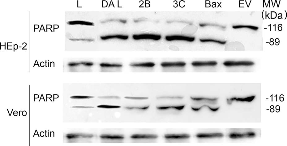
Figure 2. Western blot analysis of cleavage of poly-ADP-ribose polymerase (PARP) in HEp-2 and Vero cells expressing the L, DA L, 2B, 3C, or BAX protein at 24 h post-transfection. Membranes were probed with anti-PARP antibody. The bands represented at 89 kDa are the active form of PARP. MW, protein molecular mass in thousands Dalton (kDa). The actin staining was performed as protein loading control. The Western blot results shown here represent one of the three repeated experiments. EV, empty vector.
To confirm the apoptotic roles of SAFV L, 2B and 3C proteins, HEp-2 and Vero cells transfected with vectors encoding SAFV L, 2B, 3C, TMEV DA L or BAX protein, as well as empty vector, were fixed at 24 h and 48 h posttransfection and subjected to TUNEL assay. The apoptotic cells were labelled as the positive cells, and the percentage of the positive cells of the total transfected cells was calculated and analysed (Fig. 3). In both HEp-2 and Vero cells, the percentage of positive cells of the total transfected cells expressing SAFV 2B, 3C, TMEV DA L or BAX protein was significantly higher than that of cells transfected with empty vector at both 24 h and 48 h posttransfection. These findings confirm SAFV 2B and 3C proteins are proapoptotic, and its L protein does not have proapoptotic activity.
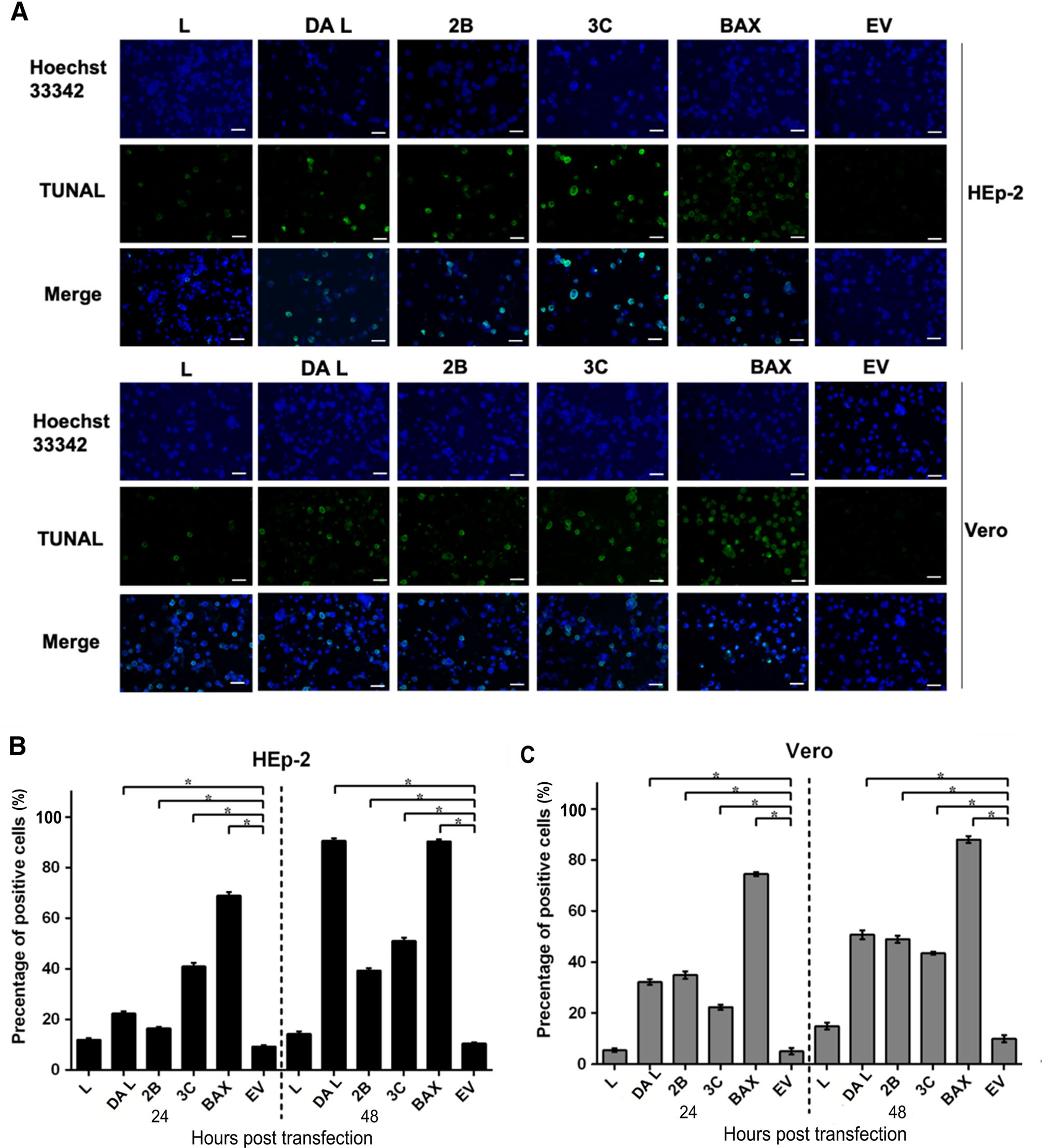
Figure 3. TUNEL assay of HEp-2 cells (A, B) and Vero cells (A, C) expressing SAFV L, 2B, 3C, TMEV DA L and BAX proteins. A TUNAL stained cells of HEp-2 and Vero cells expressing respective viral protein at 24 h post-transfection. Cell nuclei were stained with Hoechst 33258. Cells were observed with a fluorescence microscope (Leica SP8 laser scanning confocal microscope with a 63 ×/1.40 NA oil objective). Scale bar = 10 μm. B TUNAL assay of HEp-2 cells expressing respective viral protein at 24 h and 48 h posttransfection. C TUNAL assay of Vero cells expressing respective viral protein at 24 h and 48 h post-transfection. A thousand cells from at least 5 different optical fields were assessed in each experiment and the ratio of positive cells relative to total cells counted was scored. The similarities between the percentage of positive cells of the cells transfected with the pXJ40-Myc (empty vector) and that of each of other groups were compared with a paired Student's t-test, and P < 0.01 (*) was considered as significant. Results are representative of three independent experiments performed.
-
Taking into consideration that the 3C protein of SAFV was degraded by the cellular ubiquitin/26S proteasome after expression (Xu et al. 2016), we focused on the apoptotic activity of the 2B protein. SAFV 2B protein contains a transmembrane domain located between amino acid 78 and 96 of the sequence (Fig. 4A). In order to determine the function of the transmembrane domain, a plasmid encoding 2B△78-96 protein was constructed and transfected into HEp-2 and Vero cells in comparison with the wild-type 2B protein and empty vector. The cell lysates were collected at 24 h post-transfection, followed by incubation at 65 ℃ for 15 min (to reserve possible polymerization of the protein) before subjecting to Western blots analysis (Fig. 4B). The membranes were probed with anti-Myc antibody to show the expression of both 2B and 2B△78-96 proteins in their respective expected sizes, where the wild-type 2B protein lane showed four bands in one to four times of the expected size of the wild-type 2B protein. The membranes were probed with anti-PARP showing the cleavage of the PARP only in wild-type 2B protein, but not in 2B△78-96 protein. These results revealed that both 2B and 2B△78-96 proteins were expressed in HEp-2 and Vero cells, but only wildtype 2B protein formed tetramer in the transfected cells and induced the cells to undergo apoptosis.

Figure 4. Western blot analysis of the 2B and 2B△78-96 protein in HEp-2 and Vero cells at 24 h post-transfection. A Alignment of the amino acid sequences of SAFV 2B and 2B△78-96 proteins. The transmembrane domain of the 2B protein is highlighted in red. B Western blots analysis of the 2B and 2B△78-96 proteins in HEp-2 and Vero cells. Membranes were stained with anti-Myc or antiPARP antibody. The bands represent at 15 kDa is the monomer of the 2B protein, and 89 kDa is the active form of PARP. MW, protein molecular mass (in thousands Dalton, kDa). The actin staining was the protein loading control. The Western blot results shown here represent one of the three repeated experiments.
SAFV Protein(s) Responsible for Cellular Apoptosis
The Transmembrane Domain of the SAFV 2B Protein
-
In this study, we individually transfected viral proteins of SAFV in parallel with appropriate controls into HEp-2 and Vero cells to determine which of the viral protein(s) that induced apoptosis in host cells. According to our results, the expression of SAFV 2B and 3C proteins, but not the L protein as previously reported in other cardioviruses, induces both HEp-2 and Vero cells to undergo apoptosis. Apoptotic activity was not detected in both HEp-2 and Vero cells expressing viral proteins of SAFV other than the 2B and 3C proteins. The findings suggest the detected apoptotic activities in cells expressing SAFV 2B or 3C protein may not purely due to overexpression of the proteins.
The 2B protein of SAFV is a transmembrane protein with one hydrophobic domain. Unlike the 2B protein of related viruses which are antiapoptotic (Campanella et al. 2004), the SAFV 2B protein is proapoptotic. According to studies of the 2B protein of related viruses, the 2B proteins are viroporins, viral ion channel proteins. As a class of small pore-forming proteins, viroporins involve in various steps of the viral life cycle (Sze and Tan 2015). The transmembrane domain of 2B proteins involves in multimerization of the 2B proteins and formation of membraneintegral complex, which enhances membrane permeability of Golgi, ER, and plasma membrane and inhibits the efflux of ions from the ER and Golgi (Doedens and Kirkegaard 1995; Aldabe et al. 1996; van Kuppeveld et al. 1997; de Jong et al. 2002, 2003, 2006, 2008; Delhaye et al. 2004). For instance, the encephalomyocarditis virus (EMCV) 2B protein—a viroporin closely related to the SAFV 2B evolutionally—can induce the efflux of Ca2+ from ER lumen into cytosol (Ito et al. 2012). Disruption of the distribution of Ca2+ in ER may activate an ER-specific apoptotic pathway and eventually result in the cleavage of caspase-3 (Morishima et al. 2002). Our results indicate that the transmembrane domain is essential for the 2B protein to form tetramer (viral ion channel) on the ER membrane, which may function as viral ion channel and disrupt the distribution of Ca2+ in ER lumen, and subsequently may induce the host cells to undergo apoptosis. However, the exact detailed mechanism(s) of how SAFV 2B protein interacts and cleaves PARP or possibly other host factors in inducing apoptosis still need further investigation.
Unlike the DA L protein of TMEV, the L protein of SAFV does not induce apoptosis after expression. Although the TMEV DA L protein and the SAFV L protein share 62% similarity in amino acid sequence and similar patterns of cellular localization, their functions appear to be different based on our current findings. As multifunctional proteins, the L proteins play crucial roles during virus infection, and the different functions of the L protein may contribute to huge variation in virus replication and spread. Taking the L proteins of DA strain and GDVII strain of TMEV as examples for comparison, though both of the L proteins belong to the same virus type, they presented divergent cellular localizations and protein functions during virus infection, and contributed to distinct infection phenotypic features of the viruses in animal host: acute and fatal encephalomyelitis in mice without persistence in CNS by GDVII strain and subacute encephalomyelitis which progresses to a chronic inflammatory demyelinating disease with persistence in CNS by DA strain (Brahic et al. 2005). As the SAFV L protein does not involve in the apoptotic activity induced by SAFV infection, which is different from the L proteins of other cardioviruses, the SAFV L protein may contribute to the unique infection phenotypic feature of SAFV (such as incomplete apoptosis and nucleo-cytoplasmic localization in HEp-2 cells (Xu et al. 2014)), and hint to the actual diseases linked with SAFV infection.
In conclusion, the proapoptotic proteins of SAFV are the 2B and 3C proteins, while its L protein does not induce apoptosis as in the cases of the L proteins of other cardioviruses. The transmembrane domain of SAFV 2B protein is essential for its multimerization and apoptotic activity.
-
This research was fully funded by Temasek Lifesciences Laboratory, an affiliate of National University of Singapore and Nanyang Technological University, Singapore and did not receive any specific grant from any funding agency in the public, commercial, or not-for-profit sectors.
-
YX, CBLV, KBC and TYJ contributed to the acquisition, analysis and interpretation of data besides concept development and design of the work. YX, KBC, TM and QJ were responsible for drafting and revising the manuscript critically for accuracy of the intellectual content. KBC and TYJ approved the written version for publication. YX, TM, QJ, KBC and TYJ are accountable for all aspects of the work in ensuring that questions related to accuracy or integrity of any part of the work are appropriately investigated and resolved.
-
There is no competing interest in this project.
-
This article does not contain any studies with human or animal subjects performed by any of the authors.

 DownLoad: