


-
At present, repeated occurrence of disease has been the bottle-neck for sustainable development of sea cucumber (Apostichopus japonicus) culture in China, and has been the subject of multiple studies by fishery scientists. Wang et al reported that bacteria, fungi and parasites involved in the skin ulceration and edema in the sea cucumber (7); bacterial diseases in echinoderms, echinoids, holothuroids and asteroids were described by Jangoux (2). Becker et al reported a skin ulceration disease in sea cucumber Holothuria scabra caused by bacterial infection (1). The viruses were observed in the body of the sea cucumber Apostichopus japonicus by Wang et al but they did not confirm whether the viruses were pathogens (6). In this paper, for the first time, we show that the virus is a pathogen of the disease in sea cucumber Apostichopus japonicus. A disease with the symptoms of evisceration and skin ulcerations broke out and led to mass mortality in sea cucumber Apostichopus japonicus cultivated in indoor ponds near the Dalian coast from December 2004 to April 2005, and led to almost 100% mortality. Isolation and identification of the virus as a pathogen and artificial infection was conducted and is described in this paper.
HTML
-
Healthy sea cucumbers varying from 1.5 to 2.0 cm in body length were collected from the Jinzhou area of Dalian where no contagious disease had occurred, while the diseased ones, eviscerating and dying, were sampled from Dalijia area of the Dalian coast in Liaoning Province, China, where many sea cucumbers suffered from this disease. The animals were acclimatized for one week. The healthy individuals used in this experiment which were not infected by the virus were examined with the negative staining method described below.
-
The diseased animals were first fixed in 3% glutaraldehyde solution for 12 h, then in 1% osmium tetroxide solution. Samples were dehydrated through a graded series of ethanol (50%, 70%, 90% and 100%), then transferred into Epon812 resin and cut with a Leica ultracut UCT. Sections were stained with lead citrate and uranyl acetate and observed with a JEM-1200EX electron microscopy.
-
The tissue samples were centrifuged (13 000 r/min, 10 min) twice after homogenizing, freezing and thawing. The supernatant solution was dripped on to a copper net and stained with 2% PTA for 2 min and observed with electron microscopy. In addition, the remaining supernatant was conserved in a low temperature icebox for use in artificial infections. Twenty diseased sea cucumbers were examined to detect the pathogen during the disease epidemic period.
-
Tissue homogenized supernatant solution containing the virus was used as a viral pathogen. The bacterial pathogen was two dominant bacterial strains STT-041201 and STW-041202, which were isolated from diseased sea cucumbers, cultivated on 2216E medium and washed down with 0.9% physiological saline. It was confirmed that the solution contained no bacteria.
In artificial infection tests, 10 healthy sea cucumbers averaging from 1.5 to 2.0 cm in body length were stocked into a 1L column aquarium (13 cm in diameter; 19cm in height) with three control groups. In the treatment groups the healthy sea cucumbers were cultured in the sea water containing 1 mL of the tissue homogenized supernatant containing the viral particles (Group 1), STT-041201 strain (Group 2), STW-041202 strain (Group 3), and 1 mL of the tissue homogenized supernatant containing the viral particles and 0.5 mL of STT-041201 and STW-041202 strains (Group 4). The Control Group 1 contained the test sea cucumbers and 1 mL of 0.9% physiological saline, Control Group 2 contained the test sea cucumbers and 1 mL of tissue homogenized supernatant in which there were no virus and bacteria and Control Group 3 contained the test sea cucumber and 1 ml of physiological saline which was washed down from 2216E medium. The sea water used in the experiment was filtered by sand and water tem-perature was kept at 15.0 ± 0.5 ℃. The supernatant contained bacteria at a concentration of 1.0 × 10⁸ CFU/mL.
Animals
Preparation and observation of ultrathin sections
Preparation and observation of negative stained samples
Artificial infection
-
The infected sea cucumber initially becomes eviscerated, then swelling occurs around the mouth and body wall. The ambulacrum shranks, became flat, thus had no fuction of attachment, and a soft and pale body wall was common. Some individuals had ulcerations from inside to outside the body wall, which became deliquescent when touched.
-
There were no any disease symptoms in the sea cucumbers in the three control groups. On the other hand, the treated individuals exposed to the media containing viruses or viruses plus bacteria particles showed the same disease symptoms as found in naturally diseased individuals. The symptoms were observed from as early as 10 hours to as late as 5 days after administration. One hundred percent mortality occurred in the animals exposed to viruses only or viral and bacterial supernatants. The test animals exposed to the medium containing the two bacterial strains had morbidity from 30% to 80%, with 20% mortality. There was no significant difference in the repeated tests except 80% and 90% mortality in the groups infected with viruses or viruses plus bacteria (the tested individuals used in the negative staining were excluded). Negative staining revealed that the virions were detected only in the specimens in Group 1 and Group 4.
-
Shape and distribution of virus particle: The electron microscopic examinations revealed that the virus infecting the animals was an enveloped spherical virus with a nuclear capsid which varied from 60 to 110 nm in diameter. A few virions with a nuclear capsid of 200 nm in diameter were also observed. Many naked nuclear capsids and various electronic densities of virion were detected by the negative staining method, some showing almost electron transparency (Fig. 1.E). Observation of ultrathin sections showed that the virions with inclusion bodies were distributed throughout the cytoplasm. A large number of virions and inclusion bodies were also detected in the muscular cells of the water-system (such as Polian vesicle), the respiratory tree and the epithelia of the alimentary canal and in the cytoplasm of connective tissue cells in the diseased sea cucumbers. The virons had the same size, but different a electronic density (Fig. 1.F).
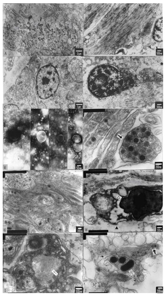
Figure 1. Electron microscopic observation of ultrathin sections and negative stained samples. A: The intestine epithelia of healthy sea cucumber. B: Showing the healthy sea cucumber coelom cells and connective tissues of Polian vesicle. C:The epithelia of respiratory tree in the healthy sea cucumber. D: The body wall cells in the healthy sea cucumber. E: Viral particles shown by negative staining. F: The inclusion body in the epidermic cells of the digestive canal. G: The disintegrating epithelia tissue of respiratory tree with many viral particles. H: The musclar cells in the water-system, showing karyopycnosis, endoplasmic reticulum and mitochondria destroyed. I: The cells in the connective tissue in the affected individual, showing myeloid, electron density material occurred in the cytoplasm. J: The bacteria (white arrow) invaded into the broken tissue where the viruses can be seen (black arrow).
Infectious extension of the virus detected by negative staining: The electron microscopic examinations of the diseased sea cucumbers from twenty aquaculture firms confirmed that the regions affected by this virus were located in part of Changxingdao island on the Dalian coast.
Tissue pathology: The comparison of the organs and tissues of body wall between the normal and diseased sea cucumbers showed that the abundant and clear organelles were distributed evenly and were observed in the cells of the intestines, the Polian vesicle, the respiratory tree, the body wall and the karyoplasm in normal individuals (Fig. 1.A, B, C, D). However, karyopycnosis, endoplasmic reticulum atrophy and the disintegration of the outer membrane and cristae in mitochondria were observed in the diseased sea cucumber. The number of ribosomes was reduced greatly or even disappeared in the cytoplasm in the diseased sea cucumber. Sometimes, virions with high electron densities appeared in the mitochondria of diseased sea cucumber. With the disintegration of organelles, the marrow presented in the cytoplasm and the cell ultimately disaggregated (Fig. 1.H, I). Many viral particles filled the disintegrating tissue (Fig. 1.G). The bacteria invasion into the tissue was expedited due to the ruptured epidermis (Fig. 1.J).
Symptoms of the progression of the disease in sea cucumber
Artificial infection
Observation under an electron microscope
-
Artificial infection indicated that a spherical virus was a pathogen causing the disease found in the sea cucumber and resulted in high mortality. There was lower infection rate in the two strains of bacteria than that observed for the virus. With a mixture of bacteria and viruses, however, the diseased sea cucumber showed an earlier onset of symptoms, indicating that these bacteria played a synergism role in the epidemiological spread of the disease. However, it is still unclear whether the virus or the bacteria, represents the original pathogen and which is present as a secondary affliction.
The serious disease in sea cucumber culture is attributed to the over-culturing and ignoring scientific management of the farms. According to present conditions, water supply, feed and larvae all could be the carriers of the pathogen. In a virtual study, control of the diseases in sea cucumber culture can be best achieved by a program of good management.
Most of the viruses found in the sea cucumber are less than 140 nm in diameter, and others which were observed with a diameter of 200 nm, may belong to another genus, which need further study. Several marine animals have been reported to be infected by a virus in the past few years, such as scallop (4, 5), and abalone (3). But the virus infecting the sea cucumber was different from the others previously identified in shellfish. This paper reports only the primary studies and the purification, identification, culture of virus and more detail research is needed to clarify the problem.

 DownLoad: