


-
Foot-and-mouth disease is a highly contagious and acute disease of cattle, goats, pigs and sheep. Its rapid spread, high morbidity and loss of productivity lead to considerable financial losses. Foot-and-mouth disease virus (FMDV) is a member of the picornavirus family. Non-enveloped FMDV has a single stranded positivesense RNA genome. The virus exists as one of seven different serotypes: O, A, Asia 1, SAT 1, SAT 2 and SAT 3. However, based on serological tests, extensive antigenic variation among FMDV isolates was recognized in the seven serotypes, resulting in a large number of subtypes that have evolved within each serotype (3, 7). Additionally, FMDV varies greatly at different antigenic sites. Consequently, many FMDV-specific antibodies bind only to homologous FMDV, and not heterologous FMDV. This is evidenced in animals that have previously been infected with one serotype, but remain susceptible to the six other serotypes. This makes FMDV diagnosis and immunifaction by immunological methods more complicated and difficult (9). Monoclonal antibodies (McAbs) have therefore played an important role in infectious disease research, diagnostic development, determination of antigenic epitope, vaccine research and studies of viral pathogenesis for many years. Therefore, production of McAbs against FMDV O/China99 would be beneficial to both basic and applied researchers.
HTML
-
BHK-21 cells, a kidney cell line from baby hamsters, were used to propagate FMDV stock and were used in the immunohistochemistry and neutralization assay. Cell lines were cultured in DMEM medium containing 10% fetal calf serum (FCS) in a 5% CO2 incubator at 37℃. The O/China99 virus was used as the immunogen, the target of immunohistochemistry and neutralization assays. The Asia 1 virus was a target in immunohistoemistry for characterizing the McAbs.
-
The O/China99 virus used as the immunogen was titrated for infectivity containing a titer of 107.5 LD50/0.1mL. Two-mouth-old female BALB/c mice kept within P3 laboratory were immunized with 107.5 LD50 infectious FMDV in 0.1mL each time at 3-to 4-week intervals except the last booster. The first and second immunizations were intraperitoneal injections. The third immunization and last injections three days before fusion were given to the anesthetic mice via intrasplenic route (1, 5). Animals were bled and sera neutralization titers were examined to monitor the individual immune response.
-
Splenocytes from mouse showing the highest virus neutralization (VN) titers to O/China99 were fused with SP2/0 myeloma cells to produce the hybridoma according to standard protocol (4). Hybridoma secreting antibodies against O/China99 virus were selected by immunohistochemistry on ethanol-fixed antigen plate described as follows. Positive hybridoma was cloned 3-to-4 times by the limiting dilution method. One monoclonal hybridoma was picked randomly from a parental to inoculate intraperitoneally into pristine-primed BALB/c mice to produce ascetic fluid containing McAb. The hybridoma cultural supernatant and ascitic fluid were collected for subsequent characterization. Isotypes of McAbs from hybridoma cultural supernatant were analyzed by a mouse monoclonal antibody isotyping kit (Sigma-Aldrich) according to the manufacturer's recommendation.
-
The immunohistochemistry was performed on antigen plates which were constituted of a BHK-21 monolayer infected by 4×103.5TCID50/well O/China99 and incubated for 6-8 h until the appearance of CPE under a microscope. These were then, were fixed by -20℃ ethanol. The preparation of antigen plates of Asia 1 was similar to that of O/China99. The antigen plates were washed three times with phosphate buffered saline (PBS) before adding 100μL hybridoma cultural supernatant or diluted mouse ascitic fluid for immunohistochemistry, meanwhile setted positive control and negative control with mouse positive antiserum to FMDV and mouse negative antiserum to FMDV, respectivly. After room temperature in-cubation for 1h, plates were washed 3 times with PBS and a horse-radish peroxides (HRP) conjugated goat anti-mouse IgG (Sigma-Aldrich) at 1: 1 000 dilution was added 100μL/well. This was followed by 1 h room temperature incubationed and the plates then were washed 3 times with PBS. Staining was performed using the 3, 3′-diaminobenzidine tetrahydrochloride substrate kit (Sigma-Aldrich) according to manucturer's instructions, then observed under an inverted microscope.
-
Virus neutralization (VN) assay was performed according to the OIE manual (10). Briefly, the titers of VN were determined in a microdilution test using the BHK-21 cells. Serial dilution of sera samples were incubated with a virus dose of 100 TCID50 of the O/China99 virus. Microplates were incubated at 37℃ for 2 days before being examined microscopically for CPE. The fifty percent end point of neutralization titers were calculated using the method of Reed-muench (11). The procedures of microneutralization (MN) assay for the detection of McAb neutralization activity were followed with modification. In principle, both of serial diluted mouse ascitic fluid and equal volume of 100 TCID50 of the O/China99 virus prepared with 2% FCS in DMEM were mixed and incubated at 37℃ for 1 h. Each virus /diluted McAb mixture was then applied to eleven wells of a 96-well microtiter plate. BHK-21 cells 5×10⁴/0.1mL/well were added as well. After incubation for 2 days at 37℃, CPE was scored under microscope. The fifty percent end points of neutralization titers for O/China99 were calculated. The amount of virus actually used per well should contain 100 TCID50 within an acceptable range (e.g.35-350 TCID50) from back titration control. Neutralization assays were repeated at least twice.
-
The VP1 capsid protein was expressed in E.coli, purified and refolded (13). Also, the polypeptide 14 (P14: RHKQKIVAPVKQLL) and polypeptide 20 (P20: VTNVRGDLQVLAQKAARTLP), representing the amino acid residues 200-213 and 141-160 of VP1 of the FMDV O/China99, were expressed and purified by the National FMD Reference Laboratory(Chinese Academy of Agricultural Science). VP1, P14 and P20 (10μg/mL) in 0.1 M bicarbonate, pH 8.3 were coated onto Microtitre plates and incubated at 4℃ overnight. The plates were blocked with 5% skim milk in PBS at 37℃. After incubation, ascitic samples in serial dilution were added to the wells, meanwhile setted positive control and negative control with mouse positive antiserum to FMDV type O and mouse negative antiserum to FMDV type O, respectivly. Then a horse-radish peroxidase (HRP) conjugated goat anti-mouse IgG (1: 3 000, Sigma-Aldrich) was added. For detection, enzyme substrate 3, 3', 5', 5-tetramethylbenzidine (TMB, Sigma-Aldrich) was added. An equal volume of 1 N H2SO4 solution sulfuric acid was added to each well to stop the color reaction. The optical absorbance was measured at 450 nm using an automated plate reader. Each incubation step was 60 min at 37℃ with gentle shaking and followed by washing three times with washing buffer (PBS with 0.05% Tween 20).
Cell line and viruses
Mice immunization
Monoclonal antibody production
Immunohistochemistry assay
Neutralization assay
Characterization McAb binding epitope
-
The BALB/c mouse which was immunized with O/China99 showed VN titer at 1 280 by virus neutralization assay, when it was sacrificed on the fusion day. Using the immunohistochemistry assay and limiting dilution, until obtained two clones. The two clones named 1A9 and 9F12. Both of the McAbs contained kappa light chains, but the McAbs 1A9 and F12 were IgG1 and IgM isotype, respectively.
-
The McAbs were examined for their specificity against O/China99 and Asia 1. The two McAbs showed reactivity with O/China99 and no reactivity with Asia 1 (Fig. 1). The O/China99 immunohisto-emistry titer of each McAb/ascites was determined based on the highest dilution of ascites to give strong immunohistochemistry reaction on a O/China99 antigen plate. The McAbs immunohistochemistry titer of O/China99 were between 20000 and 40000 fold (Table 1). The two McAbs showed neutralization activities with O/China99, and the neutralization activities of McAbs were shown as MN titer (Table 2).
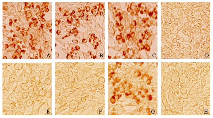
Figure 1. The reaction of McAb specificity by immunohistochemistry assay. A, B, C and D, Immunohistochemistry straining of BHK-1 cells infected O/China99 with McAb1A9, McAb9F12, Mouse positive antiserum to FMDV type O, Mouse negative antiserum to FMDV type O, respectively; E, F, G and H, Immunohistochemistry straining of BHK-21 cells infected Asia 1 with McAb 1A9, McAb 9F12, Mouse positive antiserum to FMDV type Asia 1, Mouse negative antiserum to FMDV type Asia 1, respectively.

Table 1. Detection of McAb/ascites immunohistochemistry titer

Table 2. Detection of McAbs neutralization titers by microneutralization assay
-
In order to define the binding epitopes of McAbs, the reactivity of the McAbs against VP1, P14 and P20 were examined using an indirect ELISA. The results showed that the two McAbs had reacted with VP1 and P20, but no reactivity with P14 (Table 3).

Table 3. The reaction OD450nm Values of the McAbs and the polypeptides
Production of McAbs
Specificity of the McAbs
Characterization McAb binding epitope
-
Since McAbs against type O FMDV was developed by McCullogh and Butcher(14) for the first time in 1982, many McAbs against all the serotypes FMDV had been obtained by several countries. Currently, McAbs are important tools for studies of diagnostic methods and determination of antigenic epitope on FMDV. The use of McAbs increases the specificity, accuracy and efficiency of diagnostic tests compared to polyclonal antisera and provides unlimited quantity and consistent quality of reagent supplies. Smitsaart et al reported a competitive ELISA by using the McAb specific for the 12S subunit from six of the seven serotypes to detect the serotype O, A, Asia 1, SAT 1 and SAT 3 FMDV (15). Ming et al. reported that they had obtained two McAbs with different specificities which could lead to the development of a highly specific and simple double antibody sandwich ELISA for rapid diagnosis of FMDV, and the McAb based ELISA provides many advantages such as less variation than antisera batchs, unlimited supply of reagents and unique specificity (9). Meanwhile, the two-McAbs system could also be adapted to immuno-chromatographic or flow-through rapid tests. Samir et al assessed antigenic sites of 31 FMDV type Asia-1 field isolates and two vaccine strains, using a panel of type Asia-1-specific McAbs, to reveal antigenic similarity of the virus isolates tested and absence of neutralization escape mutants (16). The developed McAbs have practical applications, especially in the manufacture of FMD vaccines, diagnosis and FMDV characterization.
Three major procedures, immunization of live virus, intrasplenic injection and screening by immunohisto-chemistry, were undertaken in this study to develop two McAbs for research and application in the future. The two McAbs, raised against a unique isolate of FMDV O/Cina99 was obtained finally in one fusion. Although BALB/c mice were not the natural host of FMD, it still replicated FMDV for a maximum of 60h which made it a perfect bio-reactor to generate McAbs against all structural and non-structural viral proteins expressed (12). Neutralizing antibody titer of the immunized BALB/c mouse showed a high titer of 1 280. Basically, the antibodies produced by live virus immunized BALB/c should be similar to the antibodies produced by the natural host. Besides, McAbs originated from live virus immunization strategy would favor the selection of escaped mutants for the purpose of mapping the neutralizing antigenic site in the long run. Therefore, the McAbs generated here would fit well to the needs of studies on viral relatedness, vaccine strain choice and so on.
To pick up the positive hybridoma secreting immunoglobulin against the immunogen, the immunohistochemistry assay on susceptible BHK monolayer infected by optimum dose of FMDV was chosen to screen the parental and monoclonal hybridoma cells. The existence of both FMDV-infected and normal BHK cells with their distinct morphology on immunohistochemistry antigen plates clearly demonstrated that antibody either bound to viral or cellular components and favored the selection of FMDV specific McAb. Moreover, the antigenicity of viral protein kept by ethanol, the fixative on the plate, was almost the same as that of the live virus, and thus could be recognized by the elicited McAbs. McAbs in ascites reported in this study were of high O/China99 immunohistochemistry titers. The McAbs showing negative immunohistochemistry result on Asia 1 antigen plate implied that these McAbs possessed an O/China99 specific binding site.
This paper describes the production of McAbs against O/China99 strain FMDV. Their reactivities with VP1 were determined primarily because VP1 possessed three out of the five neutralizing antibody binding sites identified so far (2) and attracted more interest than the other three structural proteins. N-terminal residues 43 and 44 on βB-βC loop, the βG-βH loop and the C-terminal residues on VP1 are recognized as neutralizing antigenic sit 3, 1 and 5 respectively. The βG-βH loop of VP1, including an Arg-Gly-Asp (RGD) motif at its apex, binds to RGD-dependent integrins of host cell and is involved in neutralization of viral infectivity (6, 8). It is recognized as a major antigenic and less conformation-dependent site of FMDV (17). Therefore, a well established VP1 ELISA was utilized to primarily analyze these two McAbs. Expressed peptide P14 and P20 representing the sequence of βG-βH loop was employed further to examine the activities of VP1 reacting McAbs. The positive results of two McAbs on P20 ELISA indicated that the McAbs binding sites were located on the βG-βH loop region. Then the P20 (TSRGDPSALAQ RLSGRLPTS) which represent the amino acid residues 141-160 of VP1 of Asia 1, was different from the P20 (VTNVRGDLQVLAQKAA RTLP) which represent the amino acid residues 141-160 of VP1 of the FMDV O/China99, to illustrate further the observation that both McAbs didn't react with the Asia 1 in the immunohistochemistry assay. The continuous and complete analysis on the McAbs reported here, will be beneficial to the development of the vaccine and further diagnostics.

 DownLoad: