-
Bovine immunodeficiency virus (BIV) belongs to the Lentivirus genus of the Retroviridae family. In addition to the three standard retroviral genes gag, pol and env, there are also several accessory genes, such as tat and rev [1, 19]. The proviral genome has two flanking sequences, called long terminal repeats (LTR), that are used in the regulation of viral replication and gene expression. The BIV Tat protein can transactivate its LTR and promote viral protein transcription [2, 14]. BIV is highly homologous to human immunodefi-ciency virus (HIV), the pathogen of acquired immunodeficiency syndrome (AIDS). Consequently, the replication cycle is very similar to HIV, undergoing cell entry and fusion, transcription of the RNA genome reverse into DNA and integration into the host genome. The molecular mechanism of BIV's inducing apoptosis is also very similar to HIV [23]. BIV is known to induce chronic pathological changes associated with dysfunction of the immune system in infected cattle [5]. As BIV does not infect humans, BIV is a safe model and the study of BIV has facilitated HIV research [8]. BIV infected rabbit show similar AIDS-like symptoms [10, 18]. Thus, since the animal AIDS model involving primates is very expensive, the BIV/rabbit model might be a good small animal model to study HIV/AIDS. BIV infection can also be inhibited by HIV inhibitors, indicating BIV and HIV share similar inhibitor targets [20]. Therefore BIV may also be used to screen anti-AIDS drugs.
Detection of BIV infection is an essential process in BIV research, especially in the study of the biological properties and the replication strategy of BIV. However, traditional methods cannot meet the needs of high-throughput drug screening. Observation of the cytopathic effect (CPE), or syncytia formation, is used routinely for quantification of BIV infectivity [22, 26]. Approaches based on CPE are, however, time-consuming, labor intensive, and relatively insensitive. Another frequently used method is to detect the virus capsid protein expression by Western blot. However this is not a quantitative method either and a simple and quantitative method is still required.
In order to quantify BIV infection, we established an indicator cell line (BIVL) by transecting the virus LTR promoter with the firefly luciferase gene into baby hamster kidney cells. When the BIVL was infected by BIV, the transactivator Tat protein could activate the BIV LTR promoter transcription and induce the expression of firefly luciferase. By detection of luciferase activity, the BIV infection could be quantified. This cell line has high sensitivity and specificity in detecting and quantization of active BIV infection. This quantitative and robust assay will facilitate BIV research and could also be applied to high-throughput screening of antiretroviral compounds.
HTML
-
Baby hamster kidney cell line (BHK-21) was obtained from the China Center for Type Culture Collection (CCTCC, Wuhan, China). Canine thymus cell line (Cf2Th) was kindly provided by Prof. Jin-Ming GAO (Peking Union Medical College). BHK-21 and Cf2Th cells were grown in Dulbecco's modified Eagle's medium (DMEM) supplemented with 10% (v/v) fetal calf serum at 37 ℃ in 5% CO2.
The BIV R29 strain was provided by Dr. Charles Wood (University of Nebraska Lincoln) and was cultured with Cf2Th. The BIV virus stock was added onto Cf2Th cells with a Multiplicity of Infection (MOI) of 1.0 and cultured for 2 days. At 2 days post infection, when typical syncytia could be seen, ten 100 mm-tissue culture dishes of cells were pooled and resuspended in 10 mL storage medium (10% dimethyl sulfoxide (DMSO), 20% fetal calf serum in DMEM) and then frozen at-70 ℃ with 400 μL per stock as BIV virus stock. BFV3026 is a bovine foamy virus (BFV) strain which was isolated from bovine peripheral lymphoid cells by our laboratory in an earlier study [13], and was cultured with Cf2Th. Bovine herpes virus-1 (BoHV-1) was stored in our lab, and was cultured in MDBK cells. When cell cytopathic effects (CPE) in BoHV-1 infected MDBK were seen, the medium was collected and underwent 3000×g centrifugation and then the supernatant was passed through a 0.45 μm filter, and stored in-70℃ as BoHV-1 virus stock.
-
Confluent Cf2Th cells on 24-well plates were infected with 25 μL of 10-fold serial dilutions of stock viruses and incubated at 37 ℃ in 5% CO2 for 2 h with eight parallel wells for each dilution. The cells were washed with PBS and then cultured in the maintenance medium. Examination of cytopathic effect was performed on the second day post infection by observation of syncytia formation of BIV-infected Cf2Th cells and the TCID50was calculated with the Reed-Muench method [11].
-
BIV127, a BIV provirus clone was provided by Dr Charles Wood (University of Nebraska Lincoln) [4]. The pGL3-Basic vector (Promega, USA) has the firefly luciferase (luc) gene. The pEGFP-N1 vector (Clontech Laboratories, Inc, USA) carries the enhanced green fluorescent protein (egfp)gene as a reporter gene and the neomycin-resistant gene as a selection marker.
-
A full length LTR DNA fragment of BIV127 proclone was amplified by PCR, using a sense primer with addition of an Ase I site: 5'-GTAATTAATCTT AAAAGGTGGACTTG-3' and an antisense primer with addition of a Hin dⅢ site: 5'-CGGAAGCTTTGT TGGGTGTTCTTCACCG-3'. The LTR DNA fragment of BIV was then inserted at the Ase I and Hin d Ⅲ sites of the pEGFP-N1 vector. This intermediate plasmid contained the egfp gene downstream of the LTR promoter of BIV. Then the egfp gene was replaced by the luc gene from the pGL3-Basic vector by the following steps. The intermediate plasmid was digested with Not Ⅰ, blunted by Mung bean nuclease and then digested with Hin d Ⅲ to get rid of the egfp gene; pGL3-Basic vector was digested withXbaⅠ, blunted by Mung bean nuclease and then digested with Hin d Ⅲ to get the luc gene fragment. Then the two parts were ligated by T4 DNA ligase to form the BIV reporter plasmid, containing the LTR-luc and the neomycin resistant gene.
-
The BHK-21 cells were plated at a density of 2×10⁵ cells/well in 6-well plates and allowed to grow overnight. 2 μg of BIV reporter plasmid with the luciferase gene downstream of the BIV long terminal repeat was transfected into BHK-21 cells using 12 μg polyethylenimine (PEI) (Polysciences Inc, USA) as the transfection reagent. At 48 h post transfection, the cells were cultured in a selective medium containing 600 μg/ml neomycin (G418) for an additional 3 weeks. The cells were replaced with fresh medium every 3 days. Limiting dilution onto 96-well plates was used toobtain positive stable monoclones from G418 resistant cells. The monoclones with very low constitutive luciferase expression were selected and expanded for further testing. Among the positive BHK-21 cell clones, one exhibiting a strong transactivation of the integrated BIVLTR-luc gene following BIV infection was selected to undergo a second round of monoclonization. Among the 24 monoclones obtained in the second round, the one which shows the most robust response to BIV infection was selected as a BIV indicator cell line, and designated BIVL.
-
2×10⁵ Cf2Th cells were infected with different doses of BIV stock. At 60 h post infection the cells were harvested and washed twice with PBS. After centrifugation at 3 000 × g for 3 min, the cells were resuspended in 20 μL PBS and 20 μL 2 × loading buffer containing 2% sodium dodecyl sulphate (SDS) and 5% 2-mercaptoethanol. Samples were boiled for 10 min, electrophoresed on 12% SDS-polyacrylamide gels and then transferred to nitrocellulose membranes. The membranes were blocked for 45 min at room temperature in PBS plus 0.5% Tween-20 containing 5% skimmed milk, and then incubated for 90 min at room temperature with murine polyclonal antibodies against BIV capsid protein (prepared by this laboratory; dilution 1:5 000). After being washed, the membranes were incubated for 45 min with goat anti-mouse IgG labelled with horseradish peroxidase (Santa Cruz biotechnology, CA, USA; dilution 1:5 000); bound antibody was visualized and quantified by chemiluminescence detection. β-actin was used as a loading control; the β-actin antibody was purchased from Sigma. (StLouis, MO, USA).
-
The indicator cells were plated in 96-well plates (103/well) and cultured at 37℃ for 18 h. The cells were incubated with a serial dilution of BIV stock. After replacing the virus stock with the maintenance medium, the cells were incubated at 37℃ for 60 h. Luciferase activity was measured by using a commercial assay system. 25μL of Steady-Glo luciferase substrate (Promega, USA) was added into each well, and the cultures were gently shaken for 45 min. The luciferase activity was measured for 0.5 second per well as luminescence by using a 96-channel chemiluminescense measurement machine Glomax (Promega, USA). Readout was counts per second. The relative light unit was determined automatically.
-
Sensitivities of BIV stock was measured by the end-point dilution method (CPE-based TCID50), Luciferase assay and Western blot assay was at 60 h post infection. Relative sensitivity was TCID50 of CPE assay/ minimal measure The cut-off of the BIVL-based assay was a 2.5 fold luciferase activity ratio.
The cut-off of the CPE-based assay was that at least one syncytia formation was observed in BIV-infected Cf2Th cells per well. The cut-off of the Western blot-based assay was the minimal BIV capsid protein detected from 2×10⁵ BIV-infected Cf2Th cells by antiserum against BIV capsid.
-
The Luciferase activity ratio was the luciferase activity of infected cells/ luciferase activity of mock infected cells. Luciferase activity was detected at 60 h post infection. The BIV indicator cell line was infected with BIV R29 virus stock (MOI=0.5), or co-cultured with BFV 3026 virus (MOI=0.5), or BHV-1 virus stock (MOI=0.5) respectively. At 48 h post infection, the luciferase activity ratio was calculated.
Cells and viruses
Determination of tissue culture infectious dose endpoint (TCID50) of BIV
Plasmids
Construction of BIV reporter plasmid
Construction of BIV indicator cell line
Western blot analysis
Luciferase assay
Sensitivity assay
Specificity assay
-
The BIV system has been applied to evaluate antiretroviral effects by observation of CPE [12]. However, CPE-based assays require experienced researchers to correctly identify syncytia formation and therefore are less accurate and quantitative. BIV capsid protein Western blot assay has been used to detection of BIV infection [25]. However, this method is also not quantitative. In order to develop a more robust assay to detect BIV infection which is applicable to high-throughput screening of antiretroviral compounds, a BIV indicator cell line, designated BIVL, was established and an assay was developed based on this cell line. The sensitivity of this BIVL-based assay was compared with two standard measures used to detect BIV infection. The infectious titers of a BIV stock via the BIVL-based assay, CPE-based assay and Western blot assay were determined respectively by end-point dilution method 2 days post infection. Although the sensitivity of the Western blot assay was almost the same as the BIVL-based assay, the former assay is far more labor intensive and time consuming. The BIVL-based luciferase assay was 20 times more sensitive than the syncytia formation or CPE assay. This indicates BIVL-based assay is both an efficient and sensitive method to detect BIV infection.
-
The specificity of the luciferase assay was evaluated by infecting BIVL cells with other bovine viruses. The first virus was BFV, a bovine retrovirus found to superinfect cattle with BIV. Although inoculation with BFV of high titer resulted in a cytopathic effect, no luciferase activity increase was detected. In contrast, the induction luciferase activity by BIV was very prominent (MOI=1.0, 40 fold). Bovine herpes virus (BoHV) was also found to superinfect cattle with BIV. BoHV has been reported to activate BIV LTR via its transactivator protein bICP0 [7]. Therefore, BHV was tested for its activation ability on the BIV indicator cell line, which has the LTR upstream the of the reporter gene. BoHV-1 virus stock was added on to BIVL cells and two days later, BIVL cells showed a specific cytopathic effect but only a 3.2 fold increase in luciferase activity was induced. However, the luciferase level dosen't increase as the BHV infection dose increases, because the viral protein bICP0's activation ability is limited compared with the BIV's transactivation protein Tat. This finding indicated that the specificity of the BIV LTR promoter in the BIVL cell line is good enough to identify BIV infection.
-
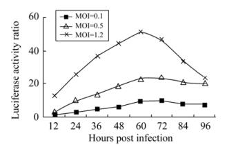
In order to determine the optimal time point to detect BIV infection using the newly established BIV indicator cell line (BIVL), the kinetics of luciferase activity of BIV infected BIVL cells was monitored. To see whether different MOI infection could also generate a similar increase in luciferase activity, the BIVL cells were infected with different MOI and the luciferase activity was detected (Fig. 1). Twelve hours after co-cultured with 1.0 MOI of BIV virus stock, BIVL showed a 10 fold increase in luciferase activity in comparison to BIVL cells alone. This increase was more pronounced after 24 h. Different MOIs similarly led to an increase in luciferase activity after being co-cultured with BIVL, which also showed a time-dependent increase before 72 h and then decreased. Of note, at high MOI ( > 1), a decrease in luciferase activity was observed at later time points, because infected BIVL cells are killed by virus-induced apoptosis [23], and BIV replication level in BHK-21 cells is not as permissive as FBL cells or Cf2Th cells. Therefore, 60 h post infection was the optimal time point to determine BIV infection in BIVL-based assay.

Figure 1. Luciferase expression in BIVL cells infected with BIV at different MOI. The luciferase expression were determined at 12, 24, 36, 48, 60, 72, 84 and 96 hours post infection by a chemiluninescence measurement machine (Glomax, Promega) in terms of luciferase activity ratio. (Luciferase activity ratio=luciferase activity of infected cells/ luciferase activity of mock infected cells.)
-
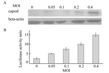
Capsid protein expression level represents a measured of viral load, therefore BIV infection was assessed by detecting BIV capsid protein through Western blot assay. It is necessary to examine whether BIV capsid expression is correlated with BIV indicator cell line luciferase activity, since BIV indicator cell line's luciferase activity depends on the expression of the BIV Tat protein. The same amount of virus stock was added to either Cf2Th cells or indicator cells, and then Western blot and luciferase activity assay were performed on the two cells respectively. Capsid expression level was dependent on the dose of BIV virus stock, whereas luciferase activity of BIV indicator cell lines was consistent with BIV Capsid expression (Fig. 2). However, Western blot assay was labor intensive and, unlike the luciferase activity, could not evaluate BIV infection quantitatively. This result indicated the correlation of BIV capsid expression with the BIV Tat protein expression, determined by BIVL luciferase activity.

Figure 2. Comparative analysis of BIV capsid protein and luciferase expression in BIVL cells infected with BIV. A: Western blot assay to detect BIV capsid protein in Cf2Th infected with diluted BIV. Beta-actin serves as internal control. B: Luciferase assay of BIVL infected with diluted BIV. The same dose was added to either Cf2Th or BIVL.
-
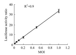
To determine the relationship between BIV virus stock amount and luciferase activity, serial dilutions of BIV stock were used to infect BIVL cells and a standard curve was plotted (Fig. 3). A standard curve was plotted. The minimal detectable concentration of BIV was 0.05 MOI. At MOI higher than 1.5, BIV virus stock and luciferase activity lost the dose dependent manner. This indicated that the linear range of quantifying BIV infection in vitro by BIVL-based assay was 0.05 MOI to 1.5 MOI. The linearity of this curve (R2=0.9) indicated that the luciferase assay may be useful in rapid detection and quantitation of BIV in vitro . Therefore, inhibition or acceleration of BIV infection might also be quantitated by BIVL-assay.

Figure 3. Determination of viral infectivity titer. BIVL cells were incubated at MOI of 0.05 to 1.0 with BIV.(tittered on Cf2Th cells by CPE assay) for 2 days. Cells were analyzed by a chemiluninescence measurement (Glomax, Promega) in terms of relative light units. Luciferase activity ratio was calculated.
Sensitivity of BIV indicator cell line
Specificity of BIV indicator cell line
Kinetics of BIV infection in BIV indicator cell line
Correlation of capsid protein expression and luciferase activity in BIV infected BIVL cells
Quantification of BIV infection with BIVL-based assay
-
Compared with current methods for detecting BIV infection, the BIVL assay is more rapid and quantitative. The CPE assay is more subjective than quantitative, and is thus both time consuming and imprecise. The Western blot assay is a labor intensive procedure and very expensive because of the reagent consumption and cost. Furthermore, we are able to carry out the luciferase assay in a 96-well plate to scan a large number of samples very easily. The BIV indicator cell line could not only be used into the basic research of BIV molecular biology, but also be applied to detect an inhibition effect. Luciferase-based TCID50 assay should be a good replacement of traditional CPE titering for BIV. Within the range of 0.05 MOI to 1.5 MOI, the BIV infection titer and indicator cell's luciferase level is dose dependent.
Modern day drug development also calls for high-throughput screening [6]. HIV-1 antiviral assay formats have been described that could be adapted for high-throughput screening, and many of these utilize either a reporter virus [3, 17] or an indicator cell line [9] to measure HIV replication. In reporter virus assays, a reporter gene is introduced into the virus genome, usually in place of a viral gene not required for replication in the target cells of interest. The cells are then infected with the recombinant reporter virus, and virus replication is quantified by measuring the expression of the virus encoded reporter gene. In indicator cell assays, the target cells of interest are engineered to contain a reporter and replication is measured by monitoring induction of the reporter gene in infected target cells.
Antiviral assays using live HIV virus have to be performed in restricted environments because of biosafety concerns; thus, they are not optimal for large-scale routine screens or structure-activity relationship studies, especially in developing countries which do not have many available P3 level laboratories. Thus, a biologically safe and high-throughput compatible surrogate antiviral assay system is would be particularly useful. Also, with regard the problem of the development of resistance in AIDS treatment this screening system could screen for inhibitors which could inhibit resistance strains, due to their broad range of inhibition [15, 16, 24]. The method reported in this study could be developed into a system for high-throughput screening of antiviral agents against HIV. Subsequently, a system screening HIV inhibitors based on BIV could also be established. The BIV indicator cell line and virus stock ensure it is a high-throughput screening system; established BIV-based assays to screen anti-AIDS drugs, use transient cell lines instead of stable cell line [21], and cannot perform high-throughput screening. Potential applica-tions of the BIVL reporter system include high-throughput assays for evaluation of anti-HIV drugs' susceptibility. The 96-well plate format permits the assay of multiple drugs at a range of concentrations. The feasibility of performing drug assays for reverse transcriptase has been demonstrated. By optimizing the conditions and using virus stock and luciferase assay reagent to detect luciferase activity, the BIV drug screening system could meet high-throughput screening quality.
In summary, the establishment of an indicator cell line expressing the BIV-inducible luciferase reporter gene has provided a robust tool for monitoring and investigating BIV infections. Study of BIV infection will be valuable for both BIV applications and basic research. Primary screening of effective anti-retroviral natural compounds via the BIV system is currently in progress.


 DownLoad: