-
Coronaviruses are vertebrate pathogens mainly associated with respiratory and enteric diseases[19]. In humans, Coronavirus infections manifest usually as mild respiratory tract disease (common cold) that may cause more severe symptoms in elderly or immune-compromised individuals[4, 18]. An international outbreak of severe acute respiratory syndrome (SARS), an atypical pneumonia, spread through more than 30 countries and caused about 8 422 cases and 916 deaths worldwide in 2002-2003[20]. A novel Coronavirus SARS-Coronavirus (SARS-CoV) that can be isolated from the SARS patients and from Vero E6 cells inoculated with clinical specimens was identified to be the causative agent of SARS [3, 10, 11]. The appearance of SARS, exemplified the potential of Coronaviruses to seriously affect human health [5, 13, 14, 15].The frequent detection of SARS-like Corona-viruses in horseshoe bats and the broad range of mammalian hosts that are susceptible to SARS-CoV infection may facilitate a potential reintroduction into the human population[12]. Therefore, the development of system for studying the SARS-CoV biology and pathogenesis is of high medical importance.
Reverse genetic system is a very important tool to the analysis of viral replication and pathogenesis. Recently, reverse genetic systems for a number of Coronaviruses have been established using non-traditional approaches which are based on the use of bacterial artificial chromosomes[1], the in vitro ligation of Coronavirus cDNA fragments[22] and the use of vaccinia virus as a vector for the propagation of Coronavirus genomic cDNA[17]. With the systems now available, it is possible to genetically modify Coronavirus genomes at will. Recombinant viruses with gene inactivations, deletions or attenuating modifications can be generated and used to study the role of specific gene products in viral replication or pathogenesis. It was reported that the expression of Coronavirus nucleocapsid protein definitely facilitated the efficient rescue of Coronaviruses in all the reverse genetic systems[2, 15, 16].
The Tet-On gene expression systems is a regulated, high-level gene expression system. Maximal expression levels in Tet-On systems are very high and compare favourably with the maximal levels obtainable from strong, constitutive mammalian promoters such as CMV[21]. In this study, we established a series of double stable SARS-CoV nucleocapsid protein-expressing cell lines derived from BHK-21, and the expression was tightly regulated in response to inducer doxycycline in a precise and dose-dependent manner. The constructed double-stable cell strains will play an important role in the reverse genetics research of SARS-CoV, it will facilitate the rescue of SARS-CoV in vitro and the analysis of SARS-CoV RNA synthesis.
HTML
-
The template plasmid p8S was kindly provided by Vorker Thiel, which contained a cDNA clone spanning nucleotides 25 641-29 734 of the SARS-CoV HKU-39849 genome. SARS-CoV nucleocapsid gene (nucleotides 28 120-29 385) was amplified using the primers: SARS-N-up (nucleotides 28 117-28 150, 5′-AAAATGgCTGATAAcGGACCCCAATCAAACCAAC-3′), SARS-N-down (nucleotides 29 368-29 394, 5′-GAGTGTTTATGCCTGAGTTGAATCAGC-3′). Two nucleotides changes were introduced into the position 28 123 (t to g) and 28 131 (t to c) where we expected to improve translation efficiency. The amplify reactions were performed with the Phusion DNA polymerase (FINNZYMES) which has proof reading ability, and carried out as the following conditions: 98℃ 2 min, then 98℃ 10 sec, 50℃ 2 min, 72℃ 1 min for 2 cycles, and followed 98℃ 10 sec, 58℃ 30 sec, 72℃ 45 sec for 28 cycles, finally extension at 72℃ for 10 min. The amplified fragment was examined by agrose gel electrophoresis.
-
The PCR product was recovered by gel extraction using the QIAquick Gel Extraction Kit (QIAGEN), and then cloned the DNA fragment into the PCR-Blunt TOPO vector using the Zero Blunt® TOPO® PCR Cloning Kits (Invitrogen). The positive clone was identified by restriction enzyme digestion and sequencing. The sequencing reactions were done with Big Dye reagents version 3.1 (Applied Biosystems). The universal primers Pcmv forward primer and PSV40 reverse primer (Invitrogen) were used as sequencing primers. Sequencing reaction was set up as follows: BigDye terminators 2.5μL, pTOPO-SARS-N DNA template 50-300ng, primers (70ng/mL) 70ng, add HiPerSolv water to 10μL. The temperature conditions for sequencing were carried out as the following: 96℃ 15 sec, 50℃ 15 sec, 60℃ 4 min for 25 cycles. The products of the 10μL reaction were precipitated with sodium acetate and ethanol in a total volume of 360μL. The pelleted samples were resuspended in 15μL HiDye formamide and separated using the ABI prism 310 sequencer. Sequences alignment was done using Seqman module of Lasergene sequence analysis software.
-
Response vector pTRE-Tight was purchased from BD Biosciences company, contained a modified TRE upstream of an altered minimal CMV promoter, which can fully minimize background expression. After digestion of plasmid pTOPO-SARS-N with the restriction enzyme BamH I and Not I, the fragment of nucleocapsid gene was purified and ligated with vector pTRE-Tight which was digested with the same restriction enzyme and dephosphated with alkaline phosphatase (Invitrogen). The mixture of ligation was transformed to the One Shot® TOP10 Chemically Competent cells (Invitrogen). The positive plasmid pTRE-Tight-SARS-N was identified by restriction enzyme digestion.
-
BHK-21 cell (Syrian golden hamster kidney cell) was purchased from ATCC and preserved by our lab. The flow-chart of double-stable cell line construction showed in Fig. 1. BHK-21 cells were cultured at 37℃ in complete medium (Dulbecco's modified eagle's medium supplemented with 5% Fetal Bovine Serum, 100U/mL penicillin, 100μg/mL streptomycin) to about 80% confluency, then were transfected with 5μg pTet-On regulator plasmid DNA (BD Biosciences) using FuGENE 6 reagent (Roche). Plate the trans-fected cells in ten 10cm culture dishes, each containing 10mL of complete medium. After cultured at 37℃ for 48 h, added different concentrations of G418 (Roche). Every four days, replace medium with fresh complete medium plus G418, split the cells when they reach confluency. After 2-4 weeks, pick large, healthy colonies using the cloning cylinders (BD Biosciences) and transfer them to individual plates.
-
The constructed Tet-on stable BHK-21 cell line (BHK-Tet) were cultured at 37℃ in complete medium, added G418 to 500μg/mL. Plasmid pTRE-Tight-SARS-N and linear selection marker pPUR (BD Biosciences) were cotransfected to 60% confluent BHK-Tet cells with FuGENE6 reagent. The method of cotransfection was done according to the manual of FuGENE6 reagent. Plate 2×10⁵ transfected cells in each 10cm dish, 48 h later, replace with fresh medium containing different concentrations selection antibiotic puromycin (BD Biosciences). After 2-4weeks, large, healthy, puromycin-resistant colonies were isolated and tranfered to individual plates.
-
Double-stable cell lines BHK-Tet-SARS-N were cultured at 37℃ in complete medium containing G418 and puromycin. To test the SASR-CoV nucleocapsid gene expression to be tightly regulated in response to varying concentrations of doxycycline (Sigma), replace the medium with the fresh complete medium containing a series of concentration of doxycycline. Return the cells to incubator at 37℃ for 48 h, remove the medium, wash the cells twice in ice-cold PBS and harvest the cells in 200 μL of 2xsample buffer. Heat the samples at 95℃ for 5min. Run 10 μL of each sample on an 10% SDS-PAGE gel; Transfer the proteins to a PDVF membrane (GE Healthcare), using the anti-SARS-N protein mouse antisera (Provided by Stuart G Siddell, 1:1000 dilution) as a primary antibody and the goat-anti-mouse IgG-HRP (1:2 000 dilution, Santa Cruz Biotechnoloy). Detect the proteins recognized by the antibody using a chemiluminescent detection kit (GE Healthcare).
PCR amplification of the SARS-CoV nucleocapsid gene
Construction and sequencing of plasmid pTOPO-SARS-N
Construction of responsive plasmid pTRE-Tight-SARS-N
Selection of stable Tet-on BHK-21 cell colonies
Establishment of the double-stable BHK-21 cell lines
SDS-PAGE and Western-blot
-
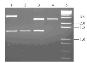
The complete sequence of SARS-CoV nucleocapsid gene is 1278 nucleotides, encode 426 amino acids. The amplified SARS-CoV nucleocapsid gene was purified and cloned into the PCR Blunt Ⅱ TOPO plasmid vector, which contained the unique BamH I and Not I restriction enzyme sites. The positive plasmid DNA was digested with BamH I and Not I, the obtained fragment is the same size (about 1.3kb) with PCR amplified SARS-CoV nucleocapsid gene in the 1% agarose gel (Fig. 2).

Figure 2. Identification of plasmid pTOPO-SARS-N. pTRE-Tight-SARS-N by BamH I and Not I digestion. Lane 1, Recombinant plasmid pTOPO-SARS-N; 2, PCR products of SARS N gene fragment; 3, Recombinant plasmid pTRE-Tight-SARS-N; 4, Plasmid vector pTRE-Tight; 5, 1kb Ladder plus DNA Marker.
The positive plasmid p-TOPO-SARS-N was sequenced by using BigDye reagents version 3.1. Sequence analysis showed the cloned SARS-CoV nucleocapsid gene was 1 278 nucleotides, nucleotide 28 123 was mutated from T to G, which will improve the context (Kozak sequence) of the AUG initiation codon; nucleotide 28 131 was mutated from T to G, which will destroy the second in frame ATG codon which is in a better context of translation initiation. These changes were expected to improve the expression level of SARS-CoV nucleocapsid gene. As reported, the cloned SARS-CoV nucleocapsid gene encoded 426 amino acids, deduced molecular weight was 46 350.46 Daltons; There are 61 strongly basic (+) amino acids (K, R), 36 strongly acidic (-) amino acids (D, E), 102 hydrophobic amino Acids (A, I, L, F, W, V), 138 polar amino acids (N, C, Q, S, T, Y).
The 1.3 kb fragment was recovered and ligated with the pTRE-Tight vector which was digested with the same restriction enzyme BamH I and Not I. The pTRE-Tight is a response plasmid that can be used to inducibly express the interest gene in the Tet-On stable cell lines[6]. The recombinant plasmid pTRE-Tight-SARS-N was constructed by transforming competent TOP10 cells with the ligation mixtures, the positive clone was selected using the antibiotic (Amp+) and the restriction enzyme digestion. As shown in Fig. 2, after digested with restriction enzyme BamH I and Not I, the positive plasmid DNA have two bands, one is the 2.6kb vector band (pTRE-Tight), and one is the 1.3kb SARS-CoV nucleocapsid gene. This result showed the SARS-CoV nucleocapsid gene was inserted into the MCS (multiple clone sites) of pTRE-Tight, which will be responsive to the regulatory proteins in the Tet-On cell line.
-
The functional double stable cell line contained the regulatory and response plasmids. The regulator plasmid pTet-On, which encoded the "reverse" Tet repressor (rTetR) also contains a neomycin-resistance gene. The response plasmids contained the intrested gene to be expressed.
After transfected the BHK-21 cells with the regulatory plasmids pTet-On, plate 2×10⁵cells in each 10cm tissue culture dish containing 10mL of the appropriate complete medium plus varying amounts (100-600μg/mL) of G418. At five days post-transfection, compared with 10%-20% cell death occured in 100-300μg/mL G418 dishes, we found more than 50% cell death in 400-600μg/mL G418 dishes. After two weeks, isolated, healthy cell clones formed in the 400-500μg/ mL G418 dishes. We selected 6 transformants from 400μg/mL G418 dish and 4 from 500 μg/mL dish. 2 stable transformants (BHK-Tet-1, BHK-Tet-2) were expanded with complete medium containing 500 μg/mL G418.
In order to construct the double-stable cell lines which express SARS-CoV nucleocapsid gene, the positive plasmid pTRE-Tight-SARS-N were cotransfected to the BHK-Tet stable cell and with the linear antibiotic marker pPUR. pPUR is a short, purified linear DNA fragments comprised of the antibiotic marker gene, an SV40 promoter, and the SV40 polyadenylation signal. Because of the small size, pPUR is highly effective at generating stable transfectants.
After 48 h of cotransfection, add 0.5-2.0μg/mL puromycin to the complete DMEM medium, examined the viable cells under microscope every two days. As shown in Fig. 3, after 144 h of cotransfection, 90% cells are viable cells without puromycin, more than 50% viable cells in 0.5μg/mL dishes and about 20% viable cells in 1μg/mL dishes, nearly all the cells are death in the dishes with 2μg/mL puromycin. Every four days, replace medium with fresh complete medium plus puromycin. After 2 weeks, only in the dishes with 1μg/mL puromycin, large, healthy colonies were formed. 30 healthy, isolated cell colonies were picked and expanded in individual plates with 1μg/mL puromycin. These expanded cell colonies were called double stable cell BHK-Tet-SARS-N.
-
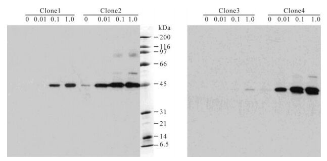
The double-stable BHK-Tet-SARS-N cells were cultured with complete medium containing 500μg/mL G418 and 1μg/mL puromycin, then added different concentrations of doxcycline (0.01-1.0mg/mL). After cultured for 48 h in 37℃, the cells were harvested and run SDS-PAGE gel. As expected, there were very low background SARS-N protein expression in the con-structed BHK-Tet-SARS-N cell strains. The molecular weight of expressed SARS-CoV nucleocapsid protein was about 46 kDa, can specifically react with the anti-SARS-N primary antibody. The expression was tightly regulated by the concentration of doxcycline. As shown in Fig. 4, the expression level of SARS-CoV nucleocapsid protein is low in the BHK-Tet-SARS-N cell clones without doxcycline, even can't be detected by Western-blot in some cell clones (such as clone 1, 3 in Fig. 4); With the improvement of different dose doxcycline, some clones exhibited at least 20-fold induction (such as clone 2, 4 in Fig. 4). Because the level of expression is profoundly affected by the site of regulator protein (rtTA) integration, we isolate and analyze as many as 30 clones to obtain two (clone 2, 4) that exhibits suitably high induction and low background. This two clones were expanded and frozen as double-stable BHK-Tet-SARS-N cell strains, can be used for the rescue of SARS-CoV RNA.

Figure 4. Western-blot analysis of four double-stable cell clones. The cells were cultured in the presence of varying concentration of doxcycline (0mg/mL, 0.01mg/mL, 0.1mg/mL, 1mg/mL). The Anti-SARS-N protein mouse antisera was used as primary antibody and the goat anti-mouse IgG-HRP as secondary antibody.
Amplification of SARS-CoV nucleocapsid gene and construction of the recombinant plasmid
Construction of double-stable cell lines which express SARS-CoV nucleocapsid gene
The tightly regulation of SARS-CoV nucleocapsid gene expression in double-stable cell lines
-
Coronaviruses are positive-sense RNA enveloped viruses that belong to the Coronaviridae family in the Nidovirales order. These viruses are found in a wide variety of animals and can cause respiratory and enteric disorders of diverse severity[11]. HCoV-229E (Human coronavirus 229E), is a prototypic group 1 Coronaviruses, works on HCoV-229E and MHV (mouse hepatitis virus) provided good models for research on SARS-CoV.
It was reported that the HCoV 229E N protein-expressing cell line, which was derived from the BHK-21 cells, designated BHK-HCoV-N, has been produced for the HCoV 229E reverse genetic system. The cell line was based on the Tet-On expression system[18] and allowed the controlled expression of N protein on induction with doxycycline. It was observed that the rescue of the recombinant HCoV 229E after transfection of full-length infectious HCoV RNA was greatly facilitated in the BHK-HCoV-N cell line. The titers of virus in the supernatant of BHK-HCoV-N cells 3 days after transfection were 104TCID50/mL[15]. Similar results had been obtained using the MHV A59 N protein-expressing cell line for the rescue of recombinant MHV A59. So we select BHK-21 cells as target cells to establish the double-stable cell lines for the inducible regulation of SARS-CoV nucleocapsid protein expression. On the other hand, BHK-21cells can be efficiently transfected with RNA, this will facilitate the rescue of the SARS-CoV RNA[16].
Our goal in this study is to generate a cell line that gives low background and high expression of SARS-CoV nucleocapsid protein. The Tet-On system has the advantages of high inducibility and fast response times. With this systems, induction can be detected within 30 minutes and induction levels up to 10 000-fold have been observed[21]. We have screened as many as 100 clones to obtain one that exhibits suitably high induction and low background.
We have shown here that the BHK-21 derived double-stable cell line (BHK-Tet-SARS-N) expressing the SARS-CoV nucleocapsid protein was established based on the Tet-On protein expression system, and the expression of SARS-CoV nucleocapsid protein was tightly controlled by the inducer with the varying concentration of doxycycline in a set of double-stable cell lines. Western-blot analysis showed there were very low background protein expression and very high SARS-CoV nucleocapsid protein expression in some of the cell strains, one of the cell lines will definitely facilitate the efficient rescue of recombinant SARS-CoV in the further research.
Using the constructed BHK-Tet-SARS-N cell lines, we expect to provide a competent host cell for the rescue of SARS-CoV in vitro, so it give the insight to the molecular biology and pathogenesis of SARS-CoV.


 DownLoad: