-
Foot-and-mouth disease virus (FMDV) is a highly infective agent of the Picornaviridae family, affecting all cloven-hoofed animals. FMDV contains a single-stranded, positive sense RNA encoding a single, long open-reading frame protein. Outbreaks of foot-and-mouth disease (FMD) in various countries in recent years have had severe economic impact on the agricultural industry worldwide[10].
In recent years, one of the most important developments in recombinant vaccines is the DNA vaccine, allowing a safe and efficient alternative to conventional vaccination. DNA vaccine technology facilitates the use of cytokines as modulators in vaccination to improve immune responses[14]. In FMD, the virus carrier state is always accompanied by FMD virus antibodies in serum and esophageal-pharyngeal fluid. Vaccinated animals with inactivated vaccine also become virus carriers after FMDV infection, to the same extent as unvaccinated animals[21]. Even in non-persistently infected cattle, viral RNA exists in the soft palate and pharynx over the normal time of virus clearance[25]. These findings indicate that the immune response induced by viral infection or traditional vaccination is not efficient for complete clearance of virus. Recent studies showed that some DNA vaccines could elicit complete protection against the challenge of FMDV[7, 22, 23], which provides us with another choice that is distinct from the traditional inactivated vaccine[24]. The aim of this study is the evaluation of a DNA immunization system using the pcDNA3.1+ plasmid containing the FMDV type O/IRN/1/2007 VP1 gene in mice and comparison with the conventional inactivated vaccine. FMDV type O/IRN/1/2007 causes severe symptoms in Iranian cattle and the goal of this research was to seek improved vaccination against this subtype. Also pCMV-SPORT vector containing the GMCSF gene (Granolocyte-Monocyte colony stimulating factor) was used as molecular adjuvant along with DNA vaccination.
HTML
-
The FMDV type O/IRN/1/2007 isolate was collected from Tehran-Ray in 2007 and cultured on pig kidney cells (IBRS-2) in the Razi Vaccine and Serum Research Institute of Iran and serotyped using polyclonal antibodies against the seven serotypes[15].
-
The first passage of the FMDV was used to extract RNA using an RNeasy Mini Kit (Qiagen) according to the manufacturer's instructions. The concentration of the total RNA was measured by a Nanodrop ND-1000 spectrophotometer.
-
The primer was designed for amplification and cloning of the VP1 gene based on the published FMDV O/2001/UKG sequence (Accession number: DQ165019.1) classified as serotype O PanAsia. The sequences of forward and reverse primers were designed by the AlleleID6 software package. The sequences of specific primers are F: 5'-CGGGGTACCACCATGGTTGACGCTCGCACGCAG-3', R:5'-CGCGGATCCCTATTACAGGTCAAAGTTCAAAAGC-3', respectively. There are Kpn I and BamH I sequences and three overhanging nucleotides at the start of forward and reverse primers, respectively. The forward primer contains the kozak consensus sequence and start codon. The reverse primer contains two stop codons. The extracted RNA was reverse transcribed and amplified using the VP1 gene, the specific primer pair and the AccuPower one-step RT-PCR kit (Bioneer). The PCR product was 699 bp in size and purified by a DNA gel extraction kit (Fermentas).
-
The purified VP1 gene was sub-cloned into the unique Kpn I and BamH I cloning sites of the pcDNA3.1+ vector (Invitrogen) to construct the VP1 gene cassette as used by other researchers and named pcDNA3.1+-VP1[9]. The DH5α strain of E.Coli was transformed with the vector using heat shock and the CaCl2 method. Positive clones were confirmed by restriction enzyme digestion and colony PCR. The confirmed clone was sequenced using the pcDNA3.1+ vector universal primer (T7 Forward)[9, 10, 11]. Also, the VP1 gene without the stop codon was sub-cloned into the unique Kpn I and BamH I cloning sites of the pEGFP-N1 vector to express the GFP-VP1 fusion protein.
-
Four hundred thousand BHKT7 cells were seeded onto cover-slips on six-well plates and incubated at 37℃ in a CO2 incubator until the cells were 50 %-80 % confluent. The following day, 10 μg of plasmid DNA in 100 μL of serum-free Dulbecco's Modified Eagle Medium (DMEM) was mixed with 7 μL of LipofectamineTM reagent (Invitrogen, USA) in 100 μL of serum-free DMEM. The mixture was then incubated at room temperature for at least 30 min before it was diluted into 800 μL reduced-serum DMEM and added to the cells. After incubation for 5 h at 37 ℃ in a humidified incubator, 1 mL of medium containing 5 % fetal calf serum was added to each well[12]. The transfected cells by pEGFP-N1-VP1 and pEGFP-N1 vectors were analyzed by Immunofluorescent microscopy (IF). Also, the transfected cells by pcDNA3.1+vector containing VP1 genes were washed by PBS buffer and harvested by lysis buffer with PMCSF (proteinase inhibitor). The harvested cells were centrifuged at 9500 rpm at 4℃ for 20 min, and supernatant was used for SDS-PAGE and Western blot analysis. The specific band of the VP1 fusion protein was detected by Guinea pig specific polyclonal antibody against FMD virus type O and conjugated rabbit anti Guinea pig antibody with HRP as the second antibody.
-
FMD virus type O/2007/IRN was propagated in BHK21 suspension culture by Earl's modified Eagle's medium (EMEM) and 0.5 % bovine serum at 37 ℃ incubator for 18 h. The Cytopathic Effect (CPE) was observed on BHK21 cells as lyses and suspended detached cells. The virus suspensions were centrifuged at 2500 rpm for 15 min, the supernatants were stored at -70 ℃[9, 11, 15].
-
The Tissue Culture Infection Dose50 /mL (TCID50 /mL) was defined as the number of virus particles per mL that can achieve CPE in 50 % of inoculated cells and was calculated by the Read and Munch method[16].
-
Virus samples were treated by 0.035 mol/L ethylenimine at 30 ℃ for 24 h for virus inactivation, then the inactivation was stopped by adding 0.04 mol/L sodium thiosulphate.
-
Infectivity of inactivated virus sample was determined by the cell culture method. The inactivated virus samples were inoculated on IBRS-2cells by Earl's modified Eagle's medium (EMEM) at 37 ℃ incubator for 18 h, then subcultured on fresh IBRS-2 cells four times. The antigenicity of inactivated FMDV samples were tested by the Complement Fixation test (CF test)[5].
-
The inactivated virus samples were treated by 6/1000 chloroform, absorbed on AL(OH)3 gel and formulated by saponin, glycine, phenol red and phosphate buffer[10]. By this method the inactivated vaccine against FMD Virus type O/IRN/1/2007 by BEI as conventional vaccine (CV) was prepared.
-
Thirty six BALB/c female mice (4-5 weeks old) were provided by the RAZI Vaccine and Serum Research Institute (Karaj, Iran). The BALB/c mice were randomly divided into six groups. Groups 1, 2 and 3 were control groups inoculated with 100 μL PBS, 100 μg of plasmid pcDNA3.1+ and 100 μg of plasmid pCMV-SPORT-GMCSF gene, respectively. Animals in groups 4 were administrated with 100 μg of DNA Vaccine and animals in group 5 were co-administered with DNA Vaccine with pCMV-SPORT-GMCSF vector. Animals in group 6 were inoculated with 100 μL inactivated conventional vaccine. All of the six groups were boosted with the same inoculation at 2 weeks intervals two times. The route of administration for all of the animals was subcutaneous. All groups of mice were bled 10 and 70 days after the last injection. The sera were separated from the blood samples. The complement factors of the sera were inactivated at 56℃ for 30 min. Then sera were tested for the presence of antibodies against FMDV type O by serum neutralization test[16].
-
The sera were diluted in Eagle's maintenance medium in a 2-fold dilution stating from 1:4 to 1:128. The Serum Neutralization Test (SNT) was carried out according to the Kraber protocol[16]. This test was performed on a monolayer of BHK21 cells in flat bottom-96 microplates. Any well in which the virus had been neutralized and for which the cells remained intact was tagged as a positive well and remaining wells in which the virus has not been neutralized and CPE could be shown were tagged as negative wells. Antibody titers were expressed as the logarithm of the reciprocal of the last serum dilution in the virus/serum mixture to neutralize 100 TCID50 of homologous FMDV at the 50 % endpoint.
-
The splenic lymphocytes were removed and cultured using a T-lymphocytes proliferation assay with MTT (3-4, 5-dimethylthiazol-2, 5-diphenyltetrazoliumbromide). Spleens of vaccinated mice were removed aseptically 10 and 70 days after last injection. The single splenic lymphocyte suspensions were prepared and incubated in 96-well plates at 5×10⁴ cells/well by RPMI 1640 plus 10 % of fetal calf serum (FCS) at 37 ℃ in a 5 % CO2. The cells were stimulated with 50 μL of Phytohemagglutinin (50 μg/mL) (positive control), 2.5 μg/mL of 146S antigen of FMDV type O IRN/1/ 2007 (specific antigen stimulation) and no antigen (negative control), in triplicates. After 48 h the MTT assay was performed using a Cell Proliferation Kit 1 (MTT) -Roche, according to manufacturer's instructions and absorbance was detected at 540 nm and stimulation index (SI) was measured.
-
For measurement of IFN-γ, IL-4 and IL-10, splenic lymphocytes were incubated as described above for the proliferation assay. After 48 h, supernatants were collected and different dilutions were assayed in duplicate using commercial ELISA kits for IFN-γ, IL-4 and IL-10 (eBioscience Mouse IFN-g, IL-4 and IL-10 kits).
-
Statistical analysis was done by analysis of variance (ANOVA-One way) followed by Duncan's multiple range test. Significance was defined at P < 0.05.
Virus isolation and serotyping of FMDV antigen
RNA extraction
Primer designing for VP1 gene and RT-PCR
Cloning and sequencing
Expression of FMDV type O IRN/1/2007 VP1 protein in BHKT7 cell
Virus propagation
Virus titration
Inactivation of FMDV type O IRN/1/2007 by Binary ethylenimine (BEI)
Safety test and Complement Fixation test (CF test)
Vaccine formulation
Vaccination of mice
Titration of neutralizing antibodies
Preparation spleen cells and T-lymphocytes proliferation assay
Cytokines assay
Statistical analysis
-
Serotyping of the isolated FMDV antigen was done by ELISA which subsequently showed serotype O. The plasmid DNA, pcDNA3.1+-VP1 cassette, was constructed and confirmed by sequencing and digestion with restriction enzymes Kpn I and BamH I.
-
There are 672 nucleotides and 224 amino acid residues in the VP1 coding region. The nucleotide sequence data was deposited in GenBank with accession number: JF288761.
-
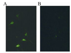
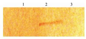
The transfected BHKT7 cells with sub-cloned pEGFP-N1-VP1 vector were expressed with GFP-VP1 fusion protein and displayed more green fluorescence spots than the transfected BHKT7 cells with pEGFP-N1 vector as shown in Fig. 1. The expression of VP1 protein was detected by specific bands in Western blotting analysis, shown in Fig. 2.
-
FMDV type O/IRN/1/2007 titration was calculated by the Read and Munch method and reported 108.5 TCID50 per mL. The Cytopathic Effect was not observed on IBRS-2 cells after three times blind culture of inactivated FMDV type O/IRN/1/2007 samples, therefore the inactivated vaccine was safe. The result of the CF test was also showed the antigenicity of the inactivated virus was not changed.
-
The anti FMDV type O/IRN/1/2007 sera titration of the vaccinated mice 10 and 70 days after last vaccination are shown in Tables 1 and 2, respectively. The mice were immunized subcutaneously using plasmids DNA to express FMDV type O/IRN/1/2007VP1 and GMCSF genes and showed significant differences compared with the negative control groups (the groups which were immunized by PBs, pcDNA3.1+ and PCMV-SPORT-GMCSF) (P < 0.01).

Table 1. The anti FMDV type O/IRN/1/2007 sera titration, MTT assay, IFN-γ, IL-4 and IL-10 concentrations of the vaccinated mice 10 days after last vaccination

Table 2. The anti FMDV type O/IRN/1/2007 sera titration, MTT assay, IFN-γ, IL-4 and IL-10 concentrations of the vaccinated mice 70 days after last vaccination
The results of Tables 1 and 2 indicate that both the mice immunized using co-administered DNA Vaccine with pCMV-SPORT-GMCSF vector and immunized with the inactivated vaccine show protective neutralizing antibody titer against FMDV type O/IRN/1/2007.However the response of vaccinated mice with PBS, pcDNA3.1+, PCMV-SPORT-GMCSF vector and DNA Vaccine were not protective.
-
The results of the MTT assay, IFN-γ, IL-4 and IL-10 concentrations in six groups of mice 10 days after last vaccination are shown in Table 1. Table 2 shows the results of the MTT assay and IFN-γ concentration after 70 days. The SI values of the vaccinated mice groups; DNA Vaccine, DNA Vaccine with PCMV-SPORT-GMCSF vector and conventional vaccine, were significantly higher than control groups (PBS and pcDNA3.1+ vector) at 10 and 70 days after the last vaccination (P < 0.05). The concentration of IFN-γ in group 5 (DNA Vaccine with PCMV-SPORT-GMCSF vector) was significantly higher than the other groups. Also, the IL-4 and IL-10 concentrations in group 6 were significantly higher than the other groups (P < 0.05).
Nucleotide sequence of FMDV/O/IRN/2007 VP1
Expression of FMDV type O IRN/1/2007 VP1 protein in BHKT7 cell
Virus titration, CF and Safety tests
Serum neutralization test
T-Lymphocytes proliferation and cytokines assay
-
Although it is generally accepted that protective immunity to FMDV is principally due to neutralizing antibody, the cellular immune response provides an essential regulatory role in the induction and expression of the serological response. Therefore, an appropriate cellular immune response is essential to resolve FMD[3, 4, 17]. DNA vaccines offer significant advantages over conventional inactivated vaccines or recombinant proteins, as the antigen is expressed intracellularly in the immunized animal.As a result, the processing and presentation of the viral epitopes occur in a way that is similar to natural infection. Indeed, DNA has been demonstrated to stimulate all effectors branches of the immune response, including antibody production, T lymphocyte proliferation response and CTL cytotoxic activity[14, 18]. However, in the absence of suitable adjuvants, DNA vaccination alone can only generate weak immune responses, particularly in the cellular response. Adjuvants, such as the Freund's complete adjuvant (FCA)[6], or QS21[20], are widely used in various vaccine formulations for the enhancement of immune responses. Some cytokines, for example, IL-1[14], IL-12[19], IFN[13], GMCSF and TNF, as immune-modulators, have been reported to be effective in animal models or clinical tests.
Ma et al . showed that DNA vaccine pVIR-P12AIL18-3C co-expressing IL-18 could elicit higher levels of T lymphocyte proliferation response and neutralizing antibody titer than DNA vaccine pVIR-P12A-3C without IL-18 in swine or mice (P < 0.05)[12]. Kim et al. intramuscularly immunized mice with a cDNA expression vector that was constructed to express the VP1 gene of FMDV and showed antibody response to VP1 was increased[10]. Park et al . constructed a plasmid DNA to expressVP1/interleukin-1α in immunized mice. The results showed that although the immunized groups did not carry a high level of neutralizing antibodies, the plasmid encoding the VP1/IL-1α was effective in inducing an enhanced immune response[14].
In the present study, interestingly, subcutaneous injection produced neutralizing antibody titres in mice immunized with plasmid vectors expressing co-administered VP1 and GMCSF genes, the VP1 gene cassette alone, and the conventional inactivated vaccine induced significant amount of antibody titres (P < 0.01). According to previous studies neutralizing antibody titres more than 1.2 are protective for FMDV[8], therefore the immunized mice with co-administered DNA vaccine and pCMV-SPORT-GMCSF vector and the conventional inactivated vaccine showed protective antibody titres, and the negative control groups (the mice which were immunized by PBs, pcDNA3.1+ and PCMV-SPORT-GMCSF vectors) showed no response. The greater neutralizing antibody response in group 5 compared to group 4 indicated that GMCSF induced an immune system as a molecular adjuvant. T-lymphocyte proliferation assay and IFN-γ concentration showed cellular immunity dependent on Th1 was the greatest in group 5 (co-administered DNA vaccine and pCMV-SPORT-GMCSF vector). The concentrations of IL-4 and IL-10 showed Th2, was not increased in groups 4 and 5 (DNA vaccine and co-administered DNA vaccine with pCMV-SPORT-GMCSF vector). Therefore in the mice group 5, which was vaccinated by DNA vaccine with pCMV-SPORT-GMCSF, neutralizing antibody response was protective and the Th1 immunity was the greatest.


 DownLoad: