Citation: Ren-rong TIAN, Qing-jiao LIAO, Xu-lin CHEN. Current Status of Targets and Assays for Anti-HIV Drug Screening .VIROLOGICA SINICA, 2007, 22(6) : 476-485.

Current Status of Targets and Assays for Anti-HIV Drug Screening

  • Corresponding author: Xu-lin CHEN, chenxl@wh.iov.cn
  • Received Date: 18 September 2007
    Accepted Date: 08 October 2007
    Available online: 01 December 2007
  • HIV/AIDS is one of the most serious public health challenges globally. Despite the great efforts that are being devoted to prevent, treat and to better understand the disease, it is one of the main causes of morbidity and mortality worldwide. Currently, there are 30 drugs or combinations of drugs approved by FDA. Because of the side-effects, price and drug resistance, it is essential to discover new targets, to develop new technology and to find new anti-HIV drugs. This review summarizes the major targets and assays currently used in anti-HIV drug screening.

  • 加载中
    1. Amit I, Yakir L, Katz M, et al.2004. Tal, a Tsg101-specific E3 ubiquitin ligase, regulates receptor endocytosis and retrovirus budding. Genes Dev, 18 (14): 1737-1752.
        doi: 10.1101/gad.294904

    2. Baba M, Nishimura O, Kanzaki N, et al.1999. A small-molecule, nonpeptide CCR5 antagonist with highly potent and selective anti-HIV-1 activity. Proc Natl Acad Sci U S A, 96 (10): 5698-5703.
        doi: 10.1073/pnas.96.10.5698

    3. Balzarini J. 2007. Carbohydrate-binding agents: a poten-tial future cornerstone for the chemotherapy of enveloped viruses?. Antivir Chem Chemother, 18 (1): 1-11.
        doi: 10.1177/095632020701800101

    4. Barbouche R, Lortat-Jacob H, Jones I M, et al.2005.Glycosaminoglycans and protein disulfide isomerase-mediated reduction of HIV Env. Mol Pharmacol, 67 (4): 1111-1118.
        doi: 10.1124/mol.104.008276

    5. Barocchi M A, Masignani V, Rappuoli R. 2005. Opinion: Cell entry machines: a common theme in nature? Nat Rev Microbiol, 3 (4): 349-358.
        doi: 10.1038/nrmicro1131

    6. Beck E J, Mandalia S, Gaudreault M, et al.2004. The cost-effectiveness of highly active antiretroviral therapy, Canada 1991-2001. AIDS, 18: 2411-2418.

    7. Bushman F D. 2002. Integration site selection by lentiviruses: biology and possible control. Curr Top Microbiol Immunol, 261: 165-177.

    8. Camarasa M J, Velázquez S, San-Félix A, et al.2006.Dimerization inhibitors of HIV-1 reverse transcriptase, protease and integrase: a single mode of inhibition for the three HIV enzymes?. Antiviral Res, 71 (2-3): 260-267.
        doi: 10.1016/j.antiviral.2006.05.021

    9. Chapman R L, Stanley T B, Hazen R, et al.2002. Small molecule modulators of HIV Rev/Rev response element interaction identified by random screening. Antiviral Res, 54 (3): 149-162.
        doi: 10.1016/S0166-3542(01)00222-4

    10. Cheng T J, Brik A, Wong C H, et al.2004. Model system for high-throughput screening of novel human immunodefi-ciency virus protease inhibitors in Escherichia coli. Antimi-crob Agents Chemother, 48(7): 2437-2447.
        doi: 10.1128/AAC.48.7.2437-2447.2004

    11. Cherepanov P. 2007. LEDGF/p75 interacts with divergent lentiviral integrases and modulates their enzymatic activity in vitro. Nucleic Acids Res, 35 (1): 113-124.
        doi: 10.1093/nar/gkl885

    12. Clapham P R, McKnight A. 2002. Cell surface receptors, virus entry and tropism of primate lentiviruses. J Gen Virol, 83: 1809-1829.
        doi: 10.1099/0022-1317-83-8-1809

    13. Craigie R, Mizuuchi K, Bushman F D, et al.1991. A rapid in vitro assay for HIV DNA integration. Nucleic Acids Research, 19: 2729-2734.
        doi: 10.1093/nar/19.10.2729

    14. Daelemans D, De Clercq E, Vandamme A M. 2001. A quantitative GFP-based bioassay for the detection of HIV-1 Tat transactivation inhibitors.J Virol Methods, 96 (2): 183-188.
        doi: 10.1016/S0166-0934(01)00330-5

    15. David C A, Middleton T, Montgomery D, et al.2002.Microarray compound screening (microARCS) to identify inhibitors of HIV integrase. J Biomol Screen, 7 (3): 259-2566.

    16. Ehrlich L S, Liu T, Scarlata S, et al.2001. HIV-1 capsid protein forms spherical (immature-like) and tubular (mature-like) particles in vitro: structure switching by pH-induced conformational changes. Biophys J, 81 (1): 586-594.
        doi: 10.1016/S0006-3495(01)75725-6

    17. Fields B M, Peter M, Howley M D, et al. 2001. Fields-Virology (4th Edition), New York: Lippincott Williams & Wilkins. 1635-1636.

    18. Franke R, Hirsch T, Eichler J. 2006. A rationally designed synthetic mimic of the discontinuous CD4-binding site of HIV-1 gp120. J Recept Signal Transduct Res, 26 (5-6): 453-460.
        doi: 10.1080/10799890600923179

    19. Freed E O. 2002. Viral late domains. J Virol, 76: 4679-4687.
        doi: 10.1128/JVI.76.10.4679-4687.2002

    20. Gabbara S, Davis W R, Hupe L, et al.1999. Inhibitors of DNA strand transfer reactions catalyzed by HIV-1 reverse transcriptase. Biochemistry, 38 (40): 13070-13076.
        doi: 10.1021/bi991085n

    21. Gallina A, Hanley T M, Mandel R, et al.2002. Inhibitors of protein-disulfide isomerase prevent cleavage of disulfide bonds in receptor-bound glycoprotein 120 and prevent HIV-1 entry. J Biol Chem, 277 (52): 50579-50588.
        doi: 10.1074/jbc.M204547200

    22. George J, Teear M L, Norey C G, et al.2003. Evaluation of an imaging platform during the development of a FRET protease assay. J Biomol Screen, 8 (1): 72-80.
        doi: 10.1177/1087057102239778

    23. Goff S P, Tachedjan G, O'Hara B M. 2004. Two hybrid assay that detects HIV-1 reverse transcriptase dimerization. US20046-812025 A1.

    24. Gottwein E, Kr usslich H G. 2004. Analysis of human immunodeficiency virus type 1 Gag ubiquitination. J Virol, 79 (14): 9134-9144.

    25. Greene W C, Stopak K S, deNoronha C M C, et al. 2005. Methods for treating lentivirus infections. US20050053977 A1.

    26. Groot F, Geijtenbeek T B, Sanders R W, et al. 2005. Lactoferrin prevents dendritic cell-mediated human imm-unodeficiency virus type 1 transmission by blocking the DC-SIGN--gp120 interaction. J Virol, 79 (5): 3009-3015.
        doi: 10.1128/JVI.79.5.3009-3015.2005

    27. Gulick R M, Mellors J W, Havlir D, et al. 1997.Treatment with indinavir, zidovudine, and lamivudine in adults with human immunodeficiency virus infection and prior antiretroviral therapy. N Engl J Med, 337: 734-739.
        doi: 10.1056/NEJM199709113371102

    28. Hammer S M, Squires K E, Hughes M D, et al.1997. A controlled trial of two nucleoside analogues plus indinavir in persons with human immunodeficiency virus infection and CD4 cell counts of 200 per cubic millimeter or less. AIDS Clinical Trials Group 320 Study Team. N Engl J Med, 337: 725-733.
        doi: 10.1056/NEJM199709113371101

    29. Hansen M S, Smith G J, Kafri T, et al.1999. Integration complexes derived from HIV vectors for rapid assays in vitro. Nat Biotechnol, 17 (6): 578-582.
        doi: 10.1038/9886

    30. Hirsch M S, Curran J. 1990. Human immunodeficiency viruses. In: Fields, editor. Virology, 4th ed. Philadelphia: Lippincott-Raven Publishers; p. 1953-1975.

    31. Hussein G, Miyashiro H, Nakamura N, et al. 1999. Inhibitory effects of Sudanese plant extracts on HIV-1 replication and HIV-1 protease. Phytother Res, 13 (1): 31-36.
        doi: 10.1002/(ISSN)1099-1573

    32. Hwang Y, Rhodes D, Bushman F. 2000. Rapid microtiter assays for poxvirus topoisomerase, mammalian type IB topoisomerase and HIV-1 integrase: application to inhibitor isolation. Nucleic Acids Res, 28 (24): 4884-4892.
        doi: 10.1093/nar/28.24.4884

    33. Jenkinson S, McCoy D C, Kerner S A, et al. 2003. Development of a novel high-throughput surrogate assay to measure HIV envelope/CCR5/CD4-mediated viral/cell fusion using BacMam baculovirus technology. J Biomol Screen, 8 (4): 463-470.
        doi: 10.1177/1087057103255747

    34. Jones A E, Saksela K, Game S M, et al.1998. Screening Assay for the Detection of the Protein-Protein Interaction Between HIV-1 Nef Protein and the SH3 Domain of Hck. J Biomol Screen, 3 (1) : 37-39.
        doi: 10.1177/108705719800300105

    35. Karvinen J, Hurskainen P, Gopalakrishnan S, et al. 2002. Homogeneous time-resolved fluorescence quenching assay (LANCE) for caspase-3. J Biomol Screen. 7 (3):223-231.
        doi: 10.1177/108705710200700306

    36. Klinger P P, Schubert U. 2005. The ubiquitin-proteasome system in HIV replication: potential targets for antiret-roviral therapy. Expert Rev Anti Infect Ther, 3 (1): 61-79.
        doi: 10.1586/14787210.3.1.61

    37. Lanman J, Sexton J, Sakalian M, et al.2002. Kinetic analysis of the role of intersubunit interactions in human immunodeficiency virus type 1 capsid protein assembly in vitro. J Virol, 76: 6900-6908.
        doi: 10.1128/JVI.76.14.6900-6908.2002

    38. Lemaitre M, Phan T, Downes M J, et al.1992. Poly r(A) reverse transcriptase (3H) SPA, a new enzyme assay system. Antiviral Res, 17 (Supp. 1): 48-54.

    39. Lindsten K, Uhlíková T, Konvalinka J, et al. 2001.Cell-based fluorescence assay for human immunodefi-ciency virus type 1 protease activity. Antimicrob Agents Chemother, 45 (9): 2616-2622.
        doi: 10.1128/AAC.45.9.2616-2622.2001

    40. Liu S, Jiang S. 2004. High throughput screening and characterization of HIV-1 entry inhibitors targeting gp41: theories and techniques. Curr Pharm Des, 10 (15): 1827-1843.
        doi: 10.2174/1381612043384466

    41. Llano M, Delgado S, Vanegas M, et al.2004. Lens epithelium-derived growth factor/p75 prevents proteasomal degradation of HIV-1 integrase. J Biol Chem, 279 (53): 55570-75557.
        doi: 10.1074/jbc.M408508200

    42. Llano M, Saenz D T, Meehan A, et al.2006. An essential role for LEDGF/p75 in HIV integration. Science, 20; 314 (5798): 461-464.

    43. Lu M, Blacklow S C, Kim P S. 1995. A trimeric structural domain of the HIV-1 transmembrane glycoprotein. Nat Struct Biol, 2 (12): 1075-1082.
        doi: 10.1038/nsb1295-1075

    44. Maertens G, Cherepanov P, Pluymers W, et al.2003.LEDGF/p75 is essential for nuclear and chromosomal targeting of HIV-1 integrase in human cells. J Biol Chem, 278 (35): 33528-33539.
        doi: 10.1074/jbc.M303594200

    45. Markovic I, Stantchev T S, Fields K H, et al.2004 Thiol/disulfide exchange is a prerequisite for CXCR4-tropic HIV-1 envelope-mediated T-cell fusion during viral entry. Blood, 103 (5): 1586-1594.
        doi: 10.1182/blood-2003-05-1390

    46. Masso M. 2003. DC-SIGN points the way to a novel mechanism for HIV-1 transmission.MedGenMed, 5 (2): 2.

    47. Matsumoto C, Hamasaki K, Mihara H, et al.2000. A high-throughput screening utilizing intramolecular fluores-cence resonance energy transfer for the discovery of the molecules that bind HIV-1 TAR RNA specifically. Bioorg Med Chem Lett, 10 (16): 1857-1861.
        doi: 10.1016/S0960-894X(00)00359-0

    48. Mei H Y, Mack D P, Galan A A, et al.1997. Discovery of selective, small-molecule inhibitors of RNA complexes-Ⅰ. The Tat protein/TAR RNA complexes required for HIV-1 transcription. Bioorg Med Chem, 5 (6): 1173-1184.
        doi: 10.1016/S0968-0896(97)00064-3

    49. Miller M D, Farnet C M, Bushman F D. 1997. Human immunodeficiency virus type 1 preintegration complexes: studies of organization and composition. J Virol, 71 (7): 5382-5390.

    50. Montagnier L. 2002. Historical essay. A history of HIV discovery. Science, 298 (5599): 1727-1728.
        doi: 10.1126/science.1079027

    51. Moore J P, Jameson B A, Weiss R A, et al. 1993. The HIV-cell fusion reaction. Viral fusion mechanisms (Bentz J. ed. ), Boca Raton, Florida: CRC; p. 233-290.

    52. Moore R D. 2000. Cost effectiveness of combination HIV therapy: 3 years later. Pharmacoeconomics, 17: 325-330.
        doi: 10.2165/00019053-200017040-00002

    53. Nguyen D H, Hildreth J E. 2000. Evidence for budding of human immunode-ficiency virus type 1 selectively from glycolipid-enriched membrane lipid rafts. J Virol, 74: 3264-3272.
        doi: 10.1128/JVI.74.7.3264-3272.2000

    54. Nobile C, Moris A, Porrot F, et al.2003. Inhibition of human immunodeficiency virus type 1 Env-mediated fusion by DC-SIGN. J Virol, 77 (9): 5313-5323.
        doi: 10.1128/JVI.77.9.5313-5323.2003

    55. Ono A, Freed E O. 2001. Plasma membrane rafts play a critical role in HIV-1 assembly and release. Proc Natl Acad Sci USA, 98: 13925-13930.
        doi: 10.1073/pnas.241320298

    56. Palella F J Jr, Delaney K M, Moorman A C, et al. 1998. Declining morbidity and mortality among patients with advanced human immunodeficiency virus infection. HIV Outpatient Study Investigators. N Engl J Med, 338 (13): 853-860.
        doi: 10.1056/NEJM199803263381301

    57. Parniak M A, Min K L, Budihas S R, et al. 2003. A fluorescence-based high-throughput screening assay for inhibitors of human immunodeficiency virus-1 reverse transcriptase-associated ribonuclease H activity. Anal Biochem, 322: 33-39.
        doi: 10.1016/j.ab.2003.06.001

    58. Piccinini M, Mostert M, Rinaudo M T. 2003. Protea-somes as drug targets. Curr Drug Targets. 4 (8): 657-671.

    59. Piccinini M, Rinaudo M T, Chiapello N, et al.2002. The human 26S proteasome is a target of antiretroviral agents. AIDS, 16 (5): 693-700.
        doi: 10.1097/00002030-200203290-00004

    60. Pommier Y, Johnson A A, Marchand C. 2005. Integrase inhibitors to treat HIV/AIDS. Nat Rev Drug Discov, 4 (3): 236-248.
        doi: 10.1038/nrd1660

    61. Rychetsky P, Hostomska Z, Hostomsky Z, et al.1996.Development of a Nonradioactive Ribonuclease H Assay. Analytical Biochemistry. 239: 113-115.
        doi: 10.1006/abio.1996.0300

    62. Schade S Z, Jolley M E, Sarauer B J, et al. 1996.BODIPY-alpha-casein, a pH-independent protein substrate for protease assays using fluorescence polarization. Anal Biochem. 243 (1): 1-7.
        doi: 10.1006/abio.1996.0475

    63. Schroeder A, Shinn P, Chen H, et al. 2002. HIV-1 integration in the human genome favors active genes and hotspots. Cell, 110: 521.
        doi: 10.1016/S0092-8674(02)00864-4

    64. Sluis-Cremer, N, Parniak M, Pelletier A, et al. 2004. Assay for identifying inhibitors of HIV RT dimerization. US 2004-6811970A1.

    65. Stebbins J, Debouck C. 1997. A microtiter colorimetric assay for the HIV-1 protease.Anal Biochem, 248 (2): 246-250.
        doi: 10.1006/abio.1997.2111

    66. Stephen A G, Worthy K M, Towler E, et al. 2002.Identification of HIV-1 nucleocapsid protein: nucleic acid antagonists with cellular anti-HIV activity. Biochem Bio-phys Res Commun, 296 (5): 1228-1237.
        doi: 10.1016/S0006-291X(02)02063-6

    67. Stricher F, Martin L, Barthe P, et al.2005. A high-throughput fluorescence polarization assay specific to the CD4 binding site of HIV-1 glycoproteins based on a fluorescein-labelled CD4 mimic. Biochem J, 390 (Pt 1): 29-39.

    68. Tang R Y, Su Y. 1997. Construction of a cell-based high-flux assay for the rev protein of HIV-1. J Virol Methods, 65 (2): 153-158.
        doi: 10.1016/S0166-0934(97)02176-9

    69. Tummino P J, Scholten J D, Harvey P J, et al.1996. The in vitro ejection of zinc from human immunodeficiency virus (HIV) type 1 nucleocapsid protein by disulfide benzamides with cellular anti-HIV activity. Proc Natl Acad Sci USA, 93 (3): 969-973.
        doi: 10.1073/pnas.93.3.969

    70. Van Maele B, Debyser Z. 2005. HIV-1 integration: an interplay between HIV-1 integrase, cellular and viral proteins. AIDS Rev, 7: 26-43.

    71. Vandekerckhove L, Christ F, Van Maele B, et al.2006. Transient and stable knockdown of the integrase cofactor LEDGF/p75 reveals its role in the replication cycle of human immunodeficiency virus. J Virol, 80(4): 1886-1889.
        doi: 10.1128/JVI.80.4.1886-1896.2006

    72. Vermeire K, Schols D. 2005. Anti-HIV agents targeting the interaction of gp120 with the cellular CD4 receptor. Expert Opin Investig Drugs, 14 (10): 1199-1212.
        doi: 10.1517/13543784.14.10.1199

    73. Weiss R A. 1993. Cellular receptors and viral glyco-proteins involved inretrovirus entry. The retroviridae (Levy J A. ed.), New York: Plenum Press; 2:1-108.

    74. Westby M, Nakayama G R, Butler S L, et al. 2005.Cell-based and biochemical screening approaches for the discovery of novel HIV-1 inhibitors. Antiviral Res, 67 (3): 121-140.
        doi: 10.1016/j.antiviral.2005.06.006

    75. Wilk T, Gross I, Gowen B E, et al.2001. Organization of immature human immunodeficiency virus type 1. J Virol, 75: 759-771.
        doi: 10.1128/JVI.75.2.759-771.2001

    76. Xu Y, Hixon M S, Dawson P E, et al.2005. Development of a FRET Assay for Monitoring of HIV gp41 Core Disruption. J Org Chem, 72 (18): 6700-6707.

    77. Xuei X, David C A, Middleton T R, et al.2003. Use of SAM2 biotin capture membrane in microarrayed compound screening (muARCS) format for nucleic acid polymeri-zation assays. J Biomol Screen, 8 (3): 273-282.
        doi: 10.1177/1087057103008003005

    78. Zhang G H, Wang Q, Chen J J, et al.2005. The anti-HIV-1 effect of scutellarin. Biochem Biophys Res Commun, 334 (3): 812-816.
        doi: 10.1016/j.bbrc.2005.06.166

    79. Zhao Q, He Y, Alespeiti G, Debnath A K. 2004. A novel assay to identify entry inhibitors that block binding of HIV-1 gp120 to CCR5. Virology, 326 (2): 299-309.
        doi: 10.1016/j.virol.2004.06.022

  • 加载中

Figures(4) / Tables(1)

Article Metrics

Article views(4233) PDF downloads(16) Cited by()

Related
Proportional views

    Current Status of Targets and Assays for Anti-HIV Drug Screening

      Corresponding author: Xu-lin CHEN, chenxl@wh.iov.cn
    • State Key Laboratory of Virology, Wuhan Institute of Virology, Chinese Academy of Sciences, Wuhan 430071, China

    Abstract: HIV/AIDS is one of the most serious public health challenges globally. Despite the great efforts that are being devoted to prevent, treat and to better understand the disease, it is one of the main causes of morbidity and mortality worldwide. Currently, there are 30 drugs or combinations of drugs approved by FDA. Because of the side-effects, price and drug resistance, it is essential to discover new targets, to develop new technology and to find new anti-HIV drugs. This review summarizes the major targets and assays currently used in anti-HIV drug screening.

    • Since the discovery of Human immunodeficiency virus (HIV) and acquired immunodeficiency syndrome (AIDS) in 1981 (50), this disease has become one of the most significant public health challenges globally. HIV/AIDS have claimed the lives of more than 25 million people worldwide. The year 2006 marks the 10th anniversity of the introduction of highly active antiretroviral therapy (HAART), a combination of three antiretrovirals (ARVs) from at least two drug classes (27, 28), that has led to significant reductions in HIV-related morbility and mortality (6, 52, 56). However, HIV virus is capable of mutating rapidly to develop drug resistance, so it is imperative to develop more effective and safe drugs to overcome the growing resistance of the virus. New molecules may block the known viral targets or other new ones. Currently there are 23 FDA-approved individual antiretroviral drugs that are classified into four categories based on their mechanism of action. There are 7 combinations of drugs currently approved by FDA. Additional agents are in various stages of clinical and preclinical development.

    • HIV-1 is an enveloped virus that contains two copies of viral genomic RNA in its core. The approximately 9 kb RNA genome encodes at least 9 proteins: Gag, Pol, Env, Tat, Rev, Nef, Vif, Vpu and Vpr, of which only the former five are essential for viral replication in vitro. The infection begins with the attachment of the virions to the cell surface mediated by an interaction among the extracellular domain of HIV-1 gp120, cellular receptors CD4 (51, 73) and chemokine coreceptors CCR5 and CXCR4 (12). After binding to coreceptor, viral and cellular membranes fuse and the viral core is released into the cytoplasm of the cell. The viral RNA genome is retrotranscribed into a full-length double-stranded DNA by the viral reverse transcriptase (RT) (30). Linear doublestranded DNA in the preintegration complex inserts into the host chromosome by the viral integrase (7, 63, 70). Expression of viral genes leads to production of precursor viral proteins. These proteins and viral RNA are assembled at the cell surface into new viral particles and leave the host cell by budding (19, 53, 55). Budding triggers the activation of the protease (PR) that autocatalytically cleaves the Gag and Gag-Pol polyprotein releasing the structural proteins and enzymes. The individual proteins undergo further interactions, with capsid and nucleocapsid protein forming the conic nucleocapsid, and matrix protein remaining associated to the viral envelope (16, 37, 75).

    • HIV entry is a critical event of HIV-1 life cycle. It is triggered by gp120 attachment to CD4, followed by gp120 engagement with a co-receptor (either CCR5 or CXCR4). Binding of HIV to co-receptors causes conformational changes in the envelope proteins, ultimately resulting in the fusion of the viral envelope and the host cytoplasmic membrane (5). As the earliest event, HIV entry represents an attractive target. In the past, this entry process between virus and host cell or transmission between cell and cell have been reestablished in vitro through using virus with reporter acceptor cell, reporter virus with reporter cells, in-fected cell and reporter cell or two reporter cell (74). But in recently years, more methods targeting one step of entry have been developed. Now, there are three types of inhibitors including: attachment inhibitors, co-receptor binding inhibitors and fusion inhibitors. Using soluble recombinant gp120 and recombinant CD4 can mock the attachment of virus to CD4+ cells in vitro, and an ELISA was developed (72). When synthetic mimetic peptides of the CD4-binding site of HIV-1 gp120 or the gp120-binding site of CD4 were used, this method was improved and new methods (such as fluorescence polarization as shown in Fig. 1) were developed with more convenience (18, 67). As one of hottest targets for entry, the binding of gp120-gp41 to co-receptor can be blocked by che-mokine antagonists. In early days, a cell-based ELISA assay using radio-labeled chemokine was widely used to measure the inhibitory activity of compounds (2). But now more other convenient methods are also used, such as baculovirus based assay and flowcytometry based assay or ELISA-based assay (33, 79). As a hottest target for entry, the process of rearrangement of gp41 to form the six-helix bundle has been investigated more intensively. Liu et al reestablished the six-helix bundles using C34 and N36 peptides in vitro (43). Based on that, many methods such as Sandwich ELISA, FLISA and FRET were developed to target this process and used in entry inhibitor screening with more convenience (76, 40).

      Figure 1.  Fluorescence polarization in screening inhibitors of gp120-CD4 binding and protease (18, 67, 62). In screening inhibitors of binding, A and B represent fluorescein-labelled CD4 peptide and mocked CD4 binding site of HIV-1 glycoproteins respectively. In screening inhibitors of protease, A and B represent fluorescein and antigen labelled peptide substrate and antidody respectively.

    • Reverse transcription is an important event of HIV life cycle. It was catalyzed by reverse transcriptase, which was encoded by HIV pol gene and consists of a 66 kDa polypeptide and a 51 kDa polypeptide with separate DNA polymerase and ribonuclease H (RNase H) domains in the 66 kDa polypeptide. Both the DNA polymerase and RNase H activities of RT are required for viral replication. In the infected cell, HIV-1 RT ultimately transcribes a single-stranded viral RNA template into double-stranded DNA (dsDNA) through a multi-step process: (ⅰ) RNA-dependent DNA polymerization to produce a (−) DNA copy, with (ⅱ) concomitant cleavage of the RNA strand of the heteroduplex by RNase H, and (ⅲ) DNA-dependent DNA polymerization to yield the dsDNA product. During this process, template switching by the newly synthesized (−) DNA takes place at least twice (21). RT inhibitors are the first HIV-1 drugs marketed and currently serve as the backbone of most frontline HIV combination therapy. Now there are three kinds of assays available for HIV inhibitor screening. First, assays for measuring DNA polymerase activity, such as Scintillation Proximity Assay(SPA)(Fig. 2), Microarray Compound Screening technology (µARCS) and enzyme immunoassays (EIAs) (38, 77, 78). In µARCS, the nucleic acid substrate was biotinylated on one end and was bound to the SAM membrane. A low melting-point agarose gel containing the rest of the reaction components was first placed on a polystyrene sheet spotted with compounds to allow passive diffusion of the compounds into the gel. The gel was removed from the compound sheet and applied to the SAM membrane with the immobilized nucleic acid template to initiate the polymerization. After the incubation, the membrane was washed with a high-salt buffer and exposed for imaging (77). Second, assays for measuring RNase H activity. In 1996, Rychetsky first development an EIA using a duplex substrate labeled with biotin and digoxigenin to high throughput screen inhibitor of RNase H (61). Recently, this method was development as a FRET assay that labels the duplex with fluorescein and Dabcyl H (Fig. 3) (57). Third, assays for measuring polymerase activity and RNase activity simultaneously. In this kind of assay, SPA assay was also used widely (20).

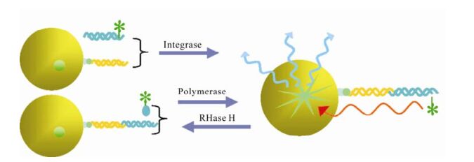
      Figure 2.  Scintillation Proximity Assay (SPA) in screening inhibitors of polymerase, RNase and integrase (5, 20, 38). When certain radioactive atoms decay, they release β-particles. The distance these particles travel through an aqueous solution is dependent on the energy of the particle. If a radioactive molecule is held in close enough proximity to a SPA Scintillation Bead or a SPA Imaging Bead, the decay particles stimulate the scintillant within the bead to emit light, which is then detected in a PMT-based scintillation counter or on a CCD-based imager respectively. However, if a radioactive molecule is free in a solution containing SPA beads, the decay particles will not have sufficient energy to reach the bead and no light will be emitted. This discrimination of binding by proximity means that no separation of bound and free ligand is required.

      Figure 3.  FRET and HTRF assays in screening inhibitor of HIV-1 protease and RNase H (22, 57). In FRET assays, F represents fluorescent donor and Q represents quenching acceptor. In HTRF assays, F represents Eu and Q represents quenching acceptor.

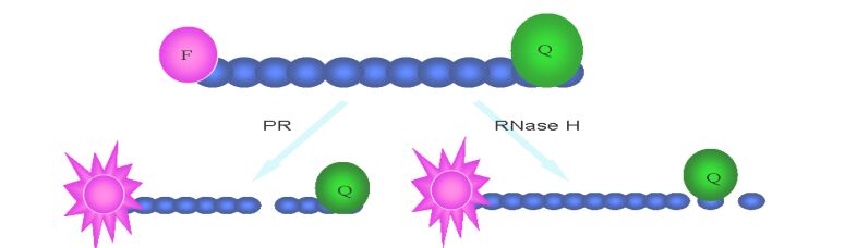
    • HIV protease is a 22 kDa homodimer aspartyl protease, which can cleave HIV Gag and Pol polypeptide precursors to form mature structural and enzymatic gene products. HIV-1 PR activity is critical for viral replication, and so it is also an ideal target for HIV therapy. In the past years, HPLC analysis of production of cut synthetic peptide substrate was used widely in protease inhibitor identification (31). But this method is too time and laborious-consuming to screen large number of samples. Subsequently, a colorimetric assay using unlabeled peptide substrate was also used with less time-cost but lower sensitivity (65). Nowadays, one synthetic peptide substrates typically consisting of a cleavage sequence flanked with fluorescent donor and acceptor labels was universally used, and the method was developed as FRET (Fig. 3). Although EDANSC/DABCYL pair is most commonly donor/acceptor labeling the peptide substrate in FRET assay, there are also other pairs with more advantage than them, such as Hilyte fluor 488/QXL 520, Hilyte Fluor TR/Qxl 620 and Cy3/ Cy5Q (22). There are also other fluorescence and label technology, such as homogeneous time-resolved fluorescence (HTRF) assays (Fig. 3), alphascreen (Fig. 4) (Perkin Elmer, Alphascreen Technical Manual) and fluorescence polarization (Fig. 1) (35, 62). In addition to enzyme assays in vitro, a number of cell-based assays have been reported for HIV-1 protease. In these assays, HIV PR expression plasmid is transfected into the reporter cell or cotransfected with plasmid in-cluding reporter gene, and then repoter gene is induced to express. Ultimately, through measuring cell viability and expression level of reporter gene, the activity of the compound is evaluated (10, 39).

      Figure 4.  Alphascreen assay in Screening inhibitors of protease (35).

    • HIV IN is a 32kDa dimmer protein encoded by 3'-end of pol gene. It comprises three structural domains: the aminal terminal domain (NTD) with HHCC motif, catalyze core domain (CCD) and carboxy terminal domain (CTD) with overall SH3 fold and nonspecifical binding activity to DNA required for process of integration. Through two consecutive steps: 3'-processing and strand transferring, it can insert the viral cDNA ends into host chromosomes. Integrase is essential for retroviral replication, and the absence of a host-cell quivalent of integrase means that the integrase inhibitors do not interfere with normal cellular processes, and therefore have a high therapeutic index (60). The screening and discovery of integrase inhibitors generally relies primarily on simple assays that use recombinant integrase and short oligonucleotide substrates that mimic the viral DNA ends. At the beginning of the screen, EIA is used commonly. It immobilized the donor dsDNA onto microplate, and then enzyme and target dsDNA being labeled were added, followed by ligated production quantified with the method compared to label (13, 32). With the same protocol, a more high throughput method was developed with donor dsDNA immobi-lized on membranes and targets DNA labeled with flurescein (15). Using this method, a library of 250, 000 compounds was screened for IN activity. In 2004 a method more compatible with HTS using robotics was established. Different from EIA, in SPA assay (Fig. 2) the median immobilized donor DNA was replaced by SPA beads, and the label was changed to isotope compared with the measurement method of scintillation counting (5). Besides recombinant enzyme based assays, there is a more authentic assay mimicing the reaction in vivo. The double-strand DNA targets are immobilized on 96-well plate, and preintegration complex (PIC) containing host and virus protein is added. After reaction is completed, the products are quantified by PCR (29, 49).

    • It has been proved that the formation of homodimer, pseudohomodimers or multimers play a central role in function of HIV RT, PR and IN and disruption of protein-protein interactions in enzymes may constitute an alternative way to achieve HIV-1 inhibition (8). Now a hybrid assay and an affinity assay were used in identifying inhibitors of HIV RT dimerization and many inhibitors have been found (23, 64). For in-tegrase and protease, only a few compounds were found with activity against formation of dimmer or multimer, but there aren't valuable assays targeting these two proteins.

    • In addition to the viral structural proteins (Gag and Env), and the pol-encoded enzymes (PR, RT, and IN), the HIV genome encodes several additional structural proteins (NC, MA), regulatory proteins (Rev and Tat) and accessory proteins, such as Vif, Vpu, Vpr, Vpx, and Nef. More recent tissue culture and in vivo experiments have revealed a strong requirement for these gene products for efficient virus replication and disease induction (17). And many assays have been developed targeting these proteins (Table 1). Using such assays more drugs with mechanisms different from recent approved drugs may be found in future.

      Table 1.  Other targets used for anti-HIV/AIDS drug screening

    • DC-SIGN, a lectin expressed on dendritic cell and macrophage subsets, binds to human immunodeficiency virus Env glycoproteins, allowing capture of viral particles. Captured virions either infect target cells or are efficiently transmitted to lymphocytes (46). It was found that competition between CD4, bovine lacto-ferrin or lactoferrin and DC-SIGN for Env binding likely can affect virus access to the cytosol, syncytium formation and prevent dendritic cellmediated human immunodeficiency virus type 1 transmission (26, 54). A group of carbohydratebinding agents (CBAs) also have this effect (3). These findings indicate that it is a potential target for anti-HIV therapy, but the side-effects resulted from blocking this receptor haven't been clarified and this may obstruct inhibitor deve-lopment.

    • Lens epithelium-derived growth factor/transcription co-activator p75 (LEDGF/p75) protein is an essential HIV integration cofactor (42). It forms a specific nuclear complex with integrase through interaction with both the N-terminal zinc binding domain and the central core domains of IN. And this interaction was conserved within and limited to lentivirus and is intimately involved in the catalysis of lentiviral DNA integration (11). This interaction with LEDGF/p75 accounts for the karyophilic properties, chromosomal targeting of HIV-1 IN, protection of HIV-1 IN from the proteasome (41, 44). The data from competition of the IBD fusion proteins with endogenous LEDGF/p75 for binding to integrase provide proof of concept for the LEDGF/p75-integrase interaction as a novel target for treating HIV-1 infection (71).

    • Conformational changes within the human immu-nodeficiency virus-1 (HIV-1) surface glycoprotein gp120 result from binding to the lymphocyte surface receptors and trigger gp41-mediated virus/cell me-mbrane fusion. The triggering of fusion requires cleavage of two of the nine disulfide bonds of gp120 by a cell-surface protein disulfide-isomerase (PDI). Using surface plasmon resonance, Barbouche et al found heparin and heparan sulfate can reduce PDI-mediated gp120 reduction by approximately 80%. And it was also found that interaction of Env with the surface of lymphocytes being treated with sodium chlorate, an inhibitor of glycosaminoglycan synthesis, led to reduction of gp120 (4). Gallina et al reported that monoclonal antibodies to protein-disulfide isom-erase (PDI) and other membrane-impermeant PDI inhibitors prevented HIV-1 infection (21). Markovic also found this phenomenon with other anti-PDI agents (45). And exogenously added PDI, in turn, can rescue fusion from this blockade (45). All these events described provide clues for identifying new potential targets for screening of new anti-HIV agents.

    • Proteasomes constitute the degradative machinery of the ubiquitin/adenosine triphosphate-dependent proteolytic pathway, which is involved in many cell functions, including immune response and apoptosis (59). Recent findings showed that proteasome inhibitors are described to interfere with HIV matu-ration, budding and aggressiveness, and cytostatic drugs, as well as antiretroviral agents used in HAART, have been shown to behave in vitro and in cultured cell lines as inhibitors of proteasome proteolytic activity at therapeutic dosages (58). In investigation of the function of the ubiquitin-proteasome system in HIV replication, Gottwein et al observed that, like several other retroviruses, HIV-1 virions contain a small amount of mono-ubiquitinylated Gag proteins (24). In another analogous investigation, Amit et al found that, in addition to one E3-like protein, two E3-type ubiquitin ligases also could act as regulators of HIV budding (1). These ligases might represent interesting targets for therapeutic intervention (36).

    • Although the future of antiretroviral therapy is challenged by side effects and drug resistance, there are prospects for novel drugs. The emerging understan-ding of viral replication may enable the development of new therapeutic targets. With more new assays and small molecule synthesis technology available, more potent new anti-HIV drugs are to be developed and used in the treatment of AIDS.

    Figure (4)  Table (1) Reference (79) Relative (20)

    目录

    /

    DownLoad:  Full-Size Img  PowerPoint
    Return
    Return