-
Dear Editor,
In this study, we aimed to develop a rescue system for genotype Ⅶ NDV by generation of a full-length cDNA comprising a longer ribozyme sequence. In addition to virus rescue, reporter gene expression from transfected mini-genome cDNAs was significantly enhanced by the optimized ribozyme. Although all isolated NDV strains belong to a single serotype, epidemiological studies have revealed that the genotype Ⅶ strain is currently the most prevalent circulating genotype worldwide and is associated with many of the most recent outbreaks in China since 1997 (Liu et al., 2007).
The standard NDV reversegenetics systembased on T7 RNA polymerase was firstreported in a lentogenic LaSota strain in 1999 (Peeters et al., 1999), followed by the successful rescue of infectious rabies viruses (RABVs) from cloned cDNA (Schnell et al., 1994). Although manyviruses within the Mononegavirales order have been recoveredfrom cloned cDNA, the efficiency of virus rescue is several orders of magnitudelower than that of positive-strand RNA viruses (Conzelmann et al., 2004). In this study, we aimed to develop a rescue system for genotypeVII NDV by generation ofa full-length cDNA comprisinga longer ribozyme sequence. Inaddition to virus rescue, reporter gene expression from transfected mini-genome cDNAs was significantly enhanced bythe optimized ribozyme.
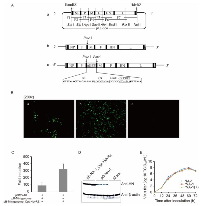
The genotype Ⅶ NDV virus, designated NA-1 (Gen-Bank accessionNo. DQ659677), was isolated from geese in Jilin Province, Northeast China in 1999 (Xu et al., 2008). The virulent strain NA-1 had been rescued from thefull-length antigenomic cDNAusing a reverse geneticssystem, as describedpreviously (Wang et al., 2014). The strategy for assembly of this full-length cDNA clone is shown in Figure 1A. In contrastto the full-length NDV antigenome-like transcript, a 5′-trailer-enhanced green fluorescentprotein (eGFP)-leader-3′ se-quence was insertedbetween the HamRz and HdvRz se-quencesat the Pme I restriction site to construct pB-Mini-genome. The HdvRz sequence wasthen replaced with a longer version (with addition of the sequence AGCCA, 89 nt), as describedpreviously (Ghanem et al., 2012), and a newly generated plasmid, designated pB-Minige-nome_Opt-HdvRz, was cloned by standard techniques. In order to check whether processing could be optimized by using the longer version of HdvRz, the open reading frame (ORF) of the eGFP gene was reverse cloned into the plasmid.Thus, no green fluorescence could be detec-ted unless the viralpolymerase was used. Importantly, pB-Minigenome expressed eGFP when cotransfected with pCI-NP, pCI-P, and pCI-L (Figure 1B-a); however, pB-Minigenome_Opt-HdvRz was found to be more effi-cient in expressingeGFP protein (Figure 1B-b). As pre-dicted, there was no fluorescence in the negative control (Figure 1B-c).

Figure 1. (A) Construction and generation of recombinant NDVs. A schematic representation of the construction of the full-length cDNA clone of pB-NA-1 is shown. Seven cDNA fragments were amplified from the genome, assembled, and subcloned stepwiseinto pCI-neo. The HamRz and HdvRz sequences were introduced into the 5′-and3′-ends ofthe viral genome, respectively (a). An artificial transcription cassette encoding eGFP was inserted into the full-length clone between the P and M genes (b, c). The full-length cDNA clone pB-NA-1was digested with Sal I and Not I and ligated in-to pB-Minigenome_Opt-HdvRz cutwith the same enzymes to generate a novel full-lengthcDNA clone, pB-NA-1_Opt-HdvRz. (B) Analysis of the expression of eGFP by Minigenome plasmids. BHK-21 cells were transfected with pB-Mini-genome (B-a) or pB-Minigenome_Opt-HdvRz (B-b) containing the "helper" plasmids, and the cells were photographed via fluorescence microscopyduring the initial 48 h post-transfection (original magnification, 200x). Cotransfection without pCI-L was performed in parallel as a negativecontrol (B-c). (C) Dual-luciferase assay. The values for fireflyluci-ferase werenormalized to their Renilla luciferase values, and the ratio for transfection with Ndivided by the ratio for transfection without N was considered the fold induction. (D) Western blot analysis of viral proteinexpression. DF1 cells were infected with cell culture supernatants. At 48 h post infection, the cells were lysed, and western blotting was per-formed using anti-HNand anti-β-actin antibodies to compare levels of HN protein.(E) Multistep growth curves of the rescued viruses. Ten-day-old embryonated eggs were inoculated with 100 TCID50 of each virus, and allantoic fluids were harvestedat 12-h intervals. The viral titers(TCID50) in DF1 cells were determined by immunofluo re scence assays. Mean values from triplicate samples are shown.
Standard transfections forminigenome assayswere performed witheither the parental pB-Minigenome or pB-Minigenome_Opt-HdvRz togetherwith the N, P, and L "helper" plasmids. Negative controls were performed by cotransfection withpCMV-RL for normalization of transfection and omission of N. A dual-luciferase assay was performedto determine the levels of firefly lucif-erase and Renilla luciferase. The values forfirefly lucif-erase were normalizedto their Renilla luciferase values, and theratio for transfection with N divided by the ratio for transfection without N was considered the fold induc-tion. The new pB-Minigenome_Opt-HdvRz wa s about four-foldmore efficient than the pB-Minigenome con-taining the common "core" HdvRz sequence (Figure 1C).
The full-length cDNA clonepB-NA-1 was digested with Sal I and Not I and ligated into the pB-Minige-nome_Opt-HdvRz plasmid cut with the same enzymesto generate a novel full-length cDNA clone pB-NA-1_Opt-HdvRz. BHK-21 cells were grown overnight to 70% confluence in 6-well plates, washed with DMEM without FBSthree times, and incubated for 1 h. Ten micrograms of pB-NA-1 or pB-NA-1_Opt-HdvRz was cotransfected with 5 μg pCI-NP, 2.5μg pCI-P, and 2.5 μgpCI-L using a calcium phosphate transfection kit (Invitrogen, Carls-bad, CA, USA). Single transfections with 10 μg pB-NA-1 or pB-NA-1_Opt-Hdv Rz were performed separately as controls.Following incubation for 6 h, the culture medium without FBS was replaced with complete DMEM. After 96 h, the supernatant from each well was harvested; half was used to infect DF-1 cells for western blotting, and the other half was injected into the allantoiccavities of 9-to 11-day-old embryonated SPF eggs. Allantoic fluid was harvestedafter 120 h, and hemagglutination assays (HAs) wereperformed according to standardmethods. The first generation of virus from the pB-NA-1 and pB-NA-1_Opt-HdvRz groups reached mean titers of 3 log2 and 6 log2, respectively, providing indirect evidence that the optimized HdvRz sequences could induce more res-cue events and thereforeincrease virus release into the cell culture supernatant.
DF1 cells were infected with the supernatant from the above wells at the same dose. Afterincubation for 48 h, the cell lysates were harvested, and proteins were separated by sodium dodecylsulfate polyacrylamide gel electrophoresis under denaturing conditions. Gels were then incubatedwith chicken serum anti-NDV or monoclonal antibodies against β-actin. Binding was visualized with 3, 3-diaminobenzidine (DAB) reagent after incubation with peroxidase-conjugated secondary antibodies. Mock-infected DF1 cells were used as the negativecontrol. The proteinlevels were able to be compared using antibodies against NDV HN and β-actin. The HN protein was not expressedat high levels in cells infected with pB-NA-1, whereas HN levels were significantly higher in cells in-fected with pB-NA-1_Opt-HdvRz (Figure 1D), indicat-ing that the full-lengthcDNA plasmid pB-NA-1_Opt-HdvRz was effectively transcribed into full-length anti-genomic RNA and then translated into HN protein.
Ten-day-old embryonated chicken eggs were inoculatedwith the recovered virusesat a 50% tissueculture infective dose (TCID50) of 100 for each egg. Viral growth was analyzed atdifferent time points afterinoculation. The TCID50 of each virus was determinedby immunofluorescence assays. Ninety-six-well plates of 80%-confluent DF1 cells were infectedwith serial 10-fold dilutions of virus (four wells perdilution). Cells wereincu-bated for 3 days and fixed with 80% cold acetone.Viral antigens were detected with chicken serum anti-NDV and FITC-conjugated goat anti-chicken antibodies. There were no discernible differences in the growth rates or maximum titers between the two recoveredviruses and the parental virus strain NA-1 (Figure 1E), indicating that the improved rescue efficiencycould be explained by the enhanced ribozyme activity of the optimized HdvRz sequences.
Previous researchhas demonstrated the importance of generating a correct 3'-endin the antigenomic RNA dur-ing rescue of NNSV from cDNA (Pattnaik et al., 1992). Thefull-length antigenome RNA with correctends could be encapsidated by N protein, and transcription and rep-licationcycles could then begin. Accordingly, incorrect ends resultedin incomplete replication such that no new progenyvirions were produced, followingthe "rule of six" (Calain et al., 1993; Peeters et al., 2000). In Para- myxovirinae, each N covers exactly six nucleotides, and RNAs that do notconsist of multiples of six nucleotides are poorly replicated. The increased rescueefficiency from pB-NA-1_Opt-HdvRz comparedwith pB-NA-1 highlighted that precise 3′-ends of the cRNA were critical for generation of functional RNP templates from naked RNA.
In contrast to the 3′-end, the RNA polymerase seemed to tolerate slight changes in the 5′-ends sequences, supported by the observation that insertion of extended G-bases in the 5′-ends of RNAs improved the efficiency in numerous virus rescue systems. These findings are most important for reverse genetics systems based on T7 RNA polymerase (Wang et al., 2014). For most standard NDV reverse genetics systems, new progeny virions are ob-served rarely in transfected cells until incubated with al-lantoic fluid for 96-120 h. Obviously, this process is la-borious, costly, and time-consuming. Recently, viral hemorrhagic septicemia virus and RABV were rescued by employingtwo separate optimized HdvRz sequences (Ammayappan et al., 2011; Ghanem et al., 2012). For RABV, cDNA clones containingthe combination of the optimized 3′-and5′-ribozymes were rescuedat an at least 100-fold increase (Ghanem et al., 2012). Our result showed that the viral protein could be detected with su-pernatantsafter cotransfection for 96 h, indicatingthat incubation in SPF eggs could be theoretically omitted. Furthermore, the first generation of virus from cDNA reached a mean HA titer of 6 log2, a significant positive signal for rescue efficiency.
To date, virulent NDV strains are still isolated fre-quently from vaccinated birds, demonstrating that NDV remains a sustainedthreat to commercial flocks. Epi-demiological studies have revealed that genotype Ⅶ NDV strains are the predominant strains circulating worldwide and are responsible for ND outbreaks in As ia, Africa, Middle East, and South America (Wang et al., 2006; Perozo et al., 2012). Antigenic differences between the prevailing andvaccine strains mayexplain current ND outbreaks in vaccinated poultry flocks (Miller et al., 2007). As werecently reported, a reverse genetics system was developed based on RNA polymerase II (Wang et al., 2014), and a recombinant genotype Ⅶ NDV ex-pressing the VP3 proteinof goose parvovirus (GPV) was generatedas a bivalent vaccine (Wang et al., 2015). In this study, substitution of the HdvRz "core" sequence with a longer optimized sequence (89 nt) resulted not only in an increasein the number of rescue events of full-length NDV cDNA clones but also in faster rescue. Therefore, this reverse genetics system will provide a powerful tool for the analysisof goose-origin NDV dis-semination and pathogenesis and facilitate the genera-tion of viral vectorsand attenuated vaccines.
HTML
-
YS and MSwere supported by the Innovation Foundation Project of China AnimalHealth and Epidemiology Center, Ministryof Agriculture, P. R. China (project no:CAHEC-2015-Y105). RY and ZD were supportedby the National Natura l ScientificFund (31272561). The funders had norole in the study design, data col-lection and analysis, decision to publish, or preparationof the manuscript. The authors declarethat they have no conflictof in-terest. This article does not contain any stu d ies with human or an-imal subjectsperformed by any of the authors.


 DownLoad: