


-
Dear Editor,
The avian influenza virus (AIV) H5N7 was first isolated from wild birds in North America in 2001(Spackman et al. 2007), and information on only 25 strains of this virus has been deposited in the Global Initiative on Sharing All Influenza Data (GISAID-http://platform.gisaid.org/epi3/frontend#493de3) database until October 30, 2018.Twenty viruses were identified in the United States from 2001 to 2017, three viruses in Denmark in 2003, and two viruses in Mongolia in 2014(GISAID-http://platform.gisaid.org/epi3/frontend#493de3).All these H5N7 viruses were low pathogenicity and were discovered in wild migratory birds.
Wild migratory birds serve as natural reservoir of AIVs and disseminate the viruses during long-distance migration. Wetlands are important for wild migratory birds as stopover for resting and breeding.In China, millions of domestic ducks are also raised near Dongting Lake Wetland Nature Reverse (Hunan Province Rural Social and Economic Investigation Team 2008), and thus, these sites play key roles in the transmission of AIVs from wild birds to domestic birds (Ma et al.2018).Moreover, domestic ducks are usually asymptomatic after AIV infection (Kuchipudi et al.2014), and these healthy appearing ducks may serve as a hidden infection source.For instance, the low-pathogenic AIVs (LPAIVs) H7N9 and H10N8, which cause human infections, were very likely transmitted from wild birds to poultry through domestic ducks (Liu et al.2013; Qi et al.2014).In particular, research has shown that the LPAIV H7N9 became highly pathogenic AIV (HPAIV) after consecutive passages within poultry.If this happened, it will not only cause economic losses to the poultry industry but also pose a great threat to human health (Zhang et al.2017).Herein, we report a novel H5N7 LPAIV that was isolated in China in 2016 from a healthy domestic duck in the Dongting Lake wetland, a key stopover site for migratory waterfowls in the East AsianAustralasian flyway.
From November 2016 to March 2017, we performed routine surveillances in Central China and collected approximately 4, 000 samples from wild birds and ~2, 400 samples from live poultry markets (LPMs).From a fecal sample collected from a healthy duck near Dongting Lake wetland, we isolated a strain of influenza virus H5N7(A/ Duck/Dongting/76-1/2016, DT-H5N7)(Detailed methods were described in the Supplementary Materials).We sequenced the entire viral genome and deposited the sequences in GenBank (accession numbers:MF362100- MF362107).A BLAST search (Detailed phylogenetic analysis was shown in the Supplementary Materials) showed that the HA gene shared 97.2%-97.3% identities with those of H5N2 LPAIVs obtained from wild waterfowls in Xianghai (China) in 2011, and the NA gene was similar to those of H10N7 and H6N7 from wild birds and H7N7 from chicken in Jiangxi Province of China (identities 97.1%-98.9%).The internal genes shared 98.2%-99.5% identities with those of the wild waterfowl viruses circulating in Eurasia during 2008-2016(Table 1).

Table 1. The highest nucleotide similarity analysis of the internal genes of DT-H5N7 isolate in this study
The H5N7 isolate possesses a pattern of PQRETRGLF at the cleavage site of HA, which is an indication of low pathogenicity (Senne et al.1996)(Table 2).At the receptor-binding sites, HA contains Q226 and G228(H3 numbering), implying a high binding affinity to avian receptors (Matrosovich et al.2000).Of note, no amino acid mutations associated with viral replication in mammalian hosts (Q591, E627, and D701 in PB2) or drug resistance (H274 in NA (N2 numbering) and S31 in M2) were observed (Table 2).

Table 2. Characterization of the key molecular markers of the DTH5N7 virus in this study
The phylogenetic analysis showed that all eight genes derived from viral genes in wild migratory waterfowls (Fig. 1, Supplementary Figure S1).The HA belonged to the Eurasian low-pathogenic H5 lineage.The most closely related strains were from wild birds along the East AsianAustralian flyway (Fig. 1A, Supplementary Figure S1D). The phylogeny of NA showed that the NA gene also belonged to the Eurasian lineage and fell within the clade of viruses from the Central Asian flyway and East AsianAustralian flyway (Fig. 1B, Supplementary Figure S1F).

Figure 1. Phylogenetic analysis of the A HA (1695 nucleotides), B NA (1416 nucleotides), C PB2 (2280 nucleotides), and D NS (858 nucleotides) genes of the DT-H5N7 isolate. The DT-H5N7 virus branch is colored in red and marked with a red five-pointed star, the isolates in the Mediterranean-Black sea flyway are marked in purple, those in the Central Asian flyway are colored in yellow, those in the Central Americas flyway are colored in orange, and those in the East Asian-Australian flyway are labeled in green
All the internal genes, except NS gene, were closely related to those of the H5N8 viruses circulating in Eurasia in 2016-2017.This was based on the finding that the PB2, PB1, and M genes clustered with strains from wild birds in the Central Asian flyway (Fig. 1C, Supplementary Figure S1A, Figure S1B and Figure S1G) and the PA and NP genes clustered with strains from wild birds in the East Asian-Australian flyway (Supplementary Figure S1C and S1E).The NS gene fell into the clade containing a mixture of genes from the East Asian-Australian, Central Asian, and MediterraneanBlack sea flyways (Fig. 1D, Supplementary Figure S1H).
We then explored the presence of molecular clock in the datasets using TempEst (Rambaut et al.2016) v1.5.1 with the Best-fitting root and estimated the potential reassortment process by molecular dating.The results showed that the most recent common ancestor (tMRCA) of HA possibly emerged in September 2007[95% highest posterior density (August 2006, August 2008)]and that the tMRCA of NA possibly emerged in July 2014(November 2013, February 2015)(Fig. 2, Supplementary Figure S2D and S2F).The internal genes probably reassorted through three independent events, as the tMRCA of the NS gene was estimated to have emerged in December 2009(July 2008, February 2011), the tMRCAs of the PB2 and PB1 genes probably emerged in August 2012(December 2011, June 2013) and January 2013(January 2012, January 2014), respectively, and those of the PA, NP, and M genes emerged in June 2015(March 2015, September 2015), September 2013(October 2012, August 2014), and March 2014(September 2012, June 2015), respectively (Fig, 2, Supplementary Figure S2).It is very likely that the H5N7 virus has experienced sequential reassortments within wild waterfowls and was then finally transmitted to domestic ducks.
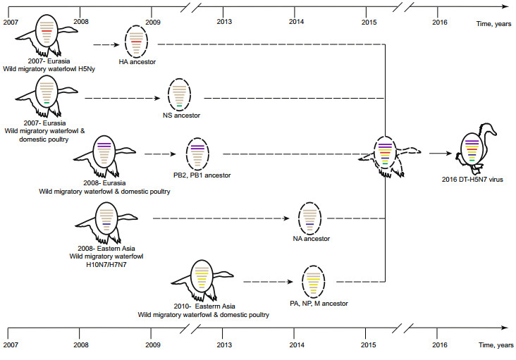
Figure 2. Hypothetical reassortment pathway of the DT-H5N7 virus. The gene segments are colored according to their ancestors
This is the first report of the isolation of an LPAI H5N7 virus in China.The most recent LPAI H5N7 virus isolates were identified in samples from ruddy turnstones in New Jersey of the United States in May 2017(GISAID-http://platform.gisaid.org/epi3/frontend#493de3).Moreover, this is the first identification of H5N7 LPAIV in the domestic duck.
Phylogenetic analysis revealed that the genome of the H5N7 isolate belonged to the Eurasian lineage and originated from wild migratory waterfowls.Notably, the H5N7 virus shared high similarity of internal genes with the Eurasian-H5N8 HPAIV in 2016-2017, which formed the prevalent AIVs in wild birds (Adlhoch et al.2016; Li et al. 2017).The process of internal genes reassortment showed extremely high complexity, possibly caused by unavailability of the surveillance data of LPAIV.Whether this combination of internal genes is adaptive to wild birds requires further investigation.
The lack of deletion in the NA stalk suggested that the H5N7 virus still possesses the adaptive feature to wild birds (Munier et al.2010).Moreover, the absence of any signs of drug resistance suggested that the transmission from wild birds to the domestic duck occurred recently. The ancestor of the circulating H5N1 HPAIV has experienced the deletion of NA stalk and became adaptive to domestic ducks (Li et al.2004).Since its emergence in 1996, 10 phylogenetic clades of H5 viruses have emerged, and two of them have crossed the barriers to infect humans (Abdel-Ghafar et al.2008).The circulating H5N1 HPAIV in ducks has become resistant to amantadine and zanamivir (Abdel-Ghafar et al.2008; Li et al.2004).Moreover, the H7N9 strain has also experienced the deletion of the NA stalk and acquisition of drug resistance in the process of ''genetic tuning''to adapt to domestic hosts and humans (Wang et al.2013).
It has been proved that LPAIVs may also serve as precursors of HPAIVs (Monne et al.2014).For instance, the A/Goose/Guangdong/1/96(H5N1) virus, the ancestor of the current H5 HPAIV, was considered to have been derived from the H5 LPAIV circulating in migratory birds (Duan et al.2007).It would follow that this H5N7 LPAIV adapted to the domestic duck after it began circulating.Our research showed that routine surveillance for influenza virus at wetlands where wild birds interact with domestic birds should not be neglected.Furthermore, the traditional breeding mode of ducks in an uncontrolled manner in wetlands should be regulated to reduce the risk of AIVs spreading from wild to domestic birds.
HTML
-
This work was funded by the National Science and Technology Major Project (2018ZX10101004), the Open Research Fund Program of Wuhan National Bio-Safety Level 4 Lab of CAS (NBL2017003), China Ministry of Science and Technology (MOST) Key Research and Development Program (2016YFC1200800), and the Shenzhen Science and Technology Research and Development Project (JCYJ20151029151932602).The funding sources had no role in the study design, data collection and analysis, decision to publish, or preparation of the manuscript.
-
The authors declare that they have no conflicts of interest.
-
This article does not contain any studies with human subjects performed by any of the authors.All studies involving animals were conducted according to the animal welfare guidelines of the World Organisation for Animal Health.
Conflict of interest
Animal and Human Rights Statement
-
Oropharyngeal, cloacal, and environmental samples were collected from resident poultry and live poultry markets (LPMs) in Hunan and Jiangxi provinces.Fresh fecal samples from migratory waterfowls were collected in Dongting Lake wetland in Hunan province.The specimens were kept in 1 mL viral-transport medium and transported to the laboratory within 24 h at 4℃.Ten-day-old specific pathogen-free (SPF) chicken embryos were used to isolate the virus, and the viral RNAs of hemagglutinin (HA)-positive samples were extracted with the Nucleic Acid Extraction System together with the matched EX-RNA/DNA viral nucleic acid extraction kits (Tianlong Science % Technology Co.Ltd.).These were then confirmed by reverse transcription polymerase chain reaction using universal primers targeting the M gene.Detection was performed using a previously described method (Bi et al., 2016).The whole genomes were obtained by next-generation sequencing on an Illumina Hiseq 4000 Sequencer (Bi et al., 2015) and were submitted to GenBank with the accession numbers MF362100-MF362107.
-
Maximum likelihood phylogenetic analysis was performed by RAxML-HPC2 on the Extreme Science and Engineering Discovery Environment (XSEDE) version 8.2.10(Towns et al., 2014) with 1000 bootstrap replicates for each tree and was based on the complete open reading frame (ORF) nucleotide sequences of PB2(2280 nucleotides), PB1(2274 nucleotides), PA (2151 nucleotides), HA (1695 nucleotides), NP (1497 nucleotides), NA (1416 nucleotides), M (982 nucleotides), and NS (858 nucleotides).The BLAST search was performed in both the Global Initiative on SharingAll Influenza Data (GISAID, http://platform.gisaid.org/epi3/frontend) and Influenza Research Database (IRD, https://www.fludb.org/brc/home.spg?decorator=influenza), and the 250 top hits from each were selected, after which the identical hits was removed to infer the overall topology. Then, we detected the existence of molecular clock in the datasets using TempEst v1.5.1 with the Best-fitting root focused on the closest clade to the DT-H5N7 virus, and molecular dating analysis was performed using Bayesian Evolutionary Analysis using Sampling Trees (BEAST) v1.8.3 under the HKY substitution model, with a strict clock and chain length of 50000000.Furthermore, Tracer v1.6 was used to confirm the reliability of the results.The trees were summarized by Tree Annotator with 10% burn-in cutoffs.All trees were visualized and labeled using FigTree v1.4.3.
-

Figure S1. Phylogenetic analysis of internal genes of the DT-H5N7 isolate. A) PB1, B) PB1, C) PA, D) HA, E) NP, F) NA, G) M and H) NS. The DT-H5N7 branch is colored red, the isolates in the Mediterranean-Black sea flyway are marked in purple, those in the East Asian-Australian flyway are labeled in green, those in the Central Asian flyway are colored yellow, and those in the Central American flyway are colored orange.

Figure S2. Maximum clade credibility (MCC) trees and root-to-tip analysis of the eight genes of the DT-H5N7 virus. A) PB2, B) PB1, C) PA, D) HA, E) NP, F) NA, G) M, and H) NS. The DT-H5N7 isolate is colored red, and the node bars are displayed with the 95% highest posterior density (HPD) of the node height.

 DownLoad: