


-
Dear Editor,
Duck Tembusu virus (DTMUV), a member of the Flavivirus genus within the Flaviviridae family, has caused huge economic losses to the poultry industry in China and even in Asia since 2010 (Zhang et al. 2017). The first strain of Tembusu virus (TMUV) MM_1775 was isolated from mosquitoes in 1955 in Malaysia (Platt et al. 1975). DTMUV CQW1 strain (GenBank: KM233707.1) was isolated from the liver tissue of Cherry Valley ducks in southwest China in 2015 (Zhu et al. 2015). The positive control rCQW1 was rescued from an infectious clone that contained the full-length cDNA of CQW1. And the complete cDNA was positioned under the control of the T7 promoter elements for in vitro transcription (Chen et al. 2018). The relationship between TMUV evolution and pathogenic variants has not been revealed. The fundamental reason is the lack of the in vitro operation platform for the prototypical strain genome.
Here, we constructed a DNA-based infectious clone for early mosquito-derived TMUV prototypical strain MM_1775. Three cDNA fragments (P1–P3) spanning the entire viral genome were synthesized and assembled into a full-length cDNA of MM_1775 (named pACNR FLMM_1775; Fig. 1A). Due to the instability of the flavivirus cDNA clone, especially the P1 fragment spanning the virus prM-E-NS1 genes is toxic to E. coli during the cloning process, it will cause the insertion fragment to be unstable. Therefore, we selected a low copy plasmid pACNR as the vector, and inserted an intron after nt position 2586 (NS1 gene) to restrict plasmid toxicity during propagation of cDNA in E. coli. In fact, there are two sequences for the prototypic TMUV strain in GenBank: named MM1775 (GenBank: MH414569.1) and MM_1775 (GenBank: JX477685.2), respectively, uploaded by different researchers. Since the former sequence was not uploaded when we synthesized the sequence fragments, we chose the latter sequence as the template. However, after comparing the two sequences of the prototype, we found a key difference at the 203 amino acid residue of NS4B (Fig. 1B). Multiple protein sequence alignment of several TMUV strains revealed that only MM1775 had the Thr at position 203, while other strains showed the Ala (Fig. 1B). Therefore, we introduced this mutation (ACA→GCA) into pACNR FL-MM_1775 to generate pACNR FL-MM_1775-m (Fig. 1C). We transfected the pACNR FL-MM_1775 plasmids into BHK-21 cells to rescue the rMM_1775 virus. As shown in Fig. 1D, 96 h after transfection, immunofluorescence assay (IFA) was used to detect protein expression. Compared with the mock-treated cells, strong green fluorescence was detected in the transfected cells, consistent with the positive control [cells infected with DTMUV rCQW1 strain at 48 h post-infection (hpi)]. This indicated that the cDNA transfected into the cells was transcribed and translated. And on the seventh day after transfection, clear cytopathic effect (CPE) was observed (Fig. 1E). The supernatant was collected, which further infected fresh BHK-21 cells for two consecutive passages. CPE was still observed on the fifth day on the F2 generation (Supplementary Fig. S1). This showed that what was produced after cDNA transfection was infectious, and we might successfully rescue the virus. Then we extracted the RNA from F0 and F2 generation. After RT-PCR identification and whole genome sequence determination, it showed that they were consistent with the MM_1775 virus sequence from GenBank (ID: JX477685.2) without any mutation. Collectively, these results indicated that rMM_1775 was successfully rescued. However, after transfecting the pACNR FL-MM_1775-m plasmid into BHK-21 cells, not only no CPE was observed (Fig. 1E), but also no fluorescence was detected by IFA (Fig. 1D), indicating this construct was nonviable.
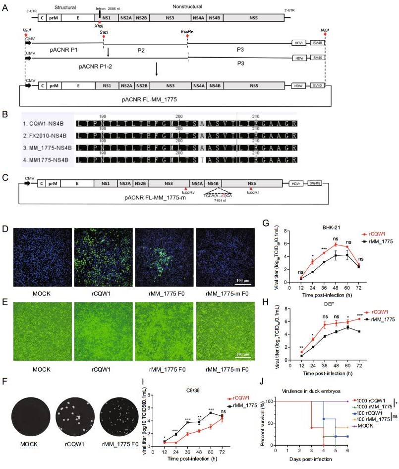
Figure 1. Infectious clone of TMUV. A The strategy for constructing the full-length cDNA clone of mosquito TMUV MM_1775. The genome sequence of mosquito TMUV MM_1775 was divided into three fragments artificially, which were finally assembled into low-copy plasmid pACNR to form full length TMUV cDNA clone (pACNR FL-MM_1775). Introns were inserted after nt position 2586 to reduce prokaryotic toxicity (black rectangle). The red arrow represents the restriction site. The complete MM_1775 cDNA is under the control of the CMV promoter element for eukaryotic transcription. A HDVr sequence was engineered at the 30 end of the viral genome to generate an authentic 30 end of viral RNA sequence. B Alignment of NS4B amino acid sequences of different TMUV. C Schematic diagram of the construction of full-length cDNA clone with mutation introduced in NS4B. D At 96 h post-transfection with plasmids pANCR FLMM_1775, strong green fluorescence was detected by IFA using mouse anti-TMUV polyclonal antibody as the primary antibody. Positive control, cells infected with rCQW1 at 48 hpi. E BHK-21 cells appeared CPE at the seven days post-transfection. Positive control, cells infected with rCQW1 at 48 hpi. F Plaque morphology of rCQW1 and rMM_1775 on BHK-21 cells. G, H and I Growth kinetics of rMM_1775 and rCQW1 in infected BHK-21, DEF and C6/36 cells. All data are presented as mean ± SD from three independent experiments (*P < 0.05, **P < 0.01, and ***P < 0.001). J Virulence of rCQW1 and rMM_1775 in duck embryos. Five 9-day-old duck embryo eggs per group were injected with 100 μL rMM_1775 or rCQW1 dilution by allantoic cavity inoculation at a dose of 100 and 1000 TCID50 (100 μL DMEMformock group). And the survival time of the inoculated eggs was recorded. Data are shown as means ± SD (*P < 0.05). TMUV, Tembusu virus; HDVr, hepatitis delta virus ribozyme; IFA, immunofluorescence assay; CPE, cytopathic effect; SD, standard deviation; ns, not significant; dpi, days post-infection; hpi, hours postinfection; TCID50, median tissue culture infectious dose.
To study the in vitro characteristics of the rMM_1775 virus, we compared the properties of rMM_1775 and rCQW1 in cell culture. As shown in Fig. 1F, rMM_1775 virus produced uniform plaque morphology on BHK-21 cells, which was smaller than that produced by rCQW1. Correspondingly, rMM_1775 virus showed weaker replication kinetics in mammalian BHK-21 and avian DEF cells, with almost two orders of magnitude lower than rCQW1 (Fig. 1G, 1H). The time required to cause CPE for the two viral strains are slightly different. And 48 h post-inoculation, DEF cells infected with rCQW1 showed obvious CPE, while those infected with rMM_1775 showed mild CPE at 60 h (Supplementary Fig. S2A). However, in Aedes albopictus C6/36 cells, rCQW1 showed weaker replication kinetics (Fig. 1I), and no obvious CPE was observed in C6/36 cells infected with rCQW1 and rMM_1775 (Supplementary Fig. S2B).
Next, we determined the virulence of rMM_1775 and rCQW1 to duck embryos. Duck embryo virulence experiments were performed on 9-day-old duck embryos. Five duck embryos within each group were injected with rMM_1775 or rCQW1, at a dose of 100 and 1000 median tissue culture infectious dose (TCID50), respectively. Three embryos inoculated with 1000 TCID50 of rCQW1 died at 3 dpi and two died at 4 dpi, while four embryos inoculated with 1000 TCID50 of rMM_1775 died at 4 dpi and one at 5 dpi. Two embryos inoculated with 100 TCID50 of rCQW1 and rMM_1775 died at 4 dpi, two and one died at 5 dpi, respectively. No embryos died in the mock group during the assay. All duck embryos inoculated with 1000 TCID50 of rMM_1775 and rCQW1 died within 120 h and 96 h, with a mortality rate of 100%. And all duck embryos inoculated with 100 TCID50 of rMM_1775 and rCQW1 did not all die within 144 h, respectively (Fig. 1J). These results indicated that mosquito-original rMM_1775 showed similar virulence on duck embryos as DTMUV rCQW1.
Previously, we have successfully constructed an infectious clone for CQW1 stain (Chen et al. 2018). The clone of prM-E-NS1 gene has potential prokaryotic toxicity to host bacteria, so the manipulation of the infectious clone requires a lot of time and manpower. Also it is easy to produce mutations. These mutations will have an important impact on the replication and transcription of the virus. To solve this problem, here, we introduced an intron into NS1 gene to interrupt potential prokaryotic toxic genes, and placed the full-length cDNA of the viral genome under the eukaryotic promoter CMV. And then the virus could be rescued by direct transfection of positive plasmid into BHK-21 cells (Fig. 1D, 1E). After sequencing, the rescued virus was exactly the same as the sequence from NCBI and did not contain any mutations (Supplementary Fig. S3). Compared to the reverse genetics system for TMUV (Li et al. 2013; Wu et al. 2016; Chen et al. 2018) constructed before, the one we constructed is more stable and is easier to modify in subsequent studies, especially when introducing a single mutation. It also avoids the problems of instability in transcription in vitro and solves the difficulty when transfecting RNA. In addition, it can reduce the cost of virus rescue.
Since the TMUV MM_1775 strain was isolated from mosquitoes in Malaysia in 1955, many strains of TMUV have been isolated in succession, but there have been few reports about animals naturally infected with TMUV. Until 2000, a virus named Sitiawan was isolated from sick chickens (Kono et al. 2000). The virus was identified to be highly homologous to the prototypical TMUV strain MM_1775, so it was classified as TMUV. This is also the first time that the virus has been reported to cause avian disease. And TMUV infection has never occurred in China for many years. Until 2010, an infectious disease mainly characterized by a severe drop in egg production, accompanied by a sudden decline in food intake, and the discharge of green faces and uncoordinated gait broke out in major duck production areas in China (Su et al. 2011). Shortly thereafter, there were reports of poultry infected with TMUV in many places in China. Although many TMUV strains have been isolated from mosquitoes and various poultry specimens, comparative studies between TMUV isolates derived from these sources are limited, especially the early mosquito-based isolate MM_1775 and duck-derived endemic strains. Here, we investigated the replication properties of MM_1775 in different cells from different sources. In both mammalian cell line BHK-21 and mosquito-derived cell line C6/36, the growth kinetics of rMM_1775 reached its peak at 60 hpi (Fig. 1G, 1I). However, rMM_1775 had a higher titer in C6/36 cells, suggesting that it replicated better in mosquito-derived cells. The reason might be that MM_1775 was isolated from mosquitoes, and C6/36 cells were its host cells, so the virus had better adaptability in C6/36 cells. As reported, the change of amino acid residue 156 in E protein of TMUV (P to S) caused the difference in transmission and pathogenicity between MM1775 and FX2010 (Yan et al. 2018). In addition, poultry-derived TMUV isolates (strain TMUV-JXSP) were more invasive than mosquito isolates (TMUV-YN12115) in mammalian cells and exerted greater expansion capacity (Lei et al. 2017). Similarly, in our research, rMM_1775 replicated in mammalian cells and DEF cells was not as well as rCQW1 (Fig. 1G, 1H). However, the results of other study were different. The infectious clone rescued strain MM1775 replicated better than FX2010 in DF-1 cells. This may be due to the different adaptability of different viruses to different cells. Moreover, the introduction of a mutation at amino acid 156 of the E protein of FX2010 (S to P) had little effect on replication. However, the replication of mosquito TMUV and DTMUV was quite different, indicating that there were more mechanisms to be revealed (Yan et al. 2018).
Here, we reported a full-length cDNA clone of mosquitoderived TMUV strain MM_1775 that could be stably inherited and easily modified. By comparing the differences of the nucleotide and amino acid sequence between MM_1775 and CQW1 strains, we can understand the molecular basis of the differences in pathogenicity of the two strains to different animals. On this basis, we can combine the reverse genetic operating systems of these two viruses, replace their genetic sequences, construct a recombinant virus, and study the effect of the related amino acid mutations on the pathogenicity of the virus. In conclusion, it will provide a complete tool platform for us to study the evolution and pathogenic mechanism of TMUV.
HTML
-
This work was funded by grants from, the National Key Research and Development Program of China (2017YFD0500800), the Sichuan-international joint research for science and technology (2018HH0098), China Agricultural Research System (CARS-42-17), the Program Sichuan Veterinary Medicine and Drug Innovation Group of China Agricultural Research System (SCCXTD-2020-18).
-
The authors declare that they have no conflict of interest.
-
All animal experimental procedures were approved by the Institutional Animal Care and Use Committee of Sichuan Agriculture University in Sichuan, China (Protocol Permit Number: SYXK(JII)2019–187).

 DownLoad: