


-
Correction to: Virologica Sinica (2021) 36:948–957
https://doi.org/10.1007/s12250-021-00352-4
Due to our negligence, the original version of this article, published online on March 15, 2021, contained a mistake in Figure 2E (The Knockdown band of Western blotting was provided incorrectly). The correct Fig. 2E is given below. We apologize for this error and state that this does not change the scientific conclusions of the article in any way.
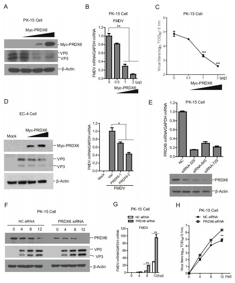
Figure 2. PRDX6 inhibited FMDV replication. A–C PK-15 cells were transfected with 0, 0.5, 1 or 2 μg of Myc-PRDX6 expressing plasmids for 24 h. The cells were then infected with FMDV (MOI = 0.5) for 12 h. The expression of viral proteins (A) and viral RNA (B) were detected by Western blotting and qPCR respectively. The virus yields were measured by TCID50 assay (C). D EC-4 cells were transfected with 0, 1 or 2 μg of Myc-PRDX6 expressing plasmids for 24 h. The cells were then mock-infected or infected with FMDV for 12 h. The expression of viral proteins and viral RNA were detected by Western blotting and qPCR respectively. E PK-15 cells were transfected with 120 nM of nontargeting control NC siRNA or PRDX6 siRNA (siRNA-329, siRNA-645 siRNA-729) for 36 h, the knockdown efficiency of each siRNA was then evaluated by qPCR and Western blotting analysis respectively. F–H PK-15 cells were transfected with 120 nM of NC siRNA or PRDX6 siRNA (siRNA-329) for 36 h, the cells were then infected with FMDV (MOI = 0.5) for 0, 4, 8 or 12 h. The expression of viral proteins (F) and viral RNA (G) were detected by Western blotting and qPCR respectively. The virus yields were measured by TCID50 assay (H).
Correction to: Porcine Picornavirus 3C Protease Degrades PRDX6 to Impair PRDX6-mediated Antiviral Function
- Congcong Wang 1, ,
- Huanhuan Feng 1, ,
- Xiangle Zhang 1 ,
- Kangli Li 1 ,
- Fan Yang 1 ,
- Weijun Cao 1 ,
- Huisheng Liu 1 ,
- Lili Gao 1 ,
- Zhaoning Xue 1 ,
- Xiangtao Liu 1 ,
-
Zixiang Zhu
1,,
 ,
, -
Haixue Zheng
1,,
- Published Date: 12 November 2021
Abstract:

 DownLoad: