


-
Correction to:
VIROLOGICA SINICA 2017, 32 (5): 404–414
https://doi.org/10.1007/s12250-017–3947-1
The original version of this article, published online on September 29, 2017, contained errors in Fig. 4B. The correct Fig. 4 is given below.
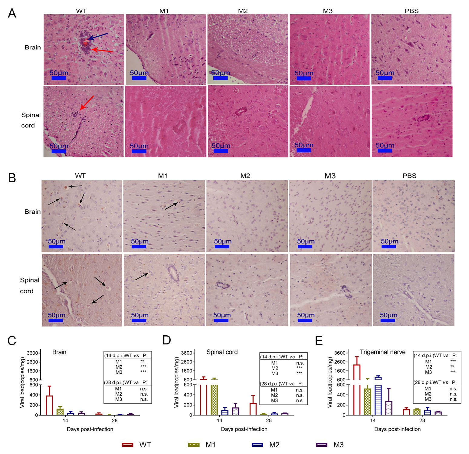
Figure 4. Histopathologic examination of CNS tissue of mice infected with the mutant and WT strains. (A) Pathological changes in the brain and spinal cords of mice infected with the WT, M1, M2 or M3 strains or PBS. The tissue sections were stained with H & E and imaged using an optical microscope at ×200 magnification. Tissue hyperemia is highlighted with blue arrows, and infiltration of inflammatory cells is highlighted with red arrows. (B) Immunohistochemical detection of HSV-1 in the brain and spinal cords from WT-, M1-, M2-, M3- or PBS-infected mice. Non-specific staining was observed in the PBSinfected mice. Positive expression of the HSV-1 antigen was detected in the brain and spinal cord tissues (arrows) of WT-, M1-, M2- and M3-infected mice. Assessment of viral load in the (C) brain, (D) spinal cord or (E) trigeminal nerve of mice challenged with WT, M1, M2 or M3, as determined by RT-qPCR. The viral copy numbers were quantified according to an HSV-1 DNA standard. The data are shown as the means ± SDs based on data from three independent mice. **P < 0.01; ***P < 0.005.
Correction to: Attenuated phenotypes and analysis of a herpes simplex virus 1 strain with partial deletion of the UL7, UL41 and LAT genes
- Xingli Xu 1, ,
- Yingqiu Guo 1, ,
- Shengtao Fan 1 ,
- Pingfang Cui 1 ,
- Min Feng 1 ,
- Lichun Wang 1 ,
- Ying Zhang 1 ,
- Yun Liao 1 ,
- Xiaolong Zhang 1 ,
-
Qihan Li
1,,

- Published Date: 19 November 2021
Abstract:

 DownLoad: