Lan-lan ZHANG, Jin-yu SHEN, Cheng-feng LEI, Xiao-ming LI and Qin FANG. High Level Expression of Grass Carp Reovirus VP7 Protein in Prokaryotic Cells[J]. Virologica Sinica, 2008, 23(1): 51-56. doi: 10.1007/s12250-008-2921-3
Citation: Lan-lan ZHANG, Jin-yu SHEN, Cheng-feng LEI, Xiao-ming LI, Qin FANG. High Level Expression of Grass Carp Reovirus VP7 Protein in Prokaryotic Cells .VIROLOGICA SINICA, 2008, 23(1) : 51-56.  http://dx.doi.org/10.1007/s12250-008-2921-3

草鱼呼肠孤病毒VP7蛋白在原核细胞中的高效表达

  • 通讯作者: 方勤, qfang@wh.iov.cn
  • 收稿日期: 2007-11-07
    录用日期: 2007-12-25
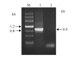
  • 序列分析表明 GCRV S10 片段长为909核苷酸,编码一个分子量为34kDa 的外衣壳蛋白VP7。为获得体外表达的外衣壳蛋白,通过RT-PCR扩增,得到一条特异的、大小约0.9 kb的草鱼呼肠孤病毒外衣壳蛋白片段;将其扩增片段克隆于含T7启动子的高效原核表达系统pRSET载体质粒,转化BL21(DE3)lysS 感受态细胞,获得重组表达菌株(pR/GCRV-VP7)。酶切重组克隆子及序列分析实验证实,所得重组克隆为目的基因插入序列。pR/GCRV-VP7表达菌株经IPTG诱导培养,SDS-PAGE结果显示,表达产物的蛋白分子量约37kDa,为目的表达融合蛋白。高效表达的融合蛋白以包含体形式存在,目的蛋白的表达量占总菌体量的60%以上。Westerblot分析表明,该表达产物与鼠抗Histag与兔抗GCRV抗体呈阳性反应。该结果为GCRV-VP7结构与功能分析奠定了基础。

High Level Expression of Grass Carp Reovirus VP7 Protein in Prokaryotic Cells

  • Corresponding author: Qin FANG, qfang@wh.iov.cn
  • Received Date: 07 November 2007
    Accepted Date: 25 December 2007

    Fund Project: National Natural Science Foundation of China 30470074Innovation Project of the Chinese Academy of Sciences KSCX2-YW-N-021Science and technology foundation of Zhejiang Province 2007C22052National Natural Science Foundation of China 30671615

  • Sequences analysis revealed Grass carp reovirus (GCRV) s10 was 909 nucleotides coding a 34 kDa protein denoted as VP7, which was determined to be a viral outer capsid protein (OCP). To obtain expressed OCP in vitro, a full length VP7 gene was produced by RT-PCR amplification, and the amplified fragment was cloned into T7 promoted prokaryotic expression vector pRSET. The recombinant plasmid,which was named as pR/GCRV-VP7,was then transformed into E.coli BL21 host cells. The data indicated that the expressed recombinant was in frame with the N-terminal fusion peptide. The over-expressed fusion protein was produced by inducing with IPTG, and its molecular weight was about 37kDa, which was consistent with its predicted size. In addition, the fusion protein was produced in the form of the inclusion body with their yield remaining steady at more than 60% of total bacterial protein. Moreover,the expressed protein was able to bind immunologically to anti-his-tag monoclonal antibody (mouse) and anti-GCRV serum (rabbit). This work provides a research basis for further structure and function studies of GCRV during entry into cells.

  • 加载中
    1. Attoui H, Fang Q, Jaafar F M, et al. 2002. Common evolutionary origin of aquareoviruses and orthoreoviruses revealed by genome characterization of Golden shiner reovirus, Grass carp reovirus, Striped bass reovirus and golden ide reovirus (genus Aquareovirus, family Reoviridae). J Gen Virol, 83: 941-1951.

    2. Chandran K, Walker S B, Chen Y, et al. 1999. In vitro recoating of reovirus cores with baculovirus-expressed outer-capsid proteins μ1 and sigma3. J Virol, 73 (5): 3941-3950.

    3. Dryden K A, Wang G J, Yeager M, et al. 1993. Early steps in reovirus infection are associated with dramatic changes in supramolecular structure and protein conformations. J Cell Biol, 122(5): 1023-1041.
        doi: 10.1083/jcb.122.5.1023

    4. Estes M K. 2001. Rotaviruses and their replication. In: Fields Virology (Knipe D M, Howley P M, Griffin D E, et al ed. ), 4 ed. Philadelphia: Lippincott Williams & Wilkins, Vol. 2, p1625-1655.

    5. Fang Q, Attoui H, Biagini P, et al. 2000. Sequence of genome segments 1, 2, and 3 of the grass carp reovirus (Genus Aquareovirus, family Reoviridae). Bioch Bioph Res Commun, 274 (3): 762-766.
        doi: 10.1006/bbrc.2000.3215

    6. Fang Q, Ke L H, Cai Y Q. 1989. Growth characterization and high titre culture of GCHV. Virologica Sinica, 3: 314-319. (in Chinese).

    7. Fang Q, Shah S, Liang Y, et al. 2005. 3D Reconstruction and Capsid Protein Characterization of Grass Carp Reovirus. Science in China Series C, 48: 593-600.
        doi: 10.1360/062004-105

    8. Ivanovic T, Agosto M A, Nibert M L. 2007. A role for molecular chaperone Hsc70 in reovirus outer capsid disassembly. J Biol Chem, 282 (16): 12210-12219.
        doi: 10.1074/jbc.M610258200

    9. Ke L H, Fang Q, Cai Y Q. 1990. Characteristics of a novel isolate of grass carp Hemorrhage Virus. Acta Hydrobiol Sinica, 14: 153-159. (in Chinese)

    10. Kim J, Zhang X, Centonze V E, et al. 2002. The hydrophilic amino-terminal arm of reovirus core-shell protein λ1 is dispensable for particle assembly. J Virol, 76: 12211-12222.
        doi: 10.1128/JVI.76.23.12211-12222.2002

    11. Liemann S, Chandran K, Baker T S, et al. 2002. Structure of the reovirus membrane-penetration protein, μ1, in a complex with its protector protein, σ3. Cell, 108: 283-295.
        doi: 10.1016/S0092-8674(02)00612-8

    12. Lupiani B, Subramanian K, Samal S K. 1995. Aquareoviruses. Ann Rev Fish Dis, 5: 175-208.
        doi: 10.1016/0959-8030(95)00006-2

    13. Mertens P P C, Arella M, Attoui H, et al. 2000. Family Reoviridae. In: Virus Taxonomy (van Regenmortel M H V, Fauguet C M, Bishop D H L, et al. ed. ), San Diego: Academic Press, CA, USA. p 395-480.

    14. Nason E L, Samal S K, Venkataram Prasad B V. 2000. Trypsin-induced structural transformation in aquareovirus. J Virol, 74(14), 6546-6555.

    15. Nibert M L, Schiff L A. 2001. Reoviruses and their replication. 4 ed. In: Fields Virology (Knipe D M, Howley P M, Griffin D E, et al. ed. ), Philadelphia: Lippincott Williams & Wilkins, Vol. 2, p1679-1728.

    16. Rangel A A, Samal S K. 1999. Identification of grass carp hemorrhage virus as a new genogroup of aquareovirus. J Gen Virol, 80: 2399-2402.
        doi: 10.1099/0022-1317-80-9-2399

    17. Reinisch K M, Nibert M L, Harrison S C. 2000. Structure of the reovirus core at 3.6 angstrom resolution. Nature, 404 (6781): 960-967.
        doi: 10.1038/35010041

    18. Shaw A L, Samal S, Subramanian K, et al. 1996. The structure of aquareovirus shows how different geometries of the two layers of the capsid are reconciled to provide symmetrical interactions and stabilization. Structure, 15: 957-968.

    19. van Regenmortel M H V, Fauquet C M, Bishop D H L, et al. 2000. Virus Taxonomy -Seventh Report of the International Committee on Taxonomy of Viruses. San Diego: Academic Press, California, USA.

    20. Zhang L, Chandran K, Nibert M L, et al. 2006. Reovirus μ1 structural rearrangements that mediate membrane penetration. J Virol, 80 (24): 12367-12376.
        doi: 10.1128/JVI.01343-06

  • 加载中

Figures(4)

Article Metrics

Article views(3472) PDF downloads(13) Cited by(0)

Related
Proportional views
    通讯作者: 陈斌, bchen63@163.com
    • 1. 

      沈阳化工大学材料科学与工程学院 沈阳 110142

    1. 本站搜索
    2. 百度学术搜索
    3. 万方数据库搜索
    4. CNKI搜索

    High Level Expression of Grass Carp Reovirus VP7 Protein in Prokaryotic Cells

      Corresponding author: Qin FANG, qfang@wh.iov.cn
    • 1. State Key Laboratory of Virology, Wuhan Institute of Virology, Chinese Academy of Sciences, Wuhan 430071, China
    • 2. Graduate School of the Chinese Academy of Sciences, Beijing 100039, China
    • 3. Zhejiang Institute of Freshwater Fisheries, Huzhou, 313001
    • 4. Basic Medical School, Wuhan University, Wuhan 430072, China
    Fund Project:  National Natural Science Foundation of China 30470074Innovation Project of the Chinese Academy of Sciences KSCX2-YW-N-021Science and technology foundation of Zhejiang Province 2007C22052National Natural Science Foundation of China 30671615

    Abstract: Sequences analysis revealed Grass carp reovirus (GCRV) s10 was 909 nucleotides coding a 34 kDa protein denoted as VP7, which was determined to be a viral outer capsid protein (OCP). To obtain expressed OCP in vitro, a full length VP7 gene was produced by RT-PCR amplification, and the amplified fragment was cloned into T7 promoted prokaryotic expression vector pRSET. The recombinant plasmid,which was named as pR/GCRV-VP7,was then transformed into E.coli BL21 host cells. The data indicated that the expressed recombinant was in frame with the N-terminal fusion peptide. The over-expressed fusion protein was produced by inducing with IPTG, and its molecular weight was about 37kDa, which was consistent with its predicted size. In addition, the fusion protein was produced in the form of the inclusion body with their yield remaining steady at more than 60% of total bacterial protein. Moreover,the expressed protein was able to bind immunologically to anti-his-tag monoclonal antibody (mouse) and anti-GCRV serum (rabbit). This work provides a research basis for further structure and function studies of GCRV during entry into cells.