


-
Post-weaning multisystemic wasting syndrome (PMWS) is considered an important and new swine viral disease, characterized clinically by fever, progressive weight loss, and respiratory and digestive disorders. The PMWS was first identified in western Canada in 1996 [3], and has spread into pig farms all over the world with the potential to cause serious economic loss for the worldwide swine industry. The disease is usually prevalent in pigs 5 to 18 weeks old, and while morbidity is ofen low, in severe cases, it has high fatality rate that is more than 50% in epidemic herds[5]. The main putative etiological agent of PMWS has been isolated, verified[1], and designated as porcine circovirus type 2 (PCV2). In contrast to PCV2, porcine circovirus type 1 (PCV1) was reported in 1982 as a persistent non-cytopathic contaminant of the continuous PK15 cell line [18], and has been demonstrated to be non-pathogenic in animal tests[19].
Although the pathogenesis of PCV2-induced PMWS is not well defined, the disease is believed to be mediated by the host immune response [7]. PCV2-infected adult pigs may recover from the disease, but they usually are virus carriers for a long time, and complicate efforts to disseminate the disease. To detect these infection persistent animals, a simple and reliable diagnostic method for monitoring the status of PCV2 infection in herds is required.
Differential reactivity with monoclonal antibodies to either PCV1 or PCV2 has revealed that they are antigenically different[1, 2]. However, a low level of cross-reactivity exists between the two types, since antibodies to PCV1 react to a low degree with PCV2. Specific tests for serologic detection are very essential to determine the prevalence of PCV2 infection and elucidate how PMWS develops in herds. Most serological diagnostic methods for detecting PCV2 antibodies, including indirect immunofluorescence assays (IIFA), immunoperoxidase monolayer assays (IPMA)[4], and various enzyme-linked immunosorbent assays (ELISAs), have been developed by preparing live virus as a diagnostic antigen in cell culture. However, these techniques are costly, and require labor-intensive and accurate interpretation of the staining reactions. Additionally, examination of IIFA and IPMA plates is tedious and time-consuming. In contrast, ELISAs can be automated, which could not only boost the processing rate, but also decrease the potential bias that may occur with the interpretation of IIFA or IPMA results.
PCV1 and PCV2 share the same genomic organization, consisting of two major open reading frames (ORFs) coding for the replicase protein (Rep, Rep') and the capsid (Cap) protein [6, 11, 12]. Recently, several studies have shown that the 28-kDa PCV2 Cap protein is the major immunogenic protein of the virus and the principal bearer of type-specific epitopes[8, 15]. The recombinant Cap protein in vitro reacts strongly with serum from PCV2-infected swine, suggesting its possible use in diagnostic assays [16]. Furthermore, a Cap epitope of PCV2 has been identified as a serological marker for viral infection [20]. The immuno-dominant epitopes of PCV2 Cap are believed to reside within amino acid residues from 47 to 84, 165 to 200, and the last 4 amino acids [9]. Therefore, the Cap protein may be a candidate PCV2 gene to provide recombinant antigen for the development a serological diagnostic method to detect PCV2 antibodies.
In order to develop a more suitable detection antibody to PCV2, we generated a prokaryotic recombinant protein as the antigen in a diagnostic ELISA. The aim of this study is to establish an indirect ELISA based on a recombinant truncated soluble Cap (rCap) protein containing a GST-fused ORF2 epitope of PCV2 expressed in E. coli and an affinity-purified recombinant protein for the serological diagnosis of PCV2 infection in swine.
HTML
-
PCV2 genomic DNA was extracted from a superficial inguinal lymph node of a pig with naturally occurring severe PMWS. A panel of PCV2 negative sera was collected from 18 uninfected 60 to 90 days old pigs from a PCV-free herd, and 42 PCV2-positive sera were used as reference sera in the assay. 113 field sera of pigs suspected to be infected with PCV2 were collected during the period 2003-2007. Antisera raised against other pig viruses, including porcine reproductive and respiratory syndrome virus (PRRSV), classical swine fever virus (CSFV), porcine parvovirus(PPV), and Pseudorabies virus (PrV), were used in the present study to determine the specificity of TcELISA (Truncated cap ELISA).
-
The truncated PCV2 Cap (rCap) coding sequence was amplified by polymerase chain reaction (PCR) using a pair of primers designed according to the Beijing strain sequence (GenBank no.EU921257). The sequence of the forward primer with a BamH Ⅰ site (underlined) was 5`-GCGGATCCGTGGACATGATGAGATTCA AT-3`, and the sequence of the reverse primer was 5`-CGCTCGAGGTATATACTGTTTTCGAACGCA GTGCC-3` with a Xhol Ⅰ site (underlined). The rCap fragment was cloned into an expression vector, pGEX-6p-1 (Pharmacia). Briefly, E. coli BL21(DE3) strain harboring the recombinant plasmids were grown in 3ml of Luria-Bertani (LB) broth at pH 7.0 containing 100 μg/mL ampicillin. The cells were induced with 0.5 mmol/L isopropyl-thio-β-D-galactoside (IPTG) for 4 h at 37 oC. The cell pellets were suspended in lysis buffer (500 mmol/L NaCl, 50 mmol/L Tris–HCl, 10 mmol/L EDTA, 5 mmol/L β-mercaptoethanol, 1.0 mg/mL lysozyme, pH 8.0) and incubated 30 min at 20 oC. Triton X-100 was added to a final concentration of 1.0% and the suspension was sonicated and centrifuged at 12 000×g min for 30 min at 4 oC. A sample of the bacterial lysate supernatant was characterized by electrophoresis in 15% SDS polyacrylamide gel (SDS-PAGE). The rCap protein was loaded onto a GSTrap affinity column (Amersham) according to the manufacturer's instructions. The purified protein was eluted and its concentration determined by the Bradford assay. GST protein was used as a control. The purified rCap and GST protein were used as antigens in Western blot (Fig. 1) and ELISA for detecting the antibodies to PCV2.
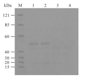
Figure 1. Western blot analysis of expressed PCV2 rCap protein. M, Protein Marker; Lane 1 and 2, The reaction of rCap protein with PCV2 positive sera; 3, The reaction of rCap protein with PCV2 negative serum; 4, The reaction of rCap protein with PCV1 positive serum.
-
The purified rCap protein was used as an indirect ELISA antigen by adding it to 96-well plates with 50 mmol/L sodium carbonate buffer (pH9.6) in a final volume of 50 μL. As a control antigen, GST protein was also added to two wells on the same plate. After incubation overnight at 4 oC, the antigen-coated plates were washed four times with phosphate-buffered saline (pH 7.3) containing 0.05% Tween20 (PBST) and blocked with PBST containing 10% (w/v) skimmed milk for 45 min at 37 oC. After washing, pig sera diluted at 1/10 with the blocking solution (2% E. coli lysis solution of PBST, ) were added to the plates at 50 μL per well, and incubated for 45 min at 37 oC. The wells were washed four times with PBST, then a rabbit anti-swine IgG conjugated with horseradish peroxidase (Sigma) diluted 1:500 in blocking solution was added and incubated for 45 min at 37oC. Reaction products were washed with PBST and 50 μL of substrate solution (TMB, Sigma) was added to each well. After incubation for 15 min at room temperature in the dark, the reaction was stopped by adding 50 μL of 2 mol/L H2SO4. The absorbance of each well was measured in a spectrophotometer at 450 nm. The results were expressed as the ratio the OD450 value generated by the serum samples compared to that from negative control serum. Sera that gave a ratio value higher than 2.1 were considered to be positive sera. The titers were expressed as the highest dilution of antibody producing a 2.1 ratio[21].
The results for each serum were obtained by calculating the ratio between the absorbance produced by the well with the recombinant Cap protein and that produced by the well with GST protein.
-
On the basis of the procedure described above, the optimal antigen concentration and sera dilution were determined through standard checkerboard titration procedures (Crowther, 2000). Briefly, the Tc protein was immobilized onto 96-well microtiter plates in serial two-fold dilution from 7.89 µg/mL to 1.34µg/mL. Standard swine PCV2-positive serum and negative control serum were also diluted in serial twofold dilutions from 1:5 to 1:640 for optimization. To determine the optimal conjugate dilution in the TcELISA, after the antigen and antisera dilutions were checked, the conditions that gave the highest OD450 ratio between positive and negative sera (P/N value) and the OD450 value of a positive serum close to 1.0 were scored as optimal working conditions.
-
To set a negative/positive cutoff value for this assay, 60 field serum samples were also tested in duplicate by the TcELISA. The positive/negative cut-off value was set at mean plus 3 standard deviations (negative serum mean + 3 S.D, NSM+ 3 S.D) of the 60 field sera obtained from the TcELISA. A cutoff point was determined such that the diagnostic sensitivity (DSN) and specificity (DSP) were maximized while the generation of false negative and false positive results was minimized, and the 95% confidence interval (CI) was calculated.
IIFA was used as the reference method to detect the presence of antibodies to PCV2. The DSN and DSP and accuracy of the TcELISA were calculated using the formulae[17]
Where TP, FN, TN, FP and TS indicated true-positive, false-negative, true-negative and false-positive, and total number of serum sample, respectively.
To detect the specificity of the TcELISA, positive sera against PCV1 (IIFA titer 1:1 026), CSFV, PPV, PrV, and PRRSV were tested according to the TcELISA procedure. Each sample was tested in triplicate, and the S/N ratios were calculated.
PCV2 genomic DNA and antisera
Antigen Preparation
Establishment an indirect ELISA using the rCap protein
Optimization of TcELISA working conditions
Validation of TcELISA
-
A truncated fragment representing the PCV2 capsid protein gene was obtained by PCR amplification. The amplified 417 bp product containing the immunoreactive ORF2 epitope of PCV2 was cloned into prokaryotic expression vector pGEX-6p-1 and the recombinant plasmid was subsequently sequenced. Recombinant rCap protein containing a GST tag was expressed as a soluble form and purified by affinity chromatography. Most rCap proteins were eluted from the Sepharose 4B resin. However, only the 42kDa fusion rCap protein could react strongly with swine anti-PCV2 serum in Western blotting analysis. Reactivity of the rCap protein with swine anti-PCV1 serum was not observed by Western blotting. All of these data confirmed that this rCap protein could be used as an antigen for the detection of specific antibodies against PCV2.
-
In the checkerboard ELISAs, the optimal antigen concentration and serum sample dilution were set at 2.68 µg/mL and 1:20, based on the standard that the OD450 value of positive serum was more than 1.0, the P/N value in the OD450 was highest, and the background was the lowest at the same conditions. The optimal dilution of the conjugate was defined cccording to the same principle, . After the above standards were tested, the coating buffer in 0.05 mol/Lcarbonate bicarbonate buffer (pH 9.6) was optimized. The optimal exposure time for serum samples and conjugate were determined to be 45 min and 15 min, respectively, at 37 oC for TcELISA.
-
The OD450 values of 18 IIFA-negative PCV2 swine sera are shown individually in Table 1. These were averaged to define a positive threshold. When the positive/negative cut-off value was set at mean plus 3 standard deviations (mean + 3 S.D, NSM+ 3 S.D), all TcELISA based on the recombinant cap protein were demonstrated to show 94.44% (17/18×100%) specificity. The specificities of TcELISA decreased to 88.33% (15/18×100%) when the cut-off value was set at mean plus 2 S.D.

Table 1. Comparison between the TcELISA and IFA of 113 field sera in pigs
Among 42 IIFA-positive field serum samples, the TcELISA revealed better sensitivities of 92.86% (39/42×100%) and 97.62% (41/42×100%) when the cut-off value was set at mean plus 2 or 3 S.D, respectively.
After the cut-off value was determined, the specificity of this assay was evaluated by testing the reactivity of antibodies against other porcine viruses with the rCap antigen. The cut-off value of standard positive sera against Classical swine fever virus (CSFV), Porcine parvovirus (PPV), Porcine pseudorrabies virus (PRV) and Porcine reproductive and respiratory syndrome virus (PRRSV) are shown in Table 2. The cut-off value of all antisera were significantly less than the NSM+ 3 S.D. This revealed that there was no cross-reactivity between the PCV2 rCap antigen and antibodies against other porcine viruses, proving that the rCap antigen was specific for the antibody to PCV2.

Table 2. Specificity of the TcELISA to antisera against other swine viruses
Compared with the TcELISA, 70 of 113 samples were detected as PCV2 antibody positive and 43 samples as negative at a cut-off value of 0.156+3SD. A total of 66 of 113 samples were judged as PCV2 antibody positive and 47 samples as negative by the IIFA assay. There were 62 samples judged positive and 39 samples judged negative by both methods. The DSN, DSP, and accuracy of the TcELISA were 88.6% (CI=79.18%-95.23%), 90.7% (CI=68.56%-92.8%) and 89.4%, respectively. The agreement between the two methods was 89.38% [(62/113+39/113)×100%].
Expression and purification of PCV2 rCap protein
Optimization of the TcELISA procedure
Validation of TcELISA
-
The incidence of PMWS in pig farms has become a potential threat to the global swine industry. As PCV2 is closely associated with PMWS, detecting antibodies to PCV2 in pig herds is essential for the control of PMWS. Therefore, PCV2-specific diagnostic tools are necessary for elucidating the course of infection.
Most serological studies have used PCV2 viral particles as an antigen for the detection of antibodies to PCV2, using procedures such as IIFA and IPMA[4]. However, complete viral particles from PCV2-infected cells lack PCV2 specificity for detection of PCV2 antibodies because of antigenic cross-reactivity between Rep proteins and of PCV1 and PCV2 [13]. In addition, it has been relatively difficult to obtain a purified antigen for ELISA from the PCV2-infected cells. These techniques also require experience when interpreting the results. Furthermore, these procedures require expensive equipment and facilities and labor-intensive and time-consuming techniques. Therefore, both IIFA and IPMA are not suitable for large-scale surveys.
The capsid protein of PCV2 was demonstrated to be a type-specific antigen that could be distinguished from PCV1. It contains a nuclear localization signal (NLS) consisting of 41 amino acid residues at the N terminus. The epitopes of the capsid protein have previously been determined at the level of amino acid residues[14]. Therefore, the capsid protein of PCV2 is a good candidate antigen for PCV2 serodiagnosis.
Recently, serodiagnostic ELISA assays based on ORF2 for specific detection of PCV2 antibodies have been developed using a recombinant capsid protein expressed in baculovirus and E. coli. The rcELISA achieved 88.5% specificity and 89.4% sensitivity for detection of PCV2 antibody in the sera[10], and the diagnositic sensitivity and specificity of the CAP ELISA were 95.3% and 95.1% respectively using the capsid protein of PCV2 produced in E.coli[17]. However, the DSN and DSP of the TcELISAwere 88.6% and 90.7%, respectively because in pig serum contains an antibody of anti-E.coli that can react with residual components from E. coli in the capsid protein and produce false positives. In the TcELISA, the blocking solution contains 2% E. coli lysis solution which can eliminate the nonspecificity to improve the accuracy of the result. Therefore, the specificity and sensitivity of the TcELISA is close to the rcELISA. In the present study, an alternative indirect ELISA for detection of antibody against PCV2 was successfully developed using an antigen in the form of a recombinant rCap protein expressed in E.coli. This technique avoided the use of a mock antigen as a negative control as described previously[16]. Furthermore, it is an ideal serodiagnostic method as it has several features including a low production cost, simpler production, ease of performance, and time-saving, making its application more suitable for large-scale clinical surveys of PCV2 infection.
In this study, a truncated gene of ORF2 was expressed successfully as a soluble protein. An indirect ELISA method was developed using the soluble and purified rCap protein expressed in E. coli as a coating antigen for the serological diagnosis of PCV2 infection in swine. A comparison of the TcELISA with IIFA indicated that the TcELISA has a relatively higher sensitivity (Table 1). One limitation of this assay is that an impure PCV2 antigen might generate a false positive. Consequently, it is essential to obtain a high purity antigen for controlling for false positives. However, the data from the TcELISA showed a good agreement (89.38%) with the IIFA for assay of the field swine sera.
The serological results of clinical sera samples by TcELISA of PCV2 specific antibodies suggested that exposure to PCV2 is widespread in different ages in the pig population in China(Table 3). The true frequency of infection may well be considerably higher, however. The growing trend of seroprevalence of PCV2 implied in this study suggests the importance of monitoring PCV2 infection in swine populations. The establishment of a TcELISA, as described in this paper, might prove to be an ideal diagnostic tool for detecting antibodies to PCV2 in suspect swine herds.

Table 3. Prevalence of PCV2 antibody in different ages by TcELISA
An indirect enzyme-linked immunosorbent assay (ELISA) for detection of PCV2 antibody was developed using a truncated soluble ORF2 protein produced in E.coli (TcELISA). The diagnostic DSN, DSP, and accuracy of the TcELISA were 88.6% (CI=79.18%-95.23%), 90.7% (CI=68.56%-92.8%) and 89.4%, respectively. The agreement rate was 89.38% between results obtained with TcELISA and IIFA on 113 field sera. A cross-reactivity assay showed that the method was PCV2-specific. The TcELISA will facilitate the development of a reliable tool for large scale detection of PCV2 antibodies and for the evaluation of vaccine against PCV2.


 DownLoad: