HTML
-
Foot-and-mouth disease (FMD) is a highly contagious and economically important disease that affects cloven-hoofed animals worldwide. The disease is caused by the foot-and-mouth disease virus (FMDV), which belongs to the genus Aphthoviruses within the Picornaviridae family (Grubman M J, et al., 2004). There are seven distinct serotypes of FMDV, namely O, A, C, Southern African Territories 1 (SAT1), SAT2, SAT3, and Asia1. The virus consists of a non-enveloped icosahedral capsid and a single-stranded positive-sense RNA genome of approximately 8, 500 nucleotides (nt). The viral RNA is translated from a single long open reading frame (ORF) into a polyprotein (Grubman M J, et al., 1984; Robertson B H, et al., 1985). The FMDV capsid is comprised of 60 copies of 4 structural proteins, VP4, VP2, VP3, and VP1 (Acharya R, et al., 1989). Cleavage of VP0 to VP2 and VP4, which is considered to be autocatalytic, is normally observed only upon encapsidation of RNA in mature virions (Grubman M J, et al., 2004; Vasquez C, et al., 1979). The whole virus, often referred to as the 146S particle on the basis of its sedimentation coefficient, is the most effective viral antigen, and the antigenic content of the vaccine is measured as the amount of 146S particles (Doel T R, 2003; Doel T R, 2005). The current FMD vaccines are produced in cell culture using virus that is chemically inactivated by using binary ethylenimine (BEI) (Barteling S J, 2004), and the maintenance of intact 146S particles during the manufacturing process is closely related to vaccine efficacy (Doel T R, 2003; Doel T R, et al., 1981). However, regarding virion integrity, the FMDV 146S is intrinsically highly acid-labile because it readily dissociates into 12S pentameric subunits in environments below pH 6.8 (Dubra M S, et al., 1982; Ehrenfeld E, et al., 2010). In addition, it has been reported that mild acid treatment largely destroys the protective capacity of experimental vaccines (Doel T R, et al., 1982). Thus, selection of acid-resistant FMDV mutant viruses is valuable for immunogenicity retention in the manufacturing process of inactivated FMD vaccines.
FMDV initiates infection when virions bind to their cellular receptors (Jackson T, et al., 1997; Monaghan P, et al., 2005). After internalization into the host cell via a clathrin-mediated mechanism, FMDV particles are delivered to endosomes, which are characterized by a mildly acidic environment (Berryman S, et al., 2005; Johns H L, et al., 2009; O'Donnell V, et al., 2005). Crystal structure analysis has revealed a high density of histidine (H) residues near the interpentameric interface; these particular H residues influence the acid lability of the virus (Acharya R, et al., 1989). The first documented acid-resistant FMDV mutant virus was selected from type A12 FMDV after exposure to an acidic environment (pH 6.4). This mutant virus has three substitutions in the capsid, including an A3S substitution in VP1 and E131K and D133S substitutions in VP2 (Twomey T, et al., 1995). Calculations of titration curves and pH stability profiles of FMDV O1BFS, A1061, and A22 Iraq revealed that H142 and H145 in VP3 contribute to FMDV acid-induced disassembly (van Vlijmen H W, et al., 1998). Moreover, it has been reported that the H142 residue located in VP3 modulates the acid sensitivity of the type A and type SAT FMDV capsid (Ellard F M, et al., 1999; Vazquez-Calvo A, et al., 2013). In addition, experimental results have revealed that VP3 A123T, VP3 A118V, and VP2 D106G in type C FMDV are involved in modulating the acid-induced dissociation of the FMDV capsid (Martin-Acebes M A, et al., 2010). Recent studies showed that a single amino acid substitution (N17D) in the VP1 protein is responsible for the increased acid resistance of type C FMDV (Liang T, et al., 2013; Martin-Acebes M A, et al., 2011).
Although several amino acids that mediate FMDV dissociation at acidic pHs have been identified, amino acid residues involved in determining the resistance of FMDV to acid-induced inactivation are different in different serotypes of FMDV, even within the same serotype (Ellard F M, et al., 1999; Martin-Acebes M A, et al., 2011; Twomey T, et al., 1995; van Vlijmen H W, et al., 1998). To characterize the molecular aspects of the acid resistance of type Asia1 FMDV, acid-screened type Asia1 FMDV mutant viruses were selected, and their resistance to acid-induced inactivation was evaluated in this study. Sequence analysis of type Asia1 FMDV acid-resistant mutants in the capsid protein-coding region revealed four amino acid substitutions (VP1 N17D, VP2 H145Y, VP2 G192D, and VP3 K153E). To investigate the role played by each substitution, a series of mutant viruses with one or two substitutions was constructed using a reverse genetic system to determine the relationship between an acid-resistant phenotype and amino acid substitution (s). Our results showed that either VP1 N17D or VP2 H145Y independently determined the acid-resistant phenotype of type Asia1 FMDV.
-
The baby hamster kidney cell line BHK-21 was maintained in Dulbecco's modified Eagle's medium (DMEM) supplemented with 10% fetal calf serum (Gibco-BRL). Serotype Asia1 FMDV Asia1/YS/CHA/05 (GenBank accession number: GU931682) was rescued from the infectious cDNA clone pAsi (Wang H, et al., 2011), and passaged on BHK-21 seven times as a viral stock.
-
The detection of FMDV structural proteins and neutralization assays were performed using mouse MAb 3E11, which recognizes a protective neutralizing epitope in the VP1 G-H loop of FMDV Asia1/YS/CHA/05 (Wang H, et al., 2011). Anti-mouse IgG (Sigma) was used as the secondary antibody in indirect immunofluorescence assays. NH4Cl (Merck) was dissolved in double distilled water.
-
A previously reported procedure (Martin-Acebes M A, et al., 2011) was followed with minor modifications. Briefly, 10 μL virus containing 1× 105.75 median tissue culture infective dose (TCID50) was mixed with 300 μL phosphate-buffered saline (PBS) solution (50 mmol/L NaH2PO4 and 140 mmol/L NaCl) with different pH values (5.0, 6.0, or 7.4) for a 30-min incubation at room temperature. The mixed solution was neutralized by the addition of 120 μL 1 mol/L Tris solution (pH 7.6), and the remaining infectious titers were determined by TCID50 assays on BHK-21 cells.
-
Viral RNA was extracted from supernatants of FMDV-infected BHK-21 cells using TRIzol Reagent (Invitrogen) and transcribed into cDNA using oligo (dT) primer. Polymerase chain reaction (PCR) was performed using Easy-A high-fidelity PCR cloning enzyme (Stratagene) with primers AR1980U (forward primer), 5'-TGAAGACCGCATTCTCACCAC-3', and AR3971L (reverse primer), 5'-TGTCGACCAGCTTGGTGAAGT-3'. The PCR products were purified and directly sequenced with four primers AR1980U, AR3971L, AR2936U (forward primer), 5'-CCGGACCCACGGATGCCAAAG-3', and AR3135L (reverse primer), 5'-CACACCCATCCCTGCACACTC-3'.
-
Plasmid pAsi (Wang H, et al., 2011), which contains the full-length cDNA of FMDV Asia1/YS/CHA/05, was used to construct the plasmids bearing nt substitutions found in the capsid-coding region of FMDV mutant viruses with increased resistance to acid-induced inactivation. For the construction of plasmids pG192D, pH145Y, and pK153E, RNA was extracted from the 10th passage of acid-selected viruses. The cDNA obtained from reverse transcription was used for PCR amplification with primers AR1980U/AR3135. The purified PCR product was subsequently digested with Nde Ⅰ and Pst Ⅰ (TaKaRa). The resultant fragment was ligated into the pAsi vector that was previously digested with the same restriction endonucleases using T4 DNA ligase (New England BioLabs). For construction of plasmid pN17D, the PCR product containing the VP1 N17D substitution was amplified with primers AR2936U/AR3971L, digested with Pst Ⅰ and Sal Ⅰ, and cloned into the previously digested plasmid pPM+S(Wang H, et al., 2012). The resulting plasmid pPM+S/N17D was digested with Pst Ⅰ and Mlu Ⅰ, and the appropriate fragment was reintroduced into pAsi, resulting in plasmid pN17D. For construction of recombinant plasmids with double amino acid substitutions, pG192D, pH145Y, or pK153E were digested with Nde Ⅰ and Pst Ⅰ and were cloned into pN17D. The resulting plasmids were respectively named pG192D/N17D, pH145Y/N17D, and pK153E/N17D. These recombinant plasmids were transformed into Escherichia coli DH5α for amplification and plasmid extraction. Nt sequences of the infectious cDNA clones were verified by DNA sequencing.
-
The recombinant plasmids were linearized by Eco RV and used as templates for in vitro RNA synthesis using the RiboMAXTM Large Scale RNA Production Systems-T7 kit (Promega). After transcription, reaction mixtures were treated with RQ1 DNase (Promega) (1 U/ μg RNA). For rescue of the infectious mutant viruses, BHK-21 cells were transfected with 5 μg RNA transcripts using Effectene® Transfection Reagent (Qiagen). Supernatants of the transfected cells were used to infect fresh BHK-21 cell monolayers. After incubation for 48 h at 37 ℃, viruses were harvested by three freeze-thaw cycles.
-
BHK-21 cells in 96-well plates were infected with FMDV mutants pretreated with PBS at pH 6.0 or 7.4 for 30 min. The parental virus Asia1/YS/CHA/05 was used as a control. Twelve hours after infection, the infected BHK-21 cells were fixed with ice-cold anhydrous ethanol for 15 min and air-dried. Dilutions of MAb 3E11 (1: 200) in PBS (50 μL/well) were added, followed by 1 h incubation at 37 ℃. After washing with PBS, the plates were incubated with fluorescein isothiocyanate (FITC)-conjugated goat anti-mouse IgG (Sigma) at a 1: 200 dilution (50 μL/well) for 1 h at 37 ℃. The plates were washed three times with PBS and were examined under an OLYMPUS microscope connected to a Leica DFC 490 digital colour camera.
-
Inhibition of endosomal acidification with NH4Cl was performed as described previously (Martin-Acebes M A, et al., 2011). Briefly, cells were treated for 1 h prior to infection with 25 mmol/L NH4Cl in cell culture medium supplemented with 25 mmol/L HEPES at pH 7.4. The NH4Cl was maintained throughout the rest of the assay.
-
The rescued acid-resistance mutant virus and its parental virus Asia1/YS/CHA/05 with a multiplicity of infection (MOI) of 0.05 were mixed and pre-incubated with buffer at pH 7.6 or 6.0. These virus mixtures were neutralized with 120 μL 1 mol/L Tris solution at pH 7.6 before infection of BHK-21 cells in a six-well dish. The viruses were collected when all the cells showed evidence of a cytopathic effect (CPE), and they were used for the next cycles of incubation, infection, and virus collection three times. Viral RNA was extracted, and the region flanking VP1 amino acid 17 or VP2 145 was amplified by RT-PCR for sequencing. The abundance of each competitor was measured as the height of the nt encoding either the WT (nt 3310A or 2383C) or each mutant (nt 3310G or 2383T) in sequencing chromatograms.
-
To model the capsid structure of FMDV Asia1/YS/CHA/05, the amino acid sequences of VP1, VP2, VP3, and VP4 were searched for homology in the Protein Data Bank (PDB). The crystal structure of template 1FOD (PDB ID) was retrieved from PDB and was used for homology modeling. The sequence identity between the 1FOD and FMDV Asia1/YS/CHA/05 capsid proteins is 86%. The Protein Modeling module of DS 2.5.5 (Discovery Studio, version 2.5.5. Accelrys, Inc.) was used to construct a three-dimensional (3D) model of the FMDV Asia1/YS/CHA/05 capsid. The best models were selected on the basis of the analysis results of the internal scoring functions of the Protein Modeling module and the profile-3D program. The chosen model was subjected to energy minimization to obtain a stable, low-energy conformation, and the best-quality model of the capsid 3-fold complex was subjected to further studies. PyMOL (http://www.pymol.org/) was used to locate the amino acid substitutions on the Asia1/YS/CHA/05 capsid structure.
Cells and viruses
Antibodies and reagents
Acid-induced inactivation assay
Viral RNA extraction, cDNA synthesis, and DNA sequencing
Manipulation of the infectious cDNA clone
Rescue of mutant viruses
Indirect immunofluorescence assay
Endosomal acidification blockage
Competition experiments
Molecular graphics
-
To screen acid-resistant mutants of type Asia1 FMDV, 105.75 TCID50 of FMDV Asia1/YS/CHA/05 was incubated in PBS at pH 5.0 for 30 min at room temperature prior to infection of BHK-21 cell monolayers. The progeny viruses were subjected to subsequent rounds of treatment for a total of 10 passages. The 10th passage (P10) viruses were evaluated for acid resistance in pH 5.0 and 6.0 acidic environments, and the results are shown in Figure 1. When incubation was performed in PBS at pH 5.0, an infectivity of more than 102 TCID50/100 μL remained for the P10, whereas infection was not detected for the parental virus Asia1/YS/CHA/05 after 30 min of incubation in PBS at pH 5.0 (Figure 1). No significant reduction in titer for P10 was observed after incubation in PBS at pH 6.0. In contrast, the titer of the parental virus decreased by more than 104.0-fold after incubation in PBS at pH 6.0 (Figure 1). These results demonstrated that the acid-selected type Asia1 FMDV P10 contains mutant viruses with increased resistance to acid-induced inactivation.
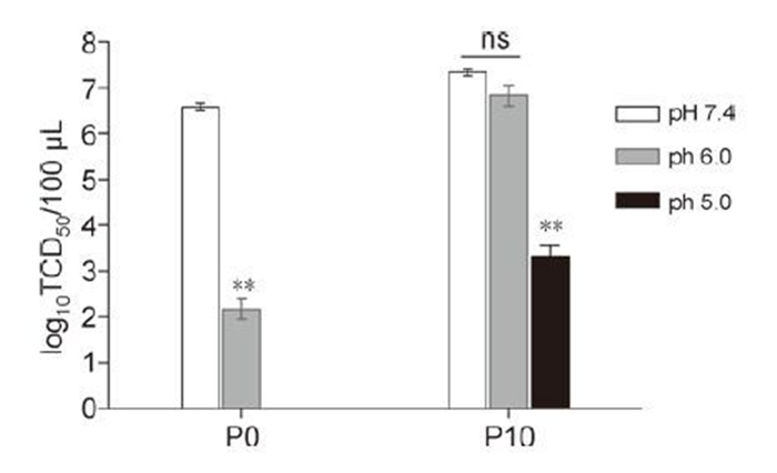
Figure 1. Resistance of the acid-selected FMDVs to acid-induced inactivation. Viral samples of P10 and their parental virus Asia1/YS/CHA/05 (P0), were treated with acidic buffers (pH 5.0 and 6.0) or neutral buffer (pH 7.4) as a control. The acidselected FMDVs were titered in TCID50 assays. Mean virus titers ± SEM are shown (two-tailed paired Student's t test, n = 4, *P < 0.05, **P < 0.01). P > 0.05 was considered nonsignificant (ns).
-
To investigate the amino acid substitutions responsible for the acid-resistant phenotype of type Asia1 FMDV, the complete capsid-coding regions of the acid-selected P10 were subjected to RT-PCR, and the purified PCR product was directly sequenced. Sequence alignment revealed that there were four amino acid substitutions in the 10th passage of the acid-selected FMDV, namely VP1 N17D (A3310G), VP2 H145Y (C2383T), VP2 G192D (G2525A) and VP3 K153E (A3061G) substitutions. These amino acid substitutions identified in the capsids were mapped on the 3D model of the FMDV Asia1/YS/CHA/05 (Figure 2). The VP1 residue N17 is located within an internal region of the capsid(Figure 2A and Figure 2B).The amino acid residue H145 of the VP2 is close to the tip of the 3-fold axis interfaces of the capsid (Figure 2A). The VP2 residue G192 is the only amino acid located at the tip of the 3-fold axis interfaces of the capsid(Figure 2A). Additionally, the VP3 residue K153 is located at the internal surface of the capsid (Figure 2A).
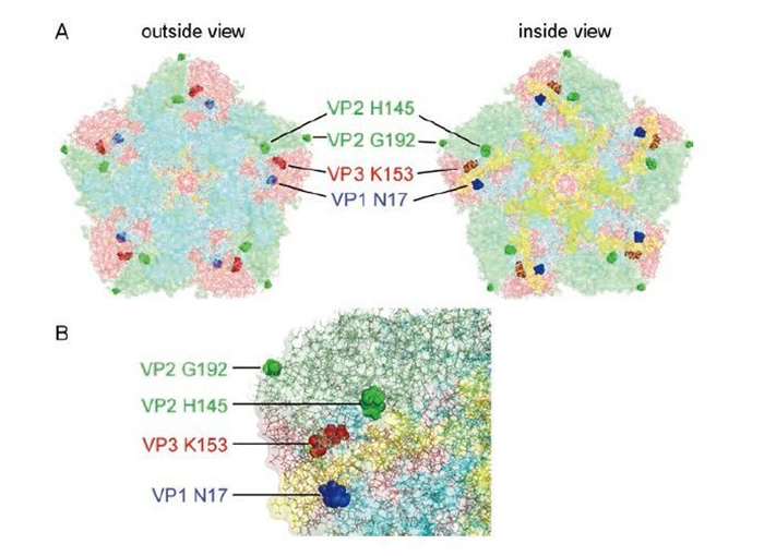
Figure 2. Location of the substituted amino acid residues found in the capsids of acid-selected FMDV mutants. (A) External and internal schematic views of a pentameric subunit in the capsid are shown. VP1 is coloured aquamarine, VP2 pale green, VP3 salmon, and VP4 yellow. The VP1 N17 position is coloured blue, VP2 H145 and G192 green, and VP3 K153 red. (B) Close-up view of the region with the amino acid substitutions in the capsid.
-
To check the effect of these amino acid changes on the acid-resistant phenotype of type Asia1 FMDV, we constructed recombinant plasmids carrying one or two of these amino acid substitutions in the capsid-coding regions. The rescued viruses were designated as rN17D, rH145Y, rG192D, rK153E, rH145Y/N17D, rG192D/N17D, and rK153E/N17D, respectively. The acid-treated mutant viruses were subjected to indirect immunofluorescence assays (IFAs), which revealed that the introduction of the VP2 H145Y substitution and/or the VP1 N17D substitution but not the VP3 K153E substitution and/or the VP2 G192D substitution conferred acid-inactivation resistance (Figure 3). These results suggest that VP1 N17D and VP2 H145Y substitutions are responsible for the acid-resistant phenotype of type Asia1 FMDV.
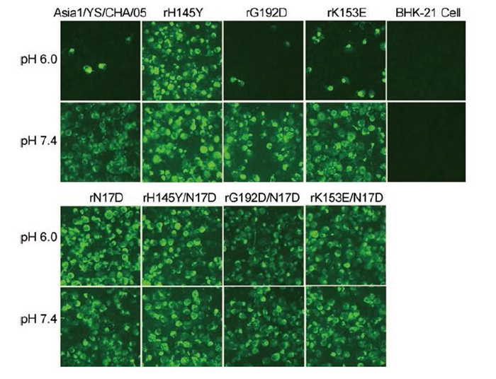
Figure 3. IFA detection of BHK-21 cells infected with FMDV mutants following acid treatment. Mutant viruses were incubated at pH 6.0 or 7.4 and subsequently used to infect BHK-21 cells grown on 96-well tissue culture plates at an initial MOI of 10. The parental virus Asia1/YS/CHA/05 was used as a control. The infected cells were fixed at 12 hours post-infection for an indirect immunofluorescence assay using MAb 3E11.
To further confirm the amino acid substitutions responsible for acid resistance of type Asia1 FDMV, the rescued mutant viruses and their parental virus Asia1/YS/CHA/05 were treated with PBS at different pH values (5.0, 6.0, or 7.4), and the remaining titers were determined on BHK-21 cells. Relative to the parental virus Asia1/YS/CHA/05, significant resistance to acid-induced inactivation (at pH 5.0 or 6.0 for 30 min) was observed for mutant viruses rN17D, rH145Y, rH145Y/N17D, rG192D/N17D and rK153E/N17D (Figure 4). In contrast, no resistance to acidic pH 5.0 was observed for rK153E and rG192D, although these two mutants exhibited slightly increased resistance to inactivation at pH 6.0 (Figure 4). These results confirmed that FMDV mutant viruses with the VP2 H145Y substitution and/or the VP1 N17D substitution were resistant to acid-induced inactivation, indicating that either substitution independently conferred the acid-resistant phenotype of type Asia1 FMDV.
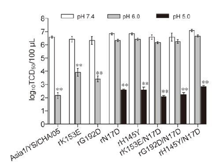
Figure 4. Effect of the VP1 N17D and VP2 H145Y substitutions on increased acid resistance in FMDV mutants. Viral samples were treated at different pH values (5.0, 6.0, or 7.4) for 30 min at room temperature, and the remaining infectious titers were determined by TCID50 assay. The parental virus Asia1/YS/CHA/05 was used as a control. Mean virus titers ± SEM are shown (two-tailed paired Student's t test, n = 4, **P < 0.01).
-
To assess the genetic stability of the mutant viruses, the rescued mutant viruses rN17D, rH145Y, and rH145Y/N17D were serially passaged in BHK-21 cells after treatment with pH 6.0 or 7.4 PBS prior to serial passages. The resulting viruses from the 5th and 10th passages were subjected to RT-PCR for sequencing to verify the stability of the acid-resistant viruses carrying the amino acid residues responsible for the acid-resistant phenotype. Sequence analysis revealed that the introduced substitutions in these acid-resistant mutants were genetically stable for at least 10 passages in either the pH 6.0 or 7.4 environment. Moreover, these mutant viruses always maintained the same acid-inactivation resistance capability over 10 passages (data not shown). Collectively, these results demonstrated that the mutants with acid-resistant properties were genetically and phenotypically stable.
-
To test the fitness of the acid-resistant mutant viruses at pH 6.0 and 7.4, Asia1/YS/CHA/05 and one of the acid-resistant mutant viruses were mixed with an initial MOI of 0.1 (0.05 for each virus), incubated at pH 6.0 or 7.4, and used to infect BHK-21 cells. Viruses recovered from these infections were incubated again at pH 6.0 or 7.4 for further passage. The percentages of competing genomes during the five serial passages were determined by sequencing (Figure 5). The experiment was repeated three times, and the results were very consistent. Any of the acid-resistant viruses of rN17D, rH145Y, and rH145Y/N17D became dominant within the population at the first passage when competing with Asia1/YS/CHA/05 at pH 6.0, driving the Asia1/YS/CHA/05 virus to near extinction. However, the abundance of Asia1/YS/CHA/05 genomes began to increase at the first passage and became the dominant population at the fifth passage in competition with rH145Y or rH145Y at pH 7.4. In contrast, the rH145Y/N17D became dominant against Asia1/YS/CHA/05 at pH 7.4 within the mixed population at the first passage, and Asia1/YS/CHA/05 almost became extinct at passage three (Figure 5C). These results showed that the mutant virus carrying double substitutions of VP1 N17D and VP2 H145Y possesses an overwhelming fitness advantage in serial passages at both tested pHs.
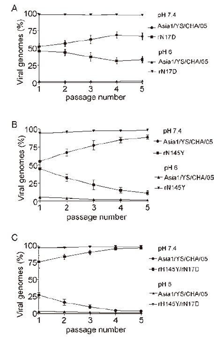
Figure 5. Competition experiments of the acid-resistant mutants and the parental virus. Viruses were mixed at a 1:1 ratio to infect BHK-21 cells in triplicate at an MOI of 0.05 for each virus. The percentages of competing genomes during serial passages were determined by sequencing. The abundances of the parental and acid-resistant viruses were measured visually as the heights of the corresponding nucleotides in sequencing chromatograms.
-
NH4Cl was used to study whether elevation of the endosomal pH decreased the infectious titers of the acid-resistant mutant viruses. In this study, differential sensitivity to the NH4Cl was observed among the rescued mutant viruses. Relative to the parental virus Asia1/YS/CHA/05, a similar sensitivity to NH4Cl treatment was observed for the rK153E and rG192D (Figure 6). However, the titers of the acid-resistant mutant viruses rN17D, rH145Y, rH145Y/N17D, rG192D/N17D, and rK153E/N17D were more effectively inhibited by NH4Cl than rK153E and rG192D (Figure 6). These results demonstrate that the increased sensitivity of type Asia1 FMDV to NH4Cl correlates with the acid-resistant phenotype.
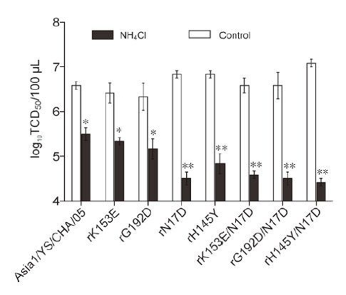
Figure 6. Sensitivity of the rescued mutants to NH4Cl treatment. BHK-21 cells with or without 25 mM NH4Cl treatment were infected with the rescued FMDV mutants and the parental virus Asia1/YS/CHA/05 as a control. Mean virus titers ± SEM and P values (two-tailed paired Student's t test, n = 3, *P < 0.05, **P < 0.01) are shown.
Generation of type Asia1 FMDV mutants with an acid-resistant phenotype
Amino acid substitutions in the capsid-coding regions of the acid-selected type Asia1 FMDV
VP1 N17D or VP2 H145Y substitution independently confers the acid-resistant phenotype of type Asia1 FMDV
Genetic stability of the mutant viruses
Biological fitness of the acid-resistant FMDV mutants
Increased resistance of the FMDV mutants to acidic pH correlates with increased sensitivity to endosomal acidification inhibition
-
Among all of the picornaviruses, FMDV is thought to be the most sensitive to acidic pH (Curry S, et al., 1995). Dissociated FMDV virions are much less immunogenic than whole viral particles (Doel T R, et al., 1982). Thus, inactivated FMD vaccines prepared using acid-stable virus could be much more effective in inducing neutralizing antibodies. The first documented acid-resistant FMDV mutant virus was selected from type A12 FMDV after exposure to an acidic environment (pH 6.4) (Twomey T, et al., 1995). Although viral RNA has been shown to be involved in the acid sensitivity of FMDV capsids (Curry S, et al., 1995), it is likely that a cluster of histidine residues (with a pKa of pH 6.8) at the pentamer interfaces is the basis of virus instability below pH 7.0 (Acharya R, et al., 1989; Ellard F M, et al., 1999; Tuthill T J, et al., 2009; van Vlijmen H W, et al., 1998). Recently, a single amino acid substitution (VP1 N17D) in the type C and type O FMDV capsid was demonstrated to confer increased acid resistance (Liang T, et al., 2013; Martin-Acebes M A, et al., 2011). In this study, type Asia1 FMDV mutants with increased resistance to acid-induced inactivation were selected through serial passages from a viral population after in vitro incubation with PBS at pH 5.0. Comparative analysis of the sequence was used to identify critical amino acid residues that confer acid resistance, followed by confirmation of molecular determinants of the acid-resistant phenotype using a reverse genetic system. Our results demonstrated that VP2 H145Y and/or VP1 N17D substitutions were responsible for the acid stability of type Asia1 FMDV. In addition, all the acid-resistant mutant viruses rescued in this study were genetically stable for at least 10 passages.
Studies have demonstrated that abrogation of the VP0 cleavage consistently correlates with loss of picornavirus infectivity (Bishop N E, et al., 1993; Lee W M, et al., 1993; Moscufo N, et al., 1993). For example, poliovirus VP2 H195 substitution results in no cleavage of VP0 into VP2 and VP4; therefore, substitutions of the H195 in poliovirus VP2 are lethal (Hindiyeh M, et al., 1999). For FMDV, VP2 H145 is the corresponding residue to the poliovirus VP2 H195. Structural studies have also implicated this equivalent H145 residue in the VP0 cleavage of FMDV (Curry S, et al., 1997), so substitution of the VP2 H145 could be lethal for FMDV. In contrast, the results of this study showed that VP2 H145Y replacement did not influence the rescue of viable rH145Y virus. More unexpectedly, the VP2 H145Y substitution conferred the acid-resistant phenotype in type Asia1 FMDV, suggesting that the H145 located closely to the 3-fold axis interfaces acts as a pH sensor. Studies have shown that the maintenance of intact 146S particles is closely related to vaccine efficacy (Doel T R, 2003; Doel T R, 2005), and dissociation of virions into pentamers leads to the loss of immunogenicity in FMD vaccines (Mateo R, et al., 2008). In this study, the VP2 H145Y substitution lowered the acid sensitivity of the type Asia1 FMDV capsid, possibly rendering the FMDV rH145Y mutant resistant to acid-induced dissociation into pentamers. Theoretically, this would aid in the rational design of an improved FMD vaccine. It has been shown that the H145 of VP2 is highly conserved in all serotypes of FMDV isolates (Carrillo C, et al., 2005; Palmenberg A C, 1989); thus, we assumed that substitution of the highly conserved VP2 H145 in other serotypes of FMDV could confer resistance to acid inactivation. Recently, Martín-Acebes's group demonstrated that VP2 H145Y conferred acid resistance to type C FMDV (Vazquez-Calvo A, et al., 2013), which is consistent with our findings.
FMDV uncoating leading to virus infection is initiated by acidification inside endosomes, especially the early endosome. Viruses exhibiting a higher acid-resistant phenotype may be less likely to survive. Therefore, although VP1 N17D functions independently of VP2 H145Y in inducing the acid-resistant phenotype of FMDV, no synergistic effect was observed for VP1 N17D and VP2 H145Y substitutions for the acid-resistance phenotype.
It is known that the antigenic site 1 (the VP1 G-H loop) is an immunodominant epitope in eliciting neutralizing antibodies (Bittle J L, et al., 1982; DiMarchi R, et al., 1986; Pfaff E, et al., 1982). Most recently, antigenic site 2 (amino acid residues at positions 70-73, 75, 77, and 131 of VP2) was shown to be important for the induction of neutralizing antibodies in cattle, sheep, and pigs (Mahapatra M, et al., 2012). For the rH145Y/N17D virus constructed in this study, neither the VP1 N17D nor the VP2 H145Y substitution was within these two major antigenic sites. In addition, the potent neutralizing MAb 3E11 prepared in our laboratory (Wang H, et al., 2011), which recognizes a dominant protective epitope in the VP1 G-H loop of FMDV Asia1/YS/CHA/05, can neutralize acid-resistant mutant viruses rN17D, rH145Y, and rH145Y/N17D at the same level as Asia1/YS/CHA/05 with a neutralizing titer of 1: 1024, indicating that the acid-resistant mutant viruses retained the type Asia1 FMDV protective neutralizing epitope recognized by MAb 3E11. Together, these findings suggested that the VP1 N17D and VP2 H145Y substitutions did not affect the antigenicity of the type Asia1 FMDV mutant rH145Y/N17D.
In conclusion, our studies showed that the single amino acid substitution VP2 H145Y is capable of conferring acid-resistance to type Asia1 FMDV Asia1/YS/CHA/05. In addition, the acid-resistant virus rH145Y/N17D, with double amino acid substitutions, was genetically stable and antigenically indistinguishable from its parental virus.
-
The authors thank Xiaochun Xie for helpful discussion regarding the study. This work was supported by grants from the National Natural Science Foundation of China (No. 31101801). We would like to express our gratitude to Yingying Guo and Professor Jingfei Wang from the Harbin Veterinary Research Institute for constructing the 3D image of the FMDV capsid.
-
All the authors declare that they have no competing interest. This article does not contain any studies with human or animal subjects performed by the any of the authors.
-
H W Wang, S S Song, and J X Zeng performed the experiments. G H Zhou, D C Yang, and T Liang participated in the experiments and data analysis. H W Wang drafted the manuscript. HW Wang and L Yu conceived of the study. All authors read and approved the final manuscript.




 DownLoad: