


HTML
-
Epstein-Barr virus(EBV) belongs to the gamma herpesvirus family that also includes the Kaposi's sarcoma-associated herpes virus(KSHV), which is more commonly associated with immune-deficient diseases. The EBV is a highly successful virus infecting the majority of the human population( < 90%) worldwide. It is also the first human tumor virus identified(Young and Rickinson, 2004). While infection of EBV is ubiquitous, tumorigenesis only occurs in a small fraction of the infected population, suggesting that the tumorigenic transformation of human cells by EBV involves complex virus-host interactions and other additional co-factors. A compromised host immune condition and a chronic inflammatory microenvironment probably play major roles in mediating the pathogenic actions of EBV in human malignancies (Rickinson, 2014).
EBV exhibits dual tropism, infecting both B cells and epithelial cells(Borza and Hutt-Fletcher, 2002). Latently infected memory B cells are believed to be the reservoir of EBV, which undergoes lytic reactivation upon stimulation to produce infectious virus. Human B cells, but not epithelial cells, are readily infected by EBV. Infection of B cells is mediated by the complement receptor type 2(CR2) on the membrane surface. EBV infection in infants and young children is generally asymptomatic; however, in adults, EBV infection induces infectious mononucleosis(glandular fever) involving a proliferation of lymphoid tissue. Symptoms include fever, sore throat, and fatigue. The lymphoproliferation process is self-limiting in healthy individuals with competent immune systems, and will eventually subside. After that, EBV establishes a lifelong infection in the body and generally remains asymptomatic. However, lymphoproliferation will take place if immune function is compromised, as in post-transplantation and HIV(human immunodeficiency virus) patients. Clearly, the body immune system plays a paramount role in checking the proliferation of EBVinfected cells. The cytotoxic T cells(CTL) are constantly checking and regulating the proliferation of EBVinfected B cells in the body.
-
The tumorigenic potential of EBV was first observed in Burkitt's lymphoma, a special type of childhood cancer common among African children. Co-factors are believed to be involved in the pathogenesis of Burkitt's lymphoma(Thorley-Lawson and Allday, 2008). The incidence of Burkitt's lymphoma is closely associated with malaria infection, though the exact contribution of malaria infection to Burkitt's lymphoma remains undefined. The chronic inflammation associated with malaria infection may promote clonal expansion of EBV-infected B cells. Malaria infection may also compromise the host immune system through unknown processes and provide a permissive environment for EBV-infected B cells to evolve into Burkitt's lymphoma cells(Moormann et al., 2011). Translocation of c-myc in infected B cells plays a key role in the etiology of Burkitt's lymphoma.
The EBV was identified directly under electron microscopic observation of cell lines established from Burkitt's lymphoma. The ability of EBV to induce proliferation in infected B cells was later demonstrated by culturing peripheral B cells with filtered supernatant harvested from Burkitt's lymphoma cells, where clusters of proliferative clones of EBV-infected B cells could be readily demonstrated (Henle et al., 1967). The ability of EBV to transform and immortalize human B cells strongly implicates the tumorigenic potential of the virus.
In addition to Burkitt's lymphoma, EBV infection was later observed in other human malignancies, including hematological and lymphatic tumours, such as Hodgkin's disease, T cell lymphoma, and NK cell lymphoma, and epithelial cancers, such as nasopharyngeal and gastric carcinomas(Young and Rickinson, 2004; Tsao et al., 2015). In all cases, the nature of EBV infection in infected cancer cells is predominantly latent.
Epstein-Barr virus infection is ubiquitous in humans
EBV infection and human cancers
-
Nasopharyngeal carcinoma(NPC) is unique in its close association(100%) with EBV infection. It is a rare cancer in Western countries, but a common cancer in the ethnic Chinese population living in the southern provinces of China. NPC is closely associated with Cantonesespeaking populations and is nicknamed "Cantonese cancer". The etiology of NPC is multifactorial, and includes genetic predisposition, EBV infection, and diet(Tsao et al., 2014). The major histological type of NPC in endemic regions is undifferentiated NPC, which is associated with EBV infection. EBV infection could be demonstrated in almost every NPC cell, distinguishing NPC from other squamous carcinomas arising in the head and neck regions, which are all EBV-negative. NPC patients have elevated serological IgA against the EBV lytic protein-viral capsid antigen(VCA) and early antigen(EA). The detection of IgA against EBV VCA and EBV DNA in plasma are important diagnostic tools of NPC, and are used extensively in early screening of NPC in high-risk populations. Clearly, the status of differentiation has a role in persistence of EBV infection in epithelial cancers. Recently, the detection of plasma EBV DNA has been shown to improve the sensitivity and specificity of the diagnosis of NPC(Le et al., 2013). The level of plasma EBV DNA also reflects faithfully the tumor burden in NPC patients, and is a powerful tool for monitoring the disease progression during treatment(To et al., 2003).
-
As previously mentioned, infection of B cells, but not epithelial cells, by EBV is a highly efficient process. The CR2 receptor, which is present on the surface of B cells and facilitates EBV entry, is generally not expressed in epithelial cells. Infection of oropharyngeal epithelial cells could be achieved via the EBV BMRF2 protein and cellular integrin receptors(Tugizov et al., 2003). Interestingly, EBV adopted an intricate cell entry mechanism by switching its envelope proteins in order to infect B cells and epithelial cells alternatively. The EBV binds to CR2 receptor present on B cell surfaces through the viral envelope protein, gp350. This interaction is augmented by the binding of another viral envelope protein, gp42, to the human leukocyte antigen(HLA) class Ⅱ protein expressed on the B cell surface, which triggers the fusion of the EBV envelope to the B cell membrane (Chesnokova et al., 2009). This process involves the viral envelope proteins gB and gHgL. Neither the CR2 and HLA class Ⅱ are expressed on the surface of epithelial cells. The EBV entry into epithelial cells involves the binding of viral envelope proteins to epithelial surface integrins, αvβ6 and αvβ8, which triggers membrane fusion and viral entry. The presence of gp42 in the viral envelope actually impedes EBV entry into epithelial cells through interaction with the integrin complex. Interestingly, EBV virions emerging from B cells that have been triggered to undergo lytic infection lack gp42, facilitating subsequent EBV binding and entry into epithelial cells. In contrast, virions released from epithelial cells are rich in gp42, which facilitates the infection of B cells. By switching its envelope proteins, EBV is able to shuttle between B cells and epithelial cells, which is crucial for its persistent infection in humans. Recently, the neuropilin1(NRP1) was identified as an entry receptor for EBV infection of epithelial cells, and found to interact with the EBV envelope protein gB to promote EBV infection of nasopharyngeal epithelial cells(Wang et al., 2015).
It remains unknown whether primary pharyngeal epithelial cells or naïve B cells are the first cellular target of EBV infection. Latently infected memory B cells are believed to be a reservoir of EBV; an estimated 1 in 106 circulating blood lymphocytes are latently infected (Babcock et al., 1998). These latently infected B lymphocytes may spontaneously reactivate into lytic cycle. The released virions may then infect a few cells in the oropharyngeal epithelium. EBV infection of primary human epithelial cells is believed to be primarily lytic in nature. A low frequency of lytic EBV infection occurring in the oropharyngeal epithelium may be responsible for the continuous release of infectious viral particles in the saliva for transmission(Hadinoto et al., 2009).
-
Similar route of infection of nasopharyngeal epithelial cells may take place in vivo. As EBV infection of normal epithelial cells normally results in a lytic infection, establishment of a latent EBV infection in epithelium may be an early step in carcinogenesis. Detection of latent EBV infection in normal epithelium is uncommon in healthy individuals. Nonetheless, a low percentage(0.005 to 0.01%) of EBV-infected cells expressing the EBV latent gene, LMP1, were present in oropharyngeal epithelium explanted to grow in culture(Pegtel et al., 2004). The status of latent infection of normal nasopharyngeal epithelium is unknown. The unique histological properties of the nasopharyngeal epithelium may support latent EBV infection, particularly in dysplastic or precancerous conditions. NPC in endemic regions is predominantly undifferentiated or poorly differentiated in nature. Inactivation of the p16 tumor suppression gene and overexpression of cyclin D1 are common events in NPC, and can be detected in premalignant and dysplastic nasopharyngeal epithelium(Lo et al., 2004b). Inactivation of p16 and activation of the cyclin D1 pathway may confer undifferentiated properties to the nasopharyngeal epithelium to support EBV infection. Using an in vitro model of immortalized nasopharyngeal epithelial cells, we showed that EBV infection readily induced growth arrest in nasopharyngeal epithelial cells. However, inactivation of p16 and /or activation of cyclin D1/cdk4/6 could override the growth arrest induced by EBV infection, and supported a stable latent EBV infection in infected nasopharyngeal epithelial cells(Tsang et al., 2012). Overexpression of cyclin D1 in immortalized nasopharyngeal epithelial cells confers resistance to stimulation of differentiation induced by serum and calcium(Tsao et al., 2002. unpublished observation). Similarly, other oncogenic alterations, including overexpression of B lymphoma Mo-MLV insertion region 1 homolog(bmi-1), may also support latent EBV infection of nasopharyngeal epithelial cells(Yip et al., 2013). Moreover, EBV-infected immortalized nasopharyngeal epithelial cells remain non-tumorigenic when injected subcutaneously in immunosuppressed mice, indicating that additional events are required for the malignant transformation of EBV-infected immortalized nasopharyngeal epithelial cells.
Route of EBV entry and infection of B cells and human epithelial cells
Establishment of latent infection in nasopharyngeal epithelial cells
-
Latent infection with EBV is commonly associated with the development of human cancers. During latent infection, EBV expresses a small number of its genes to evade detection by the host immune system. The latent gene expression in EBV-infected cells is under epigenetic regulation(Tempera and Lieberman, 2014). Several types of latent gene expression profiles have been identified in EBV-infected B cells and human cancer cells (Table 1). Type 0 latency is recognized in memory B cells, where expression of EBV genes is reduced to only EBV-encoded small RNAs(EBERs), with no EBV proteins expressed. The EBV nuclear antigen 1(EBNA1) is only expressed in memory B cells undergoing division (Hochberg et al., 2004). Type Ⅰ latency is characteristic of EBV-associated B cell lymphoma, while type Ⅱ latency is observed in nasopharyngeal and gastric carcinoma. Type Ⅲ infection represents a full-blown expression of latent EBV genes for growth promotion, and is observed during the in vitro transformation of B cells into proliferative lymphoblastoid cell lines(LCL) by EBV. Similarly, type Ⅲ latency is also observed in lymphoproliferative disorder in immunocompromised patients. The proliferative transformation of B cells during EBV infection is not observed in EBV infection of epithelial cells. EBNA2 and 3C, which are involved in cell cycle progression in EBV-transformed LCL cells are not expressed in NPC. In contrast, the BamH1 A rightward transcripts(BARTs) and their encoded EBV miRNAs are abundantly expressed in NPC(type Ⅱ latency) and Burkitt's lymphoma (type Ⅰ latency). Interestingly, the BamH1 H rightward opening reading frame 1(BHRF1) encoded miRNAs are abundantly expressed in LCL cells, a type Ⅲ latency, but not in type Ⅰ or Ⅱ latencies. The pathological role of the differential expression of EBV-encoded miRNAs in epithelial malignancies has been proposed(Lo et al., 2012). The expression profiles of EBV genes during different latency program are listed below(Table 1).

Table 1. Major EBV genes expressed in different types of latent infection.
-
A summary of the functions of EBV genes expressed in NPC(type Ⅱ latency) is reviewed here. Their potential contributions to the pathogenesis of epithelial malignancies are discussed below:
-
EBNA1 is required for the persistence of EBV genomes in latently infected cells and is expressed in all EBV-associated cancers, including NPC(Yates et al., 1984; Frappier, 2012). It is involved in the replication of EBV episomes in infected cells, and their segregation into daughter cells during mitosis. The EBNA1 protein binds to the FR element in the oriP(origin of replication) of EBV episomes, and tether them to host cell chromosomes to ensure their even segregation during cell division(Lupton and Levine, 1985; Krysan et al., 1989; Lee et al., 1999; Nanbo et al., 2007). Inactivation of EBNA1 function reduces the copy number of EBV episomes in EBVinfected B lymphoma cell lines and inhibits their growth (Kennedy et al., 2003).
EBNA1 affects multiple cellular pathways, including cell proliferation, invasion, survival, and DNA repair. Expression of EBNA1 in EBV-negative gastric carcinoma cell lines(SCM1 and TMC1) enhances their malignant properties when grown as xenografts in nude mice (Cheng et al., 2010). In HONE1 NPC cells, EBNA1 expression also promotes tumorigenicity and metastases in nude mice(Sheu et al., 1996). This is in concordance with the effects of EBNA1 to counteract the suppressive action of Nm23-H1 on cellular proliferation and migration(Murakami et al., 2005; Kaul et al., 2007). Further evidence of EBNA1 to promote metastasis was revealed by profiling the nuclear proteome of NPC cells in response to EBNA1 overexpression(Cao et al., 2012). EBNA1 increases the nuclear levels of the metastasis related proteins, including Nm23-H1, stathmin1 and maspin. Overexpression of EBNA1 has been reported to induce epithelial-mesenchymal transition in NPC cells by inhibiting the expression of miR-200a and miR-200b, hence upregulating their target genes, the zinc finger E-box binding homeobox proteins, ZEB1 and ZEB2 (Wang et al., 2014). Interestingly, EBNA1 also enhances the angiogenic properties of NPC cells by modulating the AP-1 transcriptional pathways to enhance the secretion of VEGF(O'Neil et al., 2008). EBNA1 has been suggested to contribute to epidermal hyperplasia by inhibiting NF-κB signaling through suppressing the phosphorylation of IKK alpha/beta(Valentine et al., 2010).
Another important role of EBNA1 is to promote cell survival with DNA damage. EBNA1-expressing cells have decreased levels of p53 in response to DNA damage, and therefore are more likely to survive with DNA damage. This may be related to the action of EBNA1 to disrupt the promyelocytic leukemia(PML) nuclear bodies (Sivachandran et al., 2008), which are nuclear foci containing many cellular proteins involved in cell survival, p53 activation and DNA repair(Bernardi and Pandolfi, 2007; Salomoni et al., 2008). EBNA1 induces loss of PML nuclear bodies by binding to the CK2 kinase and ubiquitin-specific protease 7(VSP7). The interaction of EBNA1 with USP7 leads to destabilization of p53. USP7 is known to bind to and stabilize p53 and Mdm2. EBNA1 outcompetes p53 and Mdm2 binding to USP7, leading to their degradation by the ubiquitin/proteasome system (Sivachandran et al., 2012). Upregulation of ROS and NADPH oxidase levels have been identified in EBNA1-expressing NPC cells(Cao et al., 2012), suggesting that EBNA1 promotes oxidative-stress induced DNA damage, but allows the survival of cells with DNA damage by destabilizing p53 via disruption of its interaction with USP7.
-
EBV encodes two small non-polyadenylated RNAs(EBER1 and EBER2), which are 167 and 172 nucleotides long, respectively, and form double-stranded RNA-like structures (Lerner et al., 1981; Takada and Nanbo, 2001). They are the most abundant viral transcripts in EBV-infected NPC and gastric cancer cells, and contribute to oncogenesis by promoting cellular growth and modulating innate immunity (Takada, 2012). A recent study has indicated that EBER may regulate LMP1/LMP2 expression and contribute to the persistence of latent EBV infection in cells (Lee et al., 2015). EBERs can induce insulin-like growth factor 1(IGF-1) to stimulate autocrine growth of NPC cells(Iwakiri et al., 2005). Induction of IGF-1 is initiated by the activation of retinotic acid inducible gene-1(RIG-1) and toll-like receptor 3 signaling, leading to the phosphorylation of downstream effector molecules, such as IRF-3, and the release of IGF-1(Yoneyama et al., 2004; Samanta et al., 2008; Liu and Gu, 2011). EBERs are responsible for the immune system activation by EBV, resulting in the production of antiviral and anti-proliferative cytokines, such as type 1 interferons(IFNs). Interestingly, EBERs can counteract the effects of INFs by inhibiting the major downstream events of IFNs and PKR signaling(Yamamoto et al., 2000; Nanbo et al., 2002; Nanbo et al., 2005). EBERs block the phosphorylation of the cellular substrate of PKR, eIF-2alpha, which signals a translational block of protein synthesis that may protect EBV-infected cells from Fas-mediated apoptosis induced by IFNs(Nanbo et al., 2005).
-
LMP1 is a transmembrane protein displaying numerous oncogenic properties in EBV-infected cells(Dawson et al., 2012). It is one of the earliest proteins identified to transform human B cells and rodent fibroblasts(Wang et al., 1985; Kaye et al., 1993). LMP1 is a potent activator of NF-κB signaling and is believed to play an essential role in promoting NPC development (Tsao et al., 2002; Dawson et al., 2012). However, expression of LMP1 alone could not transform immortalized/ premalignant nasopharyngeal epithelial cells in vitro (Tsang et al., 2010; Dawson et al., 2012; Tsang et al., 2012). LMP1 acts as a constitutively activated tumor necrosis factor receptor 1, and consists of a cytoplasmic N-terminal domain, six transmembrane spinning regions and a large cytosolic C-terminal domain(Dawson et al., 2012). The transmembrane domain has been recently reported to activate the cdc42, one of the Rho GTPases that signals cytoskeleton rearrangement and invasive properties(Liu et al., 2012). The C-terminal domain contains three activation regions, CTAR1, CTAR2 and CTAR3, which are involved in activation of a panel of signaling pathways, including NF-κB, JNK/p-38, PI3K/ AKT, ERK/MAPK and JAK/STAT, to elicit various oncogenic functions(Tsao et al., 2002; Li and Chang, 2003; Zheng et al., 2007; Morris et al., 2009). LMP1 promotes cell survival, proliferation and resistance to apoptosis in NPC cells. It upregulates the growth rate of NPC cells by enhancing the expression of EGFR, a growth-stimulating receptor frequently overexpressed in NPC tissues(Miller et al., 1995; Sheen et al., 1999). It also promotes the expression of anti-apoptotic proteins, such as survivin and Mcl-1, while inactivating pro-apoptotic proteins, such as Bad and Foxo3a(Tsao et al., 2002; Morris et al., 2009; Lo et al., 2010). LMP1-expressing cells exhibit impairment of the G2 checkpoint, which leads to unrepaired chromatid breaks after gamma-ray irradiation, and chromosome instability(Deng et al., 2012). LMP1 also resists the growth suppressive effect of TGF-beta by induction of the inhibitor of differentiation 1(Id-1) protein(Lo et al., 2010). In addition, LMP1 contributes to chemo-resistance by induction of miR-21 through activation of PI3K/AKT/FOXO3 to resist apoptotic stimuli (Yang et al., 2013). LMP1 also downregulates p16/ p21 and upregulates cyclin D1 to bypass the G1/S cell cycle checkpoint(Yang et al., 2000; Huang and Huang, 2003; Lo et al., 2004a). A recent study reported that LMP1 promotes the binding of both EGFR and STAT3 to the cyclin D1 promoter to drive the expression of cyclin D1 in NPC cells(Xu et al., 2013).
Another known function of LMP1 is to enhance the invasive and metastatic potential of NPC cells. NPC is a highly metastatic cancer(Tao and Chan, 2007). LMP1 induces epithelial-mesenchyme-transition(EMT) by downregulating E-cadherin, and upregulating Twist, Snail and Slugs(Fahraeus et al., 1992; Horikawa et al., 2007; Horikawa et al., 2011; Dawson et al., 2012). LMP1 can transcriptionally induce TNF-alpha-induced protein 2(TNFAIP2), which correlates with metastasis and poor survival in NPC patients(Chen et al., 2014). Interestingly, LMP1 increases the levels of HIF-1alpha in exosomes, which are then delivered to surrounding tumor cells for EMT induction and pro-metastatic effects (Aga et al., 2014). LMP1 can also induce the secretion of matrix metalloproteinases(MMPs) and suppress the expression of tissue inhibitor of metalloproteinases (TIMPs) to facilitate the degradation of extracellular matrix for cellular invasion or metastasis development in NPC(Horikawa et al., 2000; Yoshizaki, 2002; Lee et al., 2007; Chang et al., 2008). C-Met, an important invasive promoting protein, could be upregulated by LMP1 and has a positive association with cervical lymph node metastasis developed from primary NPC(Horikawa et al., 2001). LMP1 also regulates the expression of microRNAs, such as miRNA 203 and miRNA 10b, to promote tumor incidence and metastasis, respectively(Li et al., 2010; Yu et al., 2012). Furthermore, LMP1 induces cancer stem/progenitor cell-like properties in NPC cells, and thereby upregulates their in vitro self-renewal and in vivo tumor initiation ability(Kondo et al., 2011). LMP1 also enhances the expression of cancer stem cell-like markers, such as CD44, by activating the Hedgehog pathway and an autocrine activation of the SHH lig and (Port et al., 2013). The CD44 high cells are more radioresistant than the CD44 low cells, which may be due to the suppressed DNA damage response and p53-induced apoptosis(Yang et al., 2014). Recent publications suggest that LMP1 also modulates the cellular metabolism to promote proliferation and transformation of NPC cells(Lo et al., 2013; Xiao et al., 2014). Upregulation of hexokinase 2 and inhibition of LKB-AMPK in LMP1-expressing cells are shown to be responsible for reprogramming of glycolysis and energy metabolism, which contribute to radioresistance. Angiogeneis is another important biological process regulated by LMP1. A higher density of microvessels can be observed in NPC tumors with high expression of LMP1(Tsuji et al., 2008). This could be attributed to the reduced degradation of hypoxia inducible factor alpha (HIF-1alpha) and induced expression of VEGF by LMP1.
-
The LMP2 proteins, LMP2A and LMP2B, are transcribed from two distinct mRNAs encoding 54-kDa and 40-kDa proteins, respectively. LMP2A/B is an integral membrane protein with 12 transmembrane spanning regions. Their mRNAs share the same exons(2 to 9)(Sample et al., 1989; Pang et al., 2009; Dawson et al., 2012). While the exon 1 of LMP2B(exon 1B) is non-coding, the exon 1 of LMP2A (exon 1A) encodes an additional cytosolic N-terminus, which mediates multiple signaling processes(Sample et al., 1989; Pang et al., 2009; Dawson et al., 2012). The N-terminal domain contains multiple signaling domains, including an immunoreceptor tyrosine-based activation motif (ITAM) recognized by the Lyn/Syk kinases to transduce BCR signaling, and a PY motif that interacts with the NEDD4 family of ubiquitin ligases(Ikeda et al., 2000; Portis et al., 2002; Ikeda et al., 2003). Other signaling pathways downstream of these domains include PI3k/akt, RhoA, and MAPK/ERK(Heussinger et al., 2004; Pang et al., 2009; Dawson et al., 2012).
Genetic studies have revealed that LMP2A and LMP2B are not required for EBV-dependent transformation of B cells; however, LMP2A is required for the successful outgrowth of EBV-infected epithelial cells in vitro(Speck et al., 1999). LMP2 also induces anchorage-independent growth in soft agar and inhibits differentiation through activation of PI3 kinase and the Akt kinase(Scholle et al., 2000; Fukuda and Longnecker, 2007). In epithelial cells, LMP2 can promote β-catenin signaling through the activation of Akt and phosphorylation of GSK3 (Morrison and Raab-Traub, 2005). Activation of β-catenin is common in the development of carcinoma through genetic mutations, suggesting that activation of this pathway may mediate the effects of EBV on epithelial cell growth. LMP2A was also shown to inhibit cellular differentiation and promote cell survival through the PI3K/ Akt-mediated stabilization of ΔNp63(Fotheringham et al., 2010). Other roles of LMP2A-activated PI3K/akt signaling include the counteraction of the growth inhibitory and pro-apoptotic effects of TGF-beta1 during epithelial carcinogenesis(Fukuda and Longnecker, 2004), and the proliferation and protein synthesis in cells via the activation of mTOR pathway(Moody et al., 2005). LMP2A and LMP2B have also been shown to limit the anti-viral response against EBV-infected cells by modulating IFN signaling(Shah et al., 2009). Response to IFN was downregulated in LMP2A or LMP2B-expressing epithelial cells due to an increased turnover of IFN receptors(Shah et al., 2009). Lastly, similar to LMP1, LMP2A can promote the invasive/migratory properties of epithelial cells, which may relate to the metastatic phenotype of NPC (Allen et al., 2005; Pegtel et al., 2005; Lu et al., 2006; Kong et al., 2010). Studies have suggested that LMP2A and LMP2B modulate the interaction and focal adhesion formation with the extracellular matrix(Allen et al., 2005; Pegtel et al., 2005). Cells overexpressing LMP2A/ B have an increased rate of attachment, spreading and migratory movement on the extracellular matrix(Allen et al., 2005). This possibly involves the regulation of integrin-mediated processes. A recent study has shown that the ITAM signaling domain of LMP2A can activate the Syk tyrosine kinase and Akt to stabilize alphaV-integrin and FAK activation(Fotheringham et al., 2012). Another study showed that LMP2A expression is positively associated with integrin alpha6 in NPC biopsies(Pegtel et al., 2005). Antibodies blocking the integrins abrogate LMP2A-induced invasion(Pegtel et al., 2005). These reports suggest an interaction of LMP2A with integrins to govern the migratory, invasiveness and metastasis of the epithelial cancers. LMP2A was reported to be localized at the tumor invasive front(Kong et al., 2010). It can also potentiate cancer stem cell-like properties through activation of the Hedgehog signaling pathway(Port et al., 2013). Exogenous expression of LMP2A induces EMT, stimulates the expression of stem cell markers, and enhances the acquisition of side populations in the NPC cells(Kong et al., 2010).
-
The BARF1 is considered a lytic EBV protein and is expressed early during lytic infection. However, a high level of BARF1 expression could be detected at high levels in NPC(Decaussin et al., 2000). BARF1 is encoded in the BamH1-A fragment of EBV and is a homolog of the human colony stimulating factor 1(CSF-1) receptor (Strockbine et al., 1998). The signaling axis of CSF-1 and the CSF-1 receptor is known to be involved in promoting tumorigenicity in various types of epithelial cancer, including gastric and breast cancer(Sapi et al., 1995; Lin et al., 2001). It was also shown to have oncogenic activity, as evidenced by its malignant transforming property in rodent fibroblasts and inhibitory effects on apoptosis by activating bcl-2(Wei and Ooka, 1989; Sheng et al., 2003). Expression of BARF1, in addition to the H-Ras and SV40 T antigens, can transform nonmalignant human nasopharyngeal epithelial NP69 cells (Jiang et al., 2009). BARF1 is expressed as a latent protein in NPC and EBV-associated gastric cancer(Decaussin et al., 2000; Seto et al., 2005; Takada, 2012). It can be detected by RT-PCR and immunohistochemical assays in clinical biopsies of NPC and by a BARF1-specific nucleic acid sequence-based amplification assay in gastric tumors(Decaussin et al., 2000; zur Hausen et al., 2000). BARF1 is expressed in EBV-associated epithelial malignancies, but not in lymphoid malignancies(Takada, 2012). Detection of BARF1 mRNA in nasopharyngeal brushings has been suggested to be a promising noninvasive method for NPC diagnosis(Stevens et al., 2006).
The role of BARF1 in NPC development has been investigated by infecting CNE2 NPC cells with a recombinant EBV with BARF1 constitutively expressed under the SV40 promoter(Seto et al., 2008). Compared to control NPC cells infected with wild-type EBV, NPC cells infected with EBV constitutively expressing BARF1 have higher growth rates and are more resistant to apoptosis in serum-deprived conditions. BARF1 was detected in the culture medium, which promoted growth of the cancer cells. NPC cells infected with recombinant EBV constitutively expressing BARF1 have greater rates of tumorigenicity in the nude mice model(Seto et al., 2008). In EBV-associated gastric cancer, BARF1 enhanced the expression of cyclin D1 in vitro(Wiech et al., 2008). Analysis of a tissue array consisting of 170 gastric tumors and 11 EBV-associated gastric tumors revealed a significant overexpression of cyclin D1 in EBVassociated tumors but not in EBV-negative tumors (Wiech et al., 2008). A recent study showed that overexpression of BARF1 in gastric cancer cells promotes cellular proliferation, likely through the upregulation of expression of NF-κB, RelA, cyclin D1, and reduced expression of cell cycle inhibitor, p21(Chang et al., 2013).
-
The BARTs are multi-spliced RNAs transcribed rightwards from the BamH1 A region of EBV(Hitt et al., 1989; Smith et al., 2000; Zhang et al., 2001). The exceptional abundance of BART expression in NPC and EBV-associated gastric cancer strongly implicates an important role in these cancers (Smith et al., 2000; Al-Mozaini et al., 2009). BARTs comprise more than 96% of all EBV reads in a recent RNA-sequencing analysis of EBV-associated gastric carcinoma (Strong et al., 2013). Several ORFs in the spliced cDNA transcripts, including RPMS1, A73, BARF0, CST, vIL, and BLLF1, have been postulated to have functional roles(Kienzle et al., 1999; Al-Mozaini et al., 2009). In particular, recombinant RPMS1 and A73 expressed in E. coli were found to modulate the Notch and RACK1 signaling pathways, respectively(Smith et al., 2000; Al-Mozaini et al., 2009). RPMS1 can act as effective antagonist of Notch-IC transcription activation, and therefore may suppress the differentiation of epithelial cells(Smith et al., 2000). A73 binds with RACK1 and possibly regulates calcium release from intracellular stores by enhancing the affinity of IP3 receptor binding for IP3(Smith et al., 2000; Al-Mozaini et al., 2009). These reports suggest that BARTs may encode for proteins having biochemical functions related to oncogenesis. However, evidence for the endogenous expression of potential BART-proteins, such as RPMS1, A73 and BARFO, in EBV-infected cells is lacking(Kienzle et al., 1999; Al-Mozaini et al., 2009). Moreover, the BARTs are expressed extensively in the nucleus, but not in the cytoplasm, suggesting they are not transcribed as mRNAs(Al-Mozaini et al., 2009; Jang et al., 2011). Nevertheless, the possibility remains that these BART-proteins may be expressed under certain conditions to augment the development of cancer(Al-Mozaini et al., 2009). Another possibility is that these BARTs may act as long non-coding RNAs(lncRNA), which are involved in repressive complexes to regulate cellular gene expression(Strong et al., 2013). Interestingly, the expression patterns or levels of BARTs vary in different infection states, such as lytic and latent infections(Yamamoto and Iwatsuki, 2012). Notably, the expression of BARTs is under the regulation of c-myc and C/EBP(Chen et al., 2005) and possibly NF-κB(HL Chen, personal communication). This highlights the potential importance of local inflammation and the role of inflammatory cytokines in affecting the expression of BARTs. All this warrants the future investigation of potential functional roles of BARTs in contributing to human malignancies, particularly in NPC.
-
EBV encodes at least 44 miRNAs transcribed from the BHRF1 and BART regions(Klinke et al., 2014). miRNAs can regulate the expression of various proteins by blocking the translation of mRNAs or degrading mRNAs(Bartel, 2009). The expression pattern of miRNAs depends on the latency type and cellular context of the EBV-infected cells. BHRF1 miRNAs are only expressed in EBVinfected B cells exhibiting latency type Ⅲ infection, and are shown to mediate B cell transformation by protecting the cells from apoptosis(Amoroso et al., 2011; Vereide et al., 2014). BART miRNAs are expressed in all EBVinfected cells, but the levels are 8 to 13 fold higher in epithelial cells than in B cells(Qiu et al., 2011). In NPC and gastric cancer samples, most of the BART miRNAs are expressed. Although they are processed from the same BART transcript, they are expressed at various levels due to different biogenesis and cellular processing (Zhu et al., 2009; Chen et al., 2010; Lung et al., 2013).
The functions of BART miRNAs have been evaluated in various high-throughput studies. Recent reviews have summarized the results from different research groups in searching for high-confidence targets of EBV miRNAs (Marquitz and Raab-Traub, 2012; Cullen, 2013; Klinke et al., 2014). A few key targets of the BART miRNAs have been identified and validated for their functions. For example, miR-BART3-5p targets the tumor-suppressor gene, DICE1(Lei et al., 2013). This is in agreement with the finding that DICE1 is usually inversely correlated with the expression of miR-BART3-5p in NPC. The miR-BART2-5p targets the stress-induced immune lig and MICB to facilitate the escape from recognition by natural killer cells(Nachmani et al., 2009). In addition, BART miRNAs can target pro-apoptotic genes and thus promote host cell survival(Choy et al., 2008; Marquitz et al., 2011). Expression of PUMA-beta is regulated by miR-BART5-5p(Choy et al., 2008), and expression of Bim is regulated by miR-BART1, 3, 9, 11 and 12 (Marquitz et al., 2011). A recent study also showed that miR-BART9 promotes the invasiveness and metastatic ability of NPC cells in vivo through specific targeting of E-cadherin, a membrane protein crucial for mesenchymal-like phenotype(Hsu et al., 2014).
EBV genes can also be targets of EBV miRNAs(Lo et al., 2007; Barth et al., 2008; Lung et al., 2009). The viral DNA polymerase BALF5 is targeted by miR-BART2-5p(Barth et al., 2008), the EBV latent membrane protein 1(LMP1) by miR-BART17-5p, -1-5p or -16(Lo et al., 2007), and LMP2A by miR-BART22(Lung et al., 2009). This implicates a role for BART miRNAs in the modulation of EBV gene expression to optimize the functions of various EBV proteins in infected cells.
Contribution of EBV-encoded genes to epithelial malignancies
Epstein-Barr nuclear antigen 1(EBNA1).
EBV-encoded small RNA 1/2(EBER1/2).
Latent membrane protein 1(LMP1).
Latent membrane protein 2(LMP2).
BamH1-A fragment rightward reading frame 1(BARF1).
Bam HI A rightward transcripts(BARTs).
EBV-encoded microRNAs(miRNAs).
-
EBV infection in NPC was shown to be a clonal event and occurs during the early stages of NPC (Pathmanathan et al., 1995). Later studies have demonstrated that genetic alterations in the premalignant nasopharyngeal epithelium may precede EBV infection(Lo et al., 2004b; Tsang et al., 2012). Genetic alterations in the premalignant nasopharyngeal epithelium may confer susceptibility to latent EBV infection, which otherwise would support lytic EBV infection.
By comparing changes in EBV-infected and uninfected cancers, evidence for the pathogenic mechanism of EBV may be revealed. In NPC, this is not possible as most cases of undifferentiated NPC are associated with EBV infection. However, gastric cancer provides a unique opportunity to examine the genomic differences that may be related to EBV infection. Recent genomic profiling in EBV-associated gastric cancer reveals a distinct signature of genome wide hypermethylation compared to non-EBV gastric cancer(TCGA, 2014). Hypermethylation is commonly used in the inactivation of tumor suppressor genes. It remains to be determined if the increase of methylation in EBV-associated gastric cancer is a direct result of EBV gene expression or an adaptive response of host cells to EBV infection.
A recent genomic profile of NPC reveals multiple pathways present in NPC, and reveals a similar signature of genomic hypermethylation(Lin et al., 2014). Similar to EBV-associated gastric cancer, fewer genetic alterations were identified in NPC compared to other epithelial cancers. Presumably, EBV infection may play an important role in altering cellular pathways to promote survival of infected NPC cells, which facilitates the selection and expansion of tumorigenic clones in vivo. These reports support a causal role of EBV infection in the development of NPC.
-
It is postulated that specific EBV strain may be involved in the development of NPC. Multiple strains of EBV can be isolated from the blood and saliva of healthy individuals. Interestingly, only one strain of EBV was detected in a large cohort of NPC samples(JX Bei, Sun Yat Sen Cancer Center, personal communication). The presence of a single strain of EBV in NPC is not surprising, given the clonal origin of EBV infection at the early stages of NPC development. The NPC-associated EBV strains cluster into a distinct family that could be separated from EBV strains isolated from patients with infectious mononucleosis. In the endemic area of NPC in southern China, a specific EBV strain has been proposed to be associated with NPC. Recently, an NPC-derived EBV strain, M81, was isolated with distinct properties in host tropism and other biological properties(Tsai et al., 2013). The M81 EBV strain has a reverse tropism compared to common EBV strains, exhibiting a reduced ability to infect B cells but an increased propensity to infect epithelial cells. M81 spontaneously enters lytic replication upon infection of B cells. It remains to be determined if a specific NPC EBV strain with distinct biological properties may be involved in the pathogenesis of NPC. With further research on the genomic and biological properties of EBV isolated from NPC, the role of EBV in the development of NPC may be better understood.
-
The master switch of EBV from latent to lytic infection is triggered by the expression of BZLF1, which turns on a cascade of events that target EBV gene transcription to initiate EBV replication, packaging and release of infectious virus(Kenney and Mertz, 2014). The BRLF1 protein is also involved in the switching of EBV infection from latent to lytic mode, and may play a more important role in the lytic EBV infection of epithelial cells(Reusch et al., 2015). The physiological signals triggering lytic infection are not clearly defined, but may involve signals of differentiation and cellular stress. Lytic EBV infection was observed in non-keratinized squamous epithelial cells on the lateral side of the tongue epithelium of immunocompromised patients(Greenspan et al., 1985). Lytic replication of EBV could be demonstrated in the upper layers of the stratified squamous epithelium undergoing terminal differentiation, but not at the basal or immediate suprabasal layers, where undifferentiated epithelial cells are located. In a 3-dimensional reconstructed epithelium model of telomerase-immortalized oral keratinocytes cultured at the air-liquid phase, expression of lytic EBV genes was observed in the upper layers of differentiated epithelium but not in the undifferentiated basal layers(Kenney and Mertz, 2014). While latent EBV infection is characteristic of human malignancies, lytic EBV infection may also be involved. Interestingly, EBV defective in the lytic EBV gene, BZLF1, was found to have lower tumorigenic ability to transform B cells into lymphoma in the humanized mice model(Hong et al., 2005). A low level of expression of lytic EBV genes is often observed in NPC with predominantly latent infections (Feng et al., 2000). The significance of this low level of lytic gene expression is unclear. The late lytic genes involved in packaging of EBV for infection are not expressed, suggesting that the lytic infection is largely abortive in nature. The role of abortive lytic EBV infection in human malignancies is unclear. The BZLF1 and other lytic genes, including BGLF5, have been shown to induce DNA instability and may be involved at the initiation stage of carcinogenesis of nasopharyngeal epithelium (Sato et al., 2009; Wu et al., 2010). Promotion of premalignant nasopharyngeal epithelial cells harboring genetic mutations may be dependent on the inflammatory environment commonly present at the nasopharyngeal mucosa. It remains to be determined if these lytic genes invoke a local immune and inflammatory response to promote tumor progression in NPC(Rickinson, 2014).
-
Despite the close association of EBV infection observed in NPC, NPC cell lines established in vitro readily lose their EBV episomes upon prolonged propagation. This would suggest that EBV infection per se has no advantage in cell proliferation. Careful monitoring of replication of EBV episomes in infected cells suggests 16% of EBV episome are not replicated during each cell cycle(Nanbo et al., 2007). Hence, EBV episomes would be lost in cell culture if EBV-infected cells were not actively selected either by drugs or cell sorting. In contrast to EBV infection of human B cells, which efficiently promotes cell growth and transformation, EBV infection of primary nasopharyngeal epithelial cells induces growth arrest, probably due to the cellular stress associated with viral infection(Tsang et al., 2012). No immediate growth advantage was observed in immortalized nasopharyngeal epithelial cells after infection with EBV. The universal presence of EBV in NPC and the ability of NPC xenografts to retain their EBV episomes after repeated passage(> 25 years) in athymic nude mice suggest an advantage for EBV-infected epithelial cells grown in vivo. A recent report shows that expression of EBV-encoded miRNA was upregulated in EBV-infected NPC and gastric cancer cells grown in vivo. Furthermore, expression of BARTs enhances the growth and tumorigenicity of these cells, supporting a role for BART expression and BART-miRNA in the growth of EBV-infected epithelial cells in vivo(Qiu et al., 2015). Similarly, EBV infection and expression of EBV genes may induce inflammatory responses, which may enhance angiogenesis and growth of cancer cells in vivo. These cytokines may also attract migration of inflammatory cells, including activated macrophages and T regulatory cells, to support the growth of EBV-infected cells. Furthermore, we have also observed that stromal fibroblasts isolated from NPC synthesize and secrete IL-6 to stimulate EBV-infected NPC cells(Tsang et al., 2013; Zhang et al., 2013). EBVassociated gastric cancer is also associated with inflammatory components. A distinguishing feature of EBVassociated epithelial malignancies is the undifferentiated property of infected epithelial cells associated with the heavy infiltration of lymphoid elements; hence, the term lymphoepithelioma-like carcinoma(LELC) has been used to distinguish this group of cancer, which could also be observed in lung cancer, tonsillar and cholangiocarcinoma (Tsao et al., 2015). The stromal factors that support growth of NPC cells in vivo are not present in in vitro conditions. The postulation is that growth of NPC cells is highly dependent on the presence of inflammatory stromal elements in the nasopharyngeal mucosa, which is in a state of chronic inflammation. In concordance with this hypothesis, EBV-positive NPC cell lines are known to be difficult to establish in culture, likely due to the loss of present in NPC in vivo. The delineation and characterization of the growth requirement for these stromal elements in NPC cell lines would contribute to our understanding of the development of NPC and other EBV-infected malignancies.
EBV strains and NPC
Contribution of lytic EBV infection in human malignancies
Contribution of host stromal factors to the persistence of EBV infection in NPC
-
The role of EBV in the pathogenesis of NPC is still unknown. There are selective advantages of EBV infection in NPC in vivo, which may be facilitated by inflammatory elements in the mucosa. The understanding of these selective advantages will contribute to our undersanding of the pathogenic role of EBV infection in human malignancies. A close interaction between EBV infection, host genetic alterations, defective immune recognition and stromal inflammation are believed to be intricately involved in the pathogenesis of NPC. Defining the contribution of these parameters in the growth of NPC in vivo will provide novel therapeutic targets in the prevention and treatment of NPC. A summary of key events is included in Figure 1 to illustrate the postulated roles of EBV in NPC pathogenesis.
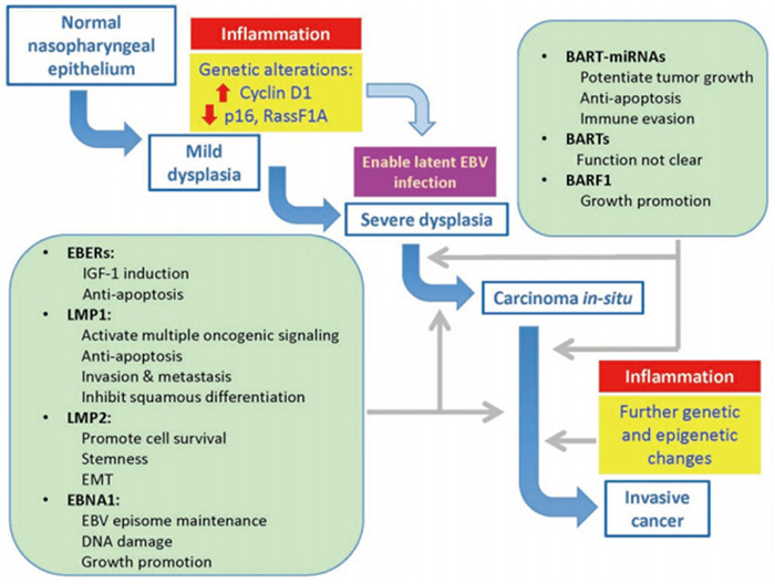
Figure 1. The role of EBV infection in the pathogenesis of NPC. The normal nasopharyngeal epithelium is refractory to EBV infection. Similar to EBV infection of oropharyngeal epithelial cells, EBV infection in the normal nasopharyngeal epithelium is presumably lytic in nature. EBV infection induces growth arrest in normal nasopharyngeal epithelial cells. Genetic alterations in the premalignant nasopharyngeal epithelium, such as cyclin D1 overexpression and p16 inactivation, override the growth arrest induced by EBV infection and support latent infection. EBV infection and expression of EBV-encoded latent genes, including BART-miRNAs, support the growth and progression of premalignant nasopharyngeal epithelial cells. Further genetic and epigenetic changes may drive the clonal expansion of EBV-infected premalignant cells and their transformation to cancer cells. Stromal inflammation is postulated to play a crucial role in modulating EBV gene expression, supporting latent EBV infection and malignant transformation of premalignant nasopharyngeal epithelial cells to cancer cells.
-
The authors acknowledge the generous funding sources for the above study: the Health and Medical Research Fund(Grant No HMRF: 12110942 and 13120872) to CMT; and GRF grants from the Hong Kong Research Grant Council(17120814, 779713, 779312, 780911, 779810), AoE NPC(Grant No. AoE/M-06/08) and the Theme-Based Research Scheme(Grant No. T12-401/13-R) to SWT.
-
The authors declare that they have no conflict of interest. This article does not contain any studies with human or animal subjects performed by any of the authors.


 DownLoad: