Wen Li, Bo Wang, Bei Li, Wei Zhang, Yan Zhu, Zheng-Li Shi and Xing-Lou Yang. Genomic Characterization of a Novel Hepatovirus from Great Roundleaf Bats in China[J]. Virologica Sinica, 2018, 33(1): 108-110. doi: 10.1007/s12250-018-0013-6
Citation: Wen Li, Bo Wang, Bei Li, Wei Zhang, Yan Zhu, Zheng-Li Shi, Xing-Lou Yang. Genomic Characterization of a Novel Hepatovirus from Great Roundleaf Bats in China .VIROLOGICA SINICA, 2018, 33(1) : 108-110.  http://dx.doi.org/10.1007/s12250-018-0013-6

蹄蝠甲肝病毒的流行病学调查及其基因组特征分析

  • 通讯作者: 杨兴娄, yangxl@wh.iov.cn, ORCID: 0000-0002-5317-8983
  • 收稿日期: 2017-11-23
    录用日期: 2018-01-04
  • 蝙蝠是多种病毒的天然存储库,近年来乙型肝炎病毒、丙型肝炎病毒、戊型肝炎病毒相继从蝙蝠中检测出,提示蝙蝠可能是多种肝炎病毒的自然宿主。2015年德国科学家从非洲刚果、加纳,欧洲德国卢森堡,拉丁美洲哥斯达黎加等地的蝙蝠、啮齿动物等多种小型哺乳动物中检测到甲型肝炎病毒存在,显示蝙蝠也携带甲型肝炎病毒。而甲型肝炎的暴发是一种对人体健康和经济发展均有严重影响的疾病,中国迄今为止还没有过关于蝙蝠携带甲型肝炎病毒的报道,本论文首次对中国各地的蹄蝠进行甲型肝炎病毒的筛查及全基因组扩增。
    本研究对2011年-2014年之间四川峨眉山、宜宾,湖北咸宁,云南墨江等地区采集的共计1266份蹄蝠粪便/肛拭子样本进行甲型肝炎病毒检测,首次从2011-2014年咸宁太乙洞的蝙蝠粪便样品中检测到9份甲型肝炎阳性样品,并获得一株长度为7,184bp的蹄蝠甲型肝炎病毒全基因组序列,命名为Hepatovirus-bat3206(HepV-bat3206)。对PCR扩增得到的部分复制酶序列进行比对,发现这些阳性序列之间的差异性很小,说明该病毒在咸宁地区的蝙蝠中可能非常保守。全基因组序列比对分析发现它与蝙蝠、刺猬和树鼩中发现的甲肝病毒同源性最高,但相似性最高为70%,其结构蛋白P1和复制酶基因相似度分析表明其属于甲肝病毒HepV-H的新的分离株。
    本研究首次从中国蝙蝠中检测到甲型肝炎病毒在,进一步丰富了中国蝙蝠携带病毒谱的知识,首次对HepV在中国蝙蝠中的流行病学进行了调查,对预防和控制该疾病提供参考。

Genomic Characterization of a Novel Hepatovirus from Great Roundleaf Bats in China

  • Corresponding author: Xing-Lou Yang, yangxl@wh.iov.cn
  • ORCID: 0000-0002-5317-8983
  • Received Date: 23 November 2017
    Accepted Date: 04 January 2018
  • The hepatitis A virus (HAV) is a positive-sense, single-stranded RNA virus of the genus Hepatovirus in the family Picornaviridae. HAV is a common agent causing acute liver disease worldwide and primarily transmitted by the fecal-oral route. Approximately 120 species of bats exist in China, but information on hepatovirus in bats is not reported. In this study, we collected 1,266 bat feces or fecal swab samples in eight places of seven provinces in China. Among these 1,266 samples, nine H. armiger feces samples were found positive in Xianning, Hubei. These nine positive samples shared 99% nucleotide identity with each other and 67%–69% nucleotide identities with other reported hepatoviruses. Meanwhile one complete genome of bat hepatovirus named HepV-bat3206 was amplified and analyzed. The complete genome length of HepV-bat3206 is 7,184 nucleotides and shared highest 70% similarity with the reported hepatoviruses. The phylogenetic tree showed that HepV-bat3206 is distantly related to human and primate hepatoviruses and but clustered together with bat, Tupia, and hedgehog hepatovirus H. The positive hepatovirus samples were collected in the same colony of bats in the years 2011, 2012 and 2014, suggesting a long history of circulation of hepatoviruses in this colony. Since the bat cave is a famous tourist site, some measures should be taken to prevent visitors are frequently exposed to the bat feces.

  • 加载中
    1. Adams MJ, Lefkowitz EJ, King AMQ, Harrach B, Harrison RL, Knowles NJ, Kropinski AM, Krupovic M, Kuhn JH, Mushegian AR, Nibert M, Sabanadzovic S, Sanfacon H, Siddell SG, Simmonds P, Varsani A, Zerbini FM, Gorbalenya AE, Davison AJ (2017) Changes to taxonomy and the international code of virus classification and nomenclature ratified by the international committee on taxonomy of viruses. Arch Virol 162:2505–2538
        doi: 10.1007/s00705-017-3358-5

    2. Aggarwal R, Goel A (2015) Hepatitis a: epidemiology in resourcepoor countries. Curr Opin Infect Dis 28:488–496
        doi: 10.1097/QCO.0000000000000188

    3. Aragones L, Guix S, Ribes E, Bosch A, Pinto RM (2010) Fine-tuning translation kinetics selection as the driving force of codon usage bias in the hepatitis a virus capsid. PLoS Pathog 6:e1000797
        doi: 10.1371/journal.ppat.1000797

    4. Dienstag JL, Davenport FM, McCollum RW, Hennessy AV, Klatskin G, Purcell RH (1976) Nonhuman primate-associated viral hepatitis type a. Serologic evidence of hepatitis a virus infection. JAMA 236:462–464

    5. Drexler JF, Corman VM, Lukashev AN, van den Brand JM, Gmyl AP, Brunink S, Rasche A, Seggewibeta N, Feng H, Leijten LM, Vallo P, Kuiken T, Dotzauer A, Ulrich RG, Lemon SM, Drosten C, Hepatovirus Ecology C (2015) Evolutionary origins of hepatitis a virus in small mammals. Proc Natl Acad Sci USA 112:15190–15195
        doi: 10.1073/pnas.1516992112

    6. He B, Zhang F, Xia L, Hu T, Chen G, Qiu W, Fan Q, Feng Y, Guo H, Tu C (2015) Identification of a novel orthohepadnavirus in pomona roundleaf bats in china. Arch Virol 160:335–337
        doi: 10.1007/s00705-014-2222-0

    7. Hollinger FB, Martin A (2013) Hepatitis a virus. In: Kluwer W (ed) Field's virology, vol 6. Lippincott Williams & Wilkins, Philadelphia, pp 550–581

    8. Jacobsen KH, Wiersma ST (2010) Hepatitis a virus seroprevalence by age and world region, 1990 and 2005. Vaccine 28:6653–6657
        doi: 10.1016/j.vaccine.2010.08.037

    9. Nie FY, Lin XD, Hao ZY, Chen XN, Wang ZX, Wang MR, Wu J, Wang HW, Zhao G, Ma RZ, Holmes EC, Zhang YZ (2017) Extensive diversity and evolution of hepadnaviruses in bats in china. Virology 514:88–97

    10. Shi Z (2013) Emerging infectious diseases associated with bat viruses. Sci China Life Sci 56:678–682
        doi: 10.1007/s11427-013-4517-x

    11. Wang B, Yang XL, Li W, Zhu Y, Ge XY, Zhang LB, Zhang YZ, Bock CT, Shi ZL (2017) Detection and genome characterization of four novel bat hepadnaviruses and a hepevirus in china. Virol J 14:40
        doi: 10.1186/s12985-017-0706-8

    12. WHO (2017) Global hepatitis report 2017. World Health Organization. http://www.who.int/hepatitis/publications/global-hepatitisreport2017/en/. Accessed 23 Nov 2017

    13. Yu JM, Li LL, Zhang CY, Lu S, Ao YY, Gao HC, Xie ZP, Xie GC, Sun XM, Pang LL, Xu JG, Lipkin WI, Duan ZJ (2016) A novel hepatovirus identified in wild woodchuck marmota himalayana. Sci Rep 6:22361
        doi: 10.1038/srep22361

  • 加载中
  • 10.1007s12250-018-0013-6.pdf

Figures(2) / Tables(2)

Article Metrics

Article views(5428) PDF downloads(29) Cited by(0)

Related
Proportional views
    通讯作者: 陈斌, bchen63@163.com
    • 1. 

      沈阳化工大学材料科学与工程学院 沈阳 110142

    1. 本站搜索
    2. 百度学术搜索
    3. 万方数据库搜索
    4. CNKI搜索

    Genomic Characterization of a Novel Hepatovirus from Great Roundleaf Bats in China

      Corresponding author: Xing-Lou Yang, yangxl@wh.iov.cn
    • 1. Key Laboratory of Special Pathogens and Biosafety, Wuhan Institute of Virology, Chinese Academy of Sciences, Wuhan 430071, China

    Abstract: The hepatitis A virus (HAV) is a positive-sense, single-stranded RNA virus of the genus Hepatovirus in the family Picornaviridae. HAV is a common agent causing acute liver disease worldwide and primarily transmitted by the fecal-oral route. Approximately 120 species of bats exist in China, but information on hepatovirus in bats is not reported. In this study, we collected 1,266 bat feces or fecal swab samples in eight places of seven provinces in China. Among these 1,266 samples, nine H. armiger feces samples were found positive in Xianning, Hubei. These nine positive samples shared 99% nucleotide identity with each other and 67%–69% nucleotide identities with other reported hepatoviruses. Meanwhile one complete genome of bat hepatovirus named HepV-bat3206 was amplified and analyzed. The complete genome length of HepV-bat3206 is 7,184 nucleotides and shared highest 70% similarity with the reported hepatoviruses. The phylogenetic tree showed that HepV-bat3206 is distantly related to human and primate hepatoviruses and but clustered together with bat, Tupia, and hedgehog hepatovirus H. The positive hepatovirus samples were collected in the same colony of bats in the years 2011, 2012 and 2014, suggesting a long history of circulation of hepatoviruses in this colony. Since the bat cave is a famous tourist site, some measures should be taken to prevent visitors are frequently exposed to the bat feces.

    • Dear Editor,

      The hepatitis A virus (HAV) is a common agent causing acute liver disease worldwide, with approximately 11, 000 deaths annually (WHO 2017). The virus is transmitted primarily by the fecal-oral route and it normally infects people living in high-density and resource-poor countries (Aggarwal and Goel 2015). Epidemics may also occur after the accidental introduction of HAV to areas with better sanitation but susceptible population (Jacobsen and Wiersma 2010). HAV is a positive-sense, single-stranded RNA virus of the genus Hepatovirus in the family Picornaviridae (Adams et al. 2017). The genome is approximately 7500 nucleotides in length and contains a single open reading frame (ORF) encoding a polyprotein, which is divided into three regions: P1 (VP1–VP4, constituting the viral capsid), P2 (2A–2C), and P3 (3A–3D, constituting the polymerase) (Hollinger and Martin 2013). HAV has been classified as the type species of Hepatovirus (HepV), and historically humans and other primates are the only natural reservoirs for HAV (Hollinger and Martin 2013). Recently, genetically diverse hepatoviruses were reported from bats, rodents, hedgehogs, and shrews worldwide. Eight novel HepV species (HepV B-I) were assigned by the International Committee on Taxonomy of Viruses under the criteria of divergent distance of P1 (> 14%) and P3CD (> 24%) (Adams et al. 2017; Drexler et al. 2015; Yu et al. 2016).

      Bats carry a range of important viruses, such as Marburg virus, Hendra virus, and severe acute respiratory syndrome coronavirus (Shi 2013). Although approximately 120 species of bats exist in China, information on bat hepatitis viruses is limited. Recently, two hepatitis agent-related viruses Orthohepadnavirus and Orthohepevirus were identified in Chinese bats (He et al. 2015; Nie et al. 2017; Wang et al. 2017). Here, we report the results of hepatovirus detection in Chinese Hipposideros bats.

      In our previous high-throughput sequencing data, we identified a short sequence related to the HAV 3Dpol gene in the feces of Hipposideros armiger in Xianning (unpublished data). Based on this sequence and the published HAV-related sequences, we designed degenerate primers targeting the conserved 3Dpol gene and detected the presence of HAV in fecal samples of Hipposideros bats collected from seven provinces between 2011 and 2014 (Supplementary Table S1). Viral RNA was extracted using the High Pure Viral RNA kit (Roche, Mannheim, Germany). Nested polymerase chain reaction (PCR) was performed using the following primers: HAV-3D-F1 50CYTATHTRAARGATGAGCTKAGA30 and HAV-3DR1 50RTCIAARACWAGRGCNATYG30 (round 1); HAV-3D-F2 50ACRTCATCICCRTARCAIAGRA30 and HAV-3D-R2 50TACCWAATCATRAATGGACT30 (round 2).

      Table Table S1 .  Hepatovirus detction in Hipposideros bat feces or fecal swab samples

      Among the 1266 samples, we detected nine hepatoviruspositive samples (Supplementary Table S1), which shared 99% nucleotide identity with each other and 67%–69% nucleotide identities with other reported hepatoviruses. All the nine sequences were detected in H. armiger feces sampled in Xianning. The sequences were submitted to GenBank under accession numbers MG559666– MG559673. We could not isolate the virus from trials on Vero (African green monkey kidney) and LLC-MK2 (Rhesus monkey kidney) cell lines. The full-length genomic sequence (GenBank accession number: MG559674) named HepV-bat3206 was obtained from a positive sample using a combination of Illumina high-throughput sequencing, genome walking, and 50 rapid amplification of cDNA ends (RACE) (Takara Bio USA, Mountain View, USA).

      The full-length genomic sequence of HepV-bat3206 was 7184 nt, which is similar to the size of other reported bat HAV genomes (Drexler et al. 2015). The HepV-bat3206 genome contained 30.44% A, 16.21% C, 22.14% G, and 31.21% U, with a highly biased purine content of 61.65% and a deoptimized codon usage similar to those of other hepatoviruses (Aragones et al. 2010). The genomic organization was identical to those of HepVs, containing a 50-UTR, 30-UTR, and a single ORF encoding the polyprotein layout as 1A–1B–1C–1D/2A–2B–2CHel/3A–3BVPg–3CPro–3DPol. A comparison of the full-length genome of HepVbat3206 with that of hepatoviruses from other organisms showed amino acid and nucleotide similarities in the range of 53%–75% and 58%–70%, respectively (Table 1). The highest amino acid and nucleotide identities were 75.3% and 70.3%, respectively, with HepV-H2_M32Eidhel2010, which was identified in Eidolon helvum from Ghana.

      Table 1.  Genome and protein similarity between HepVbat3206 and other Hepatovirus species.

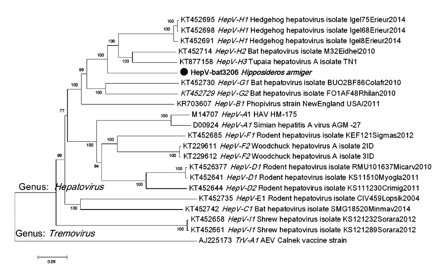
      To determine the phylogenetic relationship of HepVbat3206 with other hepatoviruses, the polyprotein sequence from representative hepatoviruses, including those from humans, primates, and other small animals, were downloaded from GenBank. Sequence alignment was performed using Clustal X and phylogenetic analysis performed using the default parameters of the MEGA7 software. The phylogenetic tree showed that HepV-bat3206 is distantly related to human and primate hepatoviruses and clustered with bat, Tupia, and hedgehog hepatovirus H (Fig. 1). The phylogenetic tree based on P1 (structural protein) and P3 (3C and 3D, polymerase) also showed a similar topology of the polyprotein tree (Supplementary Figure S1AS1B). While the P1 and P3 region of HepV-bat3206 shared 83%– 86% and 67%–70% with HepV H1-3, respectively, based on the new species criteria for hepatoviruses (distance of P1 > 14% and P3CD > 24%), HepV-bat3206 was only a new strain of HepV H.

      Figure 1.  The phylogenetic tree of hepatoviruses based on amino acid sequences of the polyprotein. The neighbor-joining phylogenetic tree was constructed using the MEGA7 with 1000 bootstrap replicates.The virus sequence obtained in this study is indicated by the black solid circle. The TrV was used as an outgroup sequence.

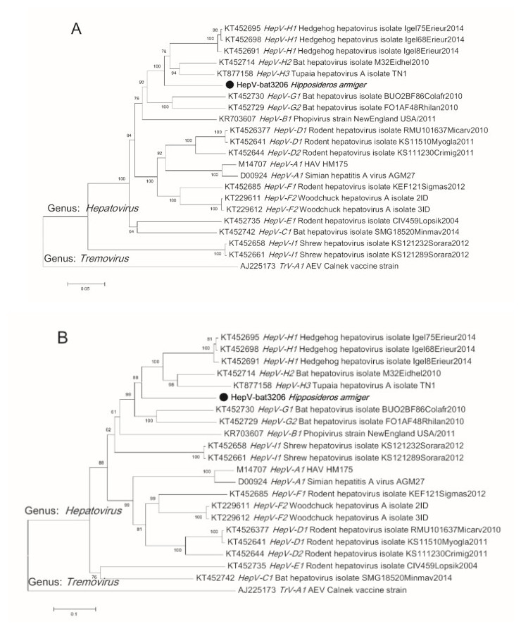
      Figure S1.  The phylogenetic tree of Hepatovirus based on amino acid sequences of P1 (capsid protein) (A) and P3CD (polymerase protein) (B). The neighbor-joining phylogenetic tree was constructed using the MEGA7 with 1000 bootstrap replicates. The virus sequence obtained in this study is indicated by the black solid circle.

      To our knowledge, this is the first report of a bat hepatovirus in China. We detected positive samples in the same colony of bats in the years 2011, 2012 and 2014, suggesting a long history of circulation of hepatoviruses in this colony. Hepatoviruses from nonhuman primates could be transmitted from chimpanzees to humans while the handlers dispose the infected animals (Dienstag et al. 1976). Since the bat cave used for sample collection is a famous tourist site, we suspect a potential risk of transmission of this bat virus to humans if visitors are frequently exposed to this bat colony. Further studies should be focused on the transmission, infection, and prevention of disease caused by these bat hepatoviruses.

    • This work was jointly funded by the National Natural Science Foundation of China (Grant No. 81290341). We also thank The Core Facility and Technical Support, Wuhan Institute of Virology for assistance in high throughput sequencing sample preparation.

    • The authors declare that they have no conflict of interest.

    • This study was approved by the Animal Ethics Committee of the Wuhan Institute of Virology (Animal ethics Approval Number: WIVA05201202).

    Figure (2)  Table (2) Reference (13) Relative (20)

    目录

    /

    DownLoad:  Full-Size Img  PowerPoint
    Return
    Return