HTML
-
The Crimean-Congo hemorrhagic fever virus (CCHFV) is a single-stranded, negative-sense RNA virus belonging to the family Nairoviridae, genus Orthonairovirus. CCHFV has a tripartite genome comprising three RNA segments: the small (S) segment, encoding the nucleocapsid protein (NP); the medium (M) segment, encoding the glycoprotein (G) precursor; the large (L) segment, encoding RNA dependent RNA polymerase (RdRp) (Anagnostou and Papa 2009). CCHFV infection in humans results in CrimeanCongo hemorrhagic fever (CCHF), a severe fatal viral disease with a worldwide mortality rate of 30%-50% (Ergonul 2006; Hoogstraal 1979; Messina et al. 2015). CCHF patients usually experience a short incubation period (3-10 d) and then develop clinical symptoms including severe fever and hemorrhage accompanied with fatigue, myalgia, oliguria, and disturbance of consciousness (Ergonul 2006; Hoogstraal 1979). Ticks are the major vectors for CCHFV transmission. CCHFV is also transmitted from animals or humans to other humans through exposure to contaminated tissues and blood from animals or through direct contact with acute-stage patients (Bente et al. 2013; Papa et al. 2017). So far, no specific drugs and/or vaccines are available for CCHF prevention and treatment. Therefore, CCHFV is a highly pathogenic virus posing a significant threat to public health.
The first CCHF outbreak was reported in 1944 in the Crimean region of the former Soviet Union, where approximately 200 Soviet troops developed a severe illness with sudden onset of fever, accompanied with a high incidence of hemorrhage (Bente et al. 2013; Hoogstraal 1979). The outbreak finally resulted in approximately 10% mortality among the diseased troops (Bente et al. 2013; Hoogstraal 1979). Before 1970, most CCHF cases were reported from countries including Uzbekistan, Kazakhstan, Tajikistan, and Bulgaria (Hoogstraal 1979). Thereafter, outbreaks and further cases of CCHF were reported in wide areas in countries in Africa (Uganda, South Africa, Sudan, and Mauritania), the Middle East (Iraq, United Arab Emirates, Iran, Oman, and Saudi Arabia), Europe (Bulgaria, Kosovo, Turkey, and Albania), and Asia (China, Pakistan, Afghanistan, and India) (Ergonul 2006). Serological evidence for CCHFV further suggested the potential prevalence of this virus in a wider geographic distribution including Malaysian, Nigeria, Tunisia, SubSaharan Africa, Iberian Peninsula (Bukbuk et al. 2014, 2016; Mohd Shukri et al. 2015; Muianga et al. 2017; Palomar et al. 2017; Wasfi et al. 2016). Hence, CCHFV may be more widely distributed worldwide than is currently known.
In China, CCHF outbreaks and epidemics and attempts at CCHFV identification and isolation have been reported from Xinjiang province for more than 50 years (Fig. 1 and Table 1). The first CCHF outbreak was reported in 1965 in Bachu County, Xinjiang province, and the first Chinese CCHFV strain was isolated from the serum of a local patient (Papa et al. 2002). In subsequent years, sporadic outbreaks were reported in Bachu County and adjacent areas (Awati, Jiashi, Kepin, Kuche, and Maigait) in and around Tarim Basin in southern Xinjiang (Liu et al. 2004; Sun et al. 2009). Most studies on Chinese CCHFV strains identified and isolated from these areas reported that CCHFV infects individuals through close contact with livestock (Bente et al. 2013). In 1997, another concentrated CCHF outbreak was reported in Bachu County, which resulted in an increased mortality of 19.4% (Papa et al. 2002). Since 2003, no cases have been reported from Xinjiang. However, a molecular epidemiological study revealed the prevalence of CCHFV in ticks collected in Tarim Basin from 2004 to 2005 (Sun et al. 2009), indicating the potential risk of CCHFV infection through tick bites in southern Xinjiang. The risk of CCHFV infection persisted beyond 10 years, since a new CCHFV strain was isolated from ticks collected from Yuli County in southern Xinjiang in 2016 (Guo et al. 2017). Cases of CCHFV infection have not been reported in northern Xinjiang; however, an etiological and serological study has suggested that areas in northern Xinjiang were also hotspots of CCHFV (Bente et al. 2013). CCHFV RNA was detected in ticks from Junggar Basin in northern Xinjiang, revealing the prevalence of CCHFV in ticks in that region (Sun et al. 2009). Five CCHFV strains have been isolated from ticks collected in Junggar Basin, of which three (Fub90009, Ml05225, and Ml05232) were identified through partial sequencing of S segments (Sun et al. 2009) and two others (FuB90-9 and FuB90-13) were isolated more than 20 years ago in suckling mice, identified through reverse possive hemagglutination (RPHA) and reverse possive hemagglutination inhibition (RPHI) tests, and cross complement fixation tests; however, the sequences have not been reported to date (Feng et al. 1991). Hence, Junggar Basin has been considered a hotspot of CCHFV in northern Xinjiang, where increasing CCHF surveillance has been suggested.
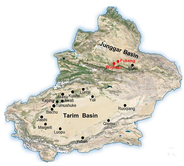
Figure 1. A map of Xinjiang province illustrating the cities and counties with previous records of CCHF epidemics and CCHFV isolates. The two cities where the two new strains derived in this study are indicated by red characters and red solid circles. Other places with the CCHF history are labeled in black.

Table 1. The recorded history related to CCHFV in Xinjiang province, China.
In this study, we isolated and identified two new CCHFV strains from H. asiaticum asiaticum ticks from Junggar Basin. We characterized the virus properties in vitro, followed by complete genome sequencing. Phylogenetic analyses were carried out to investigate evolutionary relations among Chinese strains.
-
Vero cells were provided by American Type Culture Collection (ATCC, Manassas, VA, USA) (Accession number: ATCC®CCL-81TM) and was cultured in Dulbecco's modified Eagle medium (DMEM) (Gibco) supplemented with 10% fetal bovine serum (FBS) (Gibco). The 2-day-old suckling KM mice were obtained from the Animal Center of Hubei Provincial Centers for Disease Control and Prevention and raised in specific pathogen-free (SPF) environment. A monoclonal anti CCHFV NP antibody used for an indirect immunofluorescence assay was obtained from the Xinjiang Centers for Disease Control and Prevention, as described previously (Guo et al. 2017).
-
In total, approximately 800 H. asiaticum asiaticum ticks were collected from Fukang City and Wujiaqu City in the north of Xinjiang province from April to May in 2016. The ticks were grouped in accordance with their geographic distributions (n = 50-100 ticks/group). The ticks were then washed three times with phosphate-buffered saline (PBS, pH 7.4). Homogenates were prepared using the Tissue Cell-Destroyer (D1000, Novastar, China) with 2 mL PBS in the following conditions: centrifugation at 4000 rpm for 10 s at 4 ℃, stop for 10 s, followed by one more round of centrifugation. The homogenates were clarified through centrifugation at 5000 rpm for 5 min at 4 ℃ to eliminate tissue debris. Subsequently, the clear supernatants were inoculated into KM suckling mice.
Initially, a single group of KM mice included a pregnant female mouse. Usually, a female KM mouse gives birth to 8-10 baby mice. After birth, one group would include a dam and her pups. Thereafter, suckling mice of 1-2 days of age would be inoculated with tick homogenates prepared from one tick group. In total, 75 groups of suckling mice were inoculated for the first generation. The mice were monitored daily until day 21. All diseased mice were euthanized immediately once they were noted. Brains were harvested and stored in 50% glycerol at -80 ℃. The onset of illness among more than half members of the suckling mice was found in 25 groups. The brain of one diseased suckling mouse was randomly chosen from each of the 25 groups, and homogenates were prepared. Briefly, the brain was destroyed by the Tissue Cell-Destroyer (D1000, Novastar) with 1 mL PBS and was clarified through centrifugation, as described above.
A new group of suckling mice was inoculated with the clarified supernatants as the second generation of blind passage. Although not all of them developed symptoms, diseased suckling mice were noted from all 25 groups. Their brains were harvested. Brain homogenates were prepared from one diseased mouse from each group, as mentioned above. RNA was extracted from 100-μL clarified homogenates and reverse transcription (RT-PCR)based detection of CCHFV in the 25 samples of brain homogenates was performed, as described previously (Guo et al. 2017). CCHFV-positive brain homogenates were further inoculated into suckling mice as the third generation. For the inoculation of each mouse, 30-μL homogenates were injected through both intraperitoneal and intracranial routes. Brains of other groups of healthy firstand second-generation suckling mice were also harvested on day 21 post inoculation and stored in 50% glycerol at -80 ℃ until further experiments.
-
Metagenomic sequencing was performed to identify the viruses isolated from mice after inoculation. Homogenates (1 mL) were prepared from brains (300 mg) of diseased mice, using Tissue Cell-Destroyer (Novastar). The debris were eliminated through centrifugation (4500 rpm, 5 min, 4 ℃) and total RNA was extracted from the clear supernatants, as described previously (Guo et al. 2017).
Before the preparation of RNA libraries for metagenomic sequencing, RNA quality was examined by using an Agilent 2100 Bioanalyzer (Agilent Technologies). RNA libraries were then prepared in accordance with the TruSeq Stranded Total RNA Sample Preparation Guide (Illumina). rRNA was eliminated by using VAHTSTM Total RNA-seq (H/M/R) Library Prep Kit for Illumina (Vazyme Biotech, China) in accordance with the manufacturer's instructions. Thereafter, all RNA libraries were analyzed using the Miseq platform (Illumina). The resulting reads were analyzed using the FastQC program and assembled into contigs, using the Trinity program with the default settings. Sequences of the assembled contigs were compared with virus nucleotide database (Vnt) with BLASTn and with the virus protein database (Vaa) with BLASTx to identify potential species based on the sequence analyses. The taxa of the species were identified using Megan program, and the virus-related contigs were extracted. Subsequently, virus-related contigs were compared with the whole-genome database with BLASTn and the whole protein database (nr) with BLASTx to eliminate false-positive virusrelated sequences.
-
RT-PCR analysis was performed to confirm the presence of CCHFV in the brain of infected mice, as previously described (Guo et al. 2017). PCR products were then sequenced for further analysis.
The viral loads in mouse brains were determined through real-time RT-PCR analysis. To generate the RNA standards, a partial fragment (246 nt) of the S segment in reference to CCHFV strain YL04057 from nucleotide position 450-689 was amplified using primer set 1. The PCR products were inserted into the pGEM®-T Easy vector (Promega) and sequenced to construct the recombinant plasmid pT-CCHFV-Sp. Thereafter, the fragment (348 bp) containing the T7 promoter and the S fragment were amplified using the plasmid as the template with primer set 2 and transcribed into RNA, using the MAXISCRIPT T7 KIT (AMBION, Carlsbad, USA) in accordance with the manufacturer's instructions. The RNA transcripts were purified, quantified, serially diluted by 10 folds to generate RNA standards ranging from 103 to 109 copies/μL. Realtime RT-PCR was performed with primer set 3, using the HiScript@Ⅱ One Step qRT-PCR SYBR@Green Kit (Vazyme Biotech, Nanjing, China) in accordance with the manufacturer's instructions. The reaction was carried out in a 25-μL mixture containing 1 μL RNA standards or samples, with the following conditions: reverse transcription at 50 ℃ for 3 min, 95 ℃ for 5 min, followed by 40 cycles for 10 s at 95 ℃ and 30 s at 60 ℃, and a final extension at 60 ℃ for 5 min. The sequences of primer sets are listed in Supplementary Table S1.
-
Vero cells were seeded in the 6-well plates overnight and were then incubated with the filtered supernatant (100 μL) from mouse brain homogenates at 37 ℃ for 1 h. There after, the supernatants were replaced with 2 mL fresh DMEM containing 2% FBS. At 4 d post infection, the cells were fixed with 4% Paraformaldehyde and permeabilized. Immunofluorescence assays (IFAs) then were carried out to analyze CCHFV viral protein expression in cells, as previously described (Guo et al. 2017).
To determine the viral yield after cell culture, Vero cells were infected with viruses at a multiplicity of infection (MOI) of 0.01 TCID50 units per cell. Supernatants (50 μL) were harvested at indicated time points to determine the viral titers, using the end-point dilution assay, as described previously (Guo et al. 2017). Significant differences (P values) were statistically analyzed by Student's t test. In vitro experiments on CCHFV infection in cells and in vivo experiments involving inoculation in mice were performed in a BSL-3 lab.
-
The genomic sequences (S, M, and L segments) of CCHFV strains with a complete coding sequence (CDS), which were released by GenBank before May 2017, were used for subsequent sequence analyses. Partial sequences of a 220-nt fragment (position 329-548 nt in reference to the Chinese CCHFV strain 7001) from the S segment of CCHFV strains derived from different countries were also used for phylogenetic analysis. Sequences were aligned using the ClustalW program integrated in Mega 6.0 software (Tamura et al. 2013). Maximum Likehood (ML) phylogenetic trees were constructed using Mega 6.0 software with the K2 + G model and were tested with the bootstrap method with 1000 replicates.
Cells, Mice, and Antibodies
Tick Collection and Inoculation of Mice
Metagenomic Sequencing
RT-PCR and Real-Time RT-PCR
Virus Infection Assay and End-Point Dilution Assay
Phylogenetic Analysis
-
Seventy-five groups of suckling mice were inoculated with clear homogenates from different tick pools. On 7-12 d post-inoculation (d p.i.), although not all mice experienced disease onset, there were diseased mice among all 25 groups with symptoms including hollow back, loss of balance, side-lying, anorexia, limb paralysis, and articulo mortis. These mice were euthanized and the brains were harvested. One brain from each group was homogenized and CCHFV were detected through RT-PCR analysis. CCHFV RNA was detected in the brains from two different groups, one inoculated with tick homogenates derived from Wujiaqu City. The CCHFV positive homogenates (Wujiaqu group) were inoculated into a group of suckling mice as the third generation. All suckling mice in this group developed illness with similar symptoms on 4-6 d p.i. after inoculation, which indicated that the mouse pathogen(s) could be transferred to mouse neonates. Hence, we harvested brains from these mice and one of them was prepared for RNA-seq to detect the potential pathogen(s). Moreover, because less than half of the suckling mice in the some groups experienced disease onset in the first generation, the second inoculation was not conducted before. One other library for RNA-seq was prepared with the homogenate mixture of three brains from the first generation of three different groups, which included all samples from Fukang City. In silico analyses of the resulted contigs revealed a high abundance of the CCHFV genome in the infected brains from the two libraries, suggesting that CCHFV isolated from ticks and inoculated in mice were associated with the onset of illness in the two mouse groups. RT-PCR analyses confirmed the isolation of CCHFV in the Wujiaqu group and the mixed Fukang group. Further, the presence of CCHFV was identified in homogenates from one of the three brains derived from Fukang City as the S, M, and L segments were all detected from this sample (data not shown). Furthermore, real-time RT-PCR revealed comparable viral loads in both brain samples (Table 2).

Table 2. Viral loads, titers, and virulence of CCHFV strains FK16116 and WJQ16206 in homogenates of respective mouse brain samples.
-
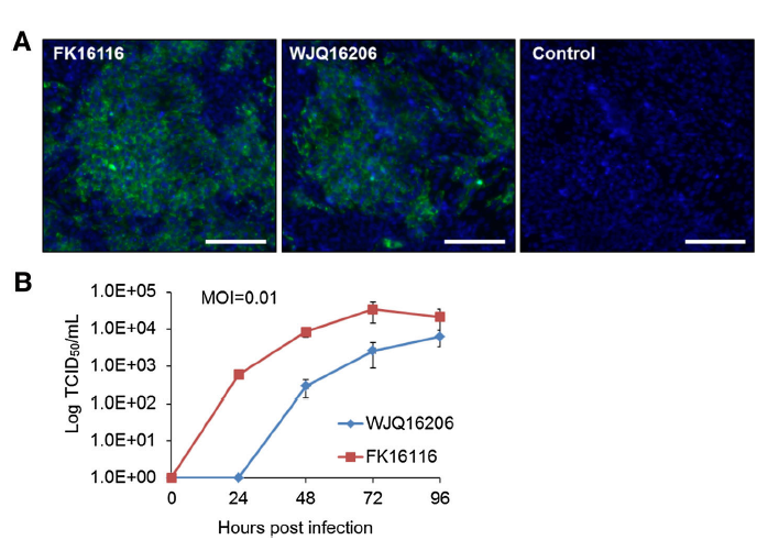
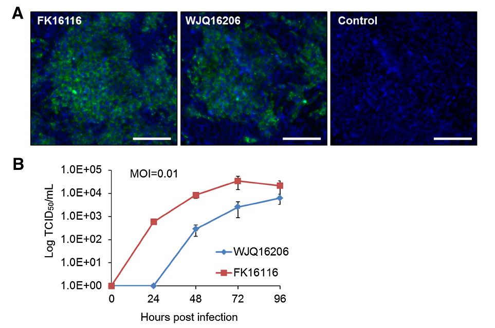
The sequences of three segments of the genome of the CCHFV strains isolated from two different tick groups were obtained through RNA-seq. Sequence comparison revealed that they were two different strains sharing very high nucleotide and amino acid similarities in the S, M, and L segments (Table 3). The two CCHFV strains were named FK16116 and WJQ16206 in accordance with the site of collection (Fukang City and Wujiaqu City) of ticks. BLASTn analysis revealed that both strains shared very high nucleotide sequence similarities with the strain SK 2015/human isolated from human serum in southern Kazakhstan in 2015 (98% for FK16116 and 97% for WJQ16206). Furthermore, their sequences were deposited in GenBank under the accession numbers as WJQ16206 (MG659722, MG659723, and MG659724); FK16116 (MG659725, MG659726, and MG659727). Subsequently, Vero cells were incubated with homogenates from the two CCHFV-infected brain homogenates. CCHFV NP expression in Vero cells was detected on 5 d p.i. through IFAs (Fig. 2A), indicating that the isolated CCHFV strains in mouse brains could infect and replicate in Vero cells. Furthermore, the rate of viral generation from infected Vero cells was determined. The viral yield of strain WJQ16206 was significantly lesser than that of strain FK16116 at 24, 48, and 72 h p.i. (P < 0.01 at 24 and 48 h p.i., and P < 0.05 at 72 h p.i.). Although no significant differences were noted at 96 h p.i. (P > 0.05), the virus yields for strain WJQ16206 (6.3 ± 2.9 × 10³ TCID50/mL) was slightly less than that of strain FK16116 (2.1 ± 1.4 × 10⁴ TCID50/mL) (Fig. 2B). The results might indicate the differences in infectivity and replication between the two strains, especially at the early stage of infection in Vero cells.

Table 3. Nucleotide and amino acid similarities of strains FK16116 and WJQ16206.
-
Phylogenetic analyses were performed with CCHFV strains with complete sequences of the three segments. FK16116 and WJQ16206 clustered together but presented different phylogenetic relationships with other Chinese strains in the S, M, and L trees (Fig. 3). In the S tree, CCHFV strains divided into eight genotypes in accordance with their geographic distributions, namely Asia 1 and 2, Africa 1-3, and Europe 1-3 groups. The previously reported Chinese strains all clustered in Asia 2 group, except for strains FK16116 and WJQ16206, both belonging to Asia 1 group (Fig. 3A). The CCHFV strains in the M tree were also divided into eight groups; however, these groups were different from those of the S tree (Fig. 3B). The Europe 3 group was not included, and the Chinese strains were divided into three Asian groups. FK16116 and WJQ162062 together with two other Chinese strains (7803 and 75024) identified in the 1970s were included in Asia 2 group; however, they were present in different subbranches (Fig. 3B). In the L tree, CCHFV strains were divided into six genotypes: Asia 1/2, Asia 3, Africa 2 and 3, and Europe 1 and 2 groups. Three Chinese strains constituted the Asia 3 group, and FK16116 and WJQ162062 together with strain C-68031 were identified in 1968 clustered in Asia 1/2 group (Fig. 3C). To further identify the phylogenetic relationships among Chinese CCHFV strains from different locations in Xinjiang province, the ML tree was constructed with all reported Chinese strains available in GenBank with a 220-nt fragment of CCHFV S segment (Fig. 4). Most Chinese strains from cities and counties in southern Xinjiang and two strains from Mulei County in northern Xinjiang belonged to the Asia 2 group, while the two new strains (FK16116 and WJQ16206) and one other strain from Fukang City (Fub90009) belonged to the Asia 1 group. There were differences in CCHFV genotypes from Fukang City and Wujiaqu City with distant genetic relations to Chinese strains from other areas in Xinjiang province.

Figure 3. Maximum likelihood trees of CCHFV strains based on the complete sequences of S (A), M (B), and L (C) segments. The strains are presented in the form of GenBank accession number_strain name_isolated country_isolated year. All Chinese strains are labeled in red characters, and the two new isolated strains in this study are highlighted with a black solid circle.
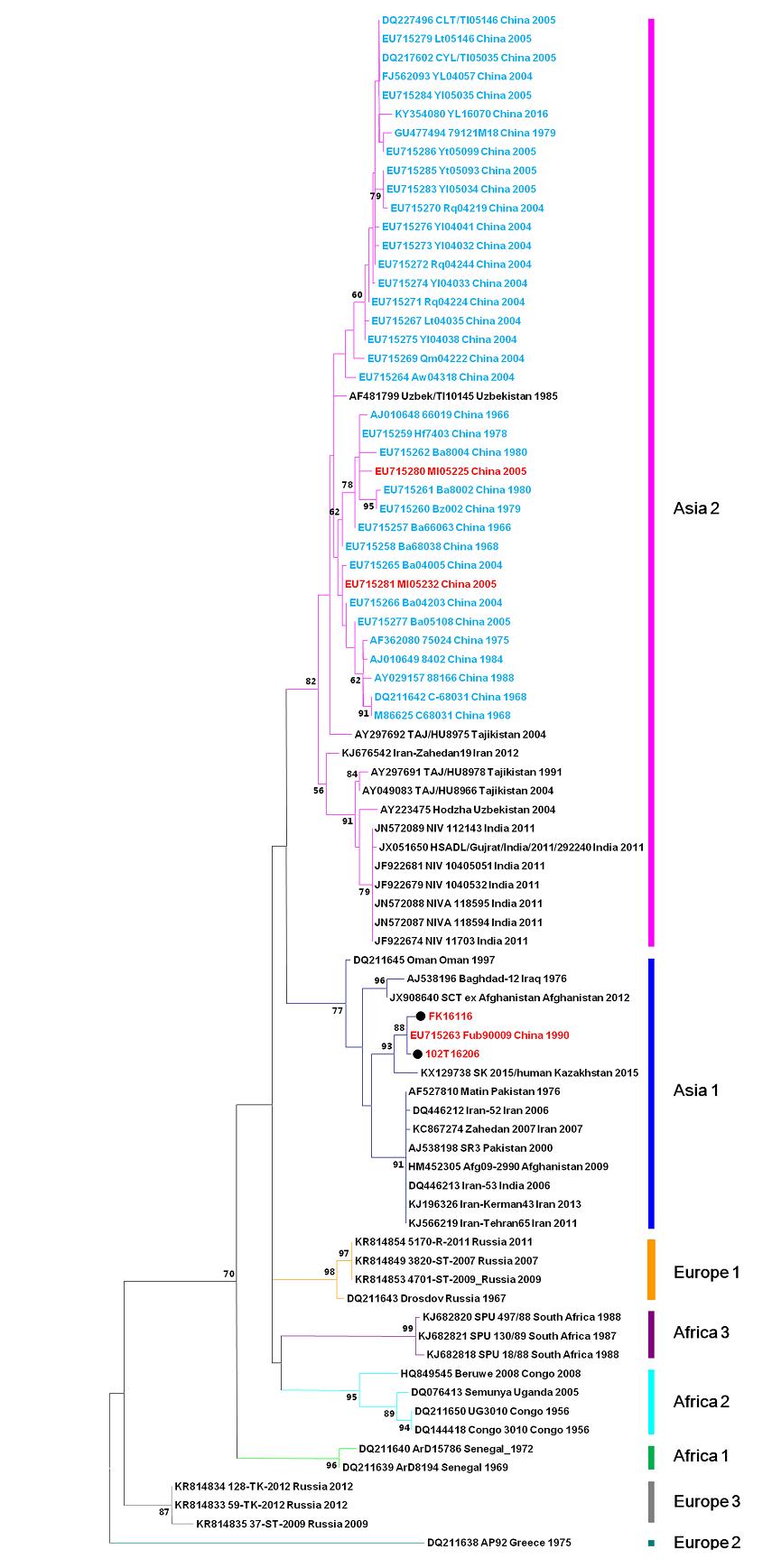
Figure 4. The maximum likelihood tree of partial sequences of 220 nt from S segments of CCHFV strains. The strains were shown in the form of "GenBank accession number_strain name_isolated country_isolated year. The Chinese strains from areas in northern regions of Xinjiang province are shown in red characters, and the Chinese strains from areas in southern regions of Xinjiang province are shown in cyan. The two new strains isolated from Fukang City and Wujiaqu City are labeled with a black solid circle.
Isolation and Identification of Two New CCHFV Strains from Ticks Collected in the Northern Region of Xinjiang Province, China
Characterization of the Two New CCHFV Isolates
Phylogenetic Analysis of the Two New CCHFV Strains
-
In the present study, two new CCHFV strains were isolated from H. asiaticum asiaticum ticks collected from Fukang City and Wujiaqu City. The viral properties of the two strains were analyzed. Viral antigen expression was detected in infected cells, and both strains were found to infect and replicate in Vero cells. However, they displayed different growth properties despite being phylogenetically clustered together. Strain WJQ16206 produced infectious progeny viruses at a significantly lower rate than strain FK16116 (Fig. 2B), indicating differences in virulence of the two strains. The titer of FK16116 (2 × 10⁵ TCID50/ mL) in clear supernatants from mouse brain homogenates was approximately two folds that of WJQ16206 (1 × 10⁵ TCID50/mL). Real-time RT-PCR analysis revealed no significant differences in viral RNA copies of the two strains (2.74 ± 1.92 × 10⁹ copies/mL for FK16116 and 3.41 ± 1.91 × 10⁹ copies/mL for WJQ16206). Hence, one TCID50 is equal to 1.37 ± 0.96 × 10⁴ copies for FK16116 and 3.41 ± 1.91 × 10⁴ copies for WJQ16206 (P > 0.05). Therefore, strain WJQ16206 is slightly, but not significantly, less infectious than strain FK16116. We also investigated the amino acid (aa) sequences of the glycoprotein precursors (G) and RdRp and found aa differences at 5 positions in Gn, 1 in Nsm, 2 in Gc, and 7 positions in RdRp between the two strains (Table 4). These differences might be related to the different abilities of the strains in binding to cellular receptors and cellular penetration, along with differences in replication efficiency, which requires further investigation.

Table 4. Amino acid shifts in the glycoprotein precursor and RdRp between FK16116 and WJQ16206.
Vero/Vero E6 cells were widely used to investigate the roles of CCHFV infection in innate immune response and cell apoptosis, the mechanisms of CCHFV entry, transcription, and replication in host cells, and CCHFV identification and isolation and the development of antiCCHFV drugs and techniques for CCHFV detection (Andersson et al. 2008, 2012; Connolly-Andersen et al. 2011; Ferraris et al. 2015; Guo et al. 2017; Karlberg et al. 2011; Rodrigues et al. 2012; Simon et al. 2009a, b; Spengler et al. 2015). Although SW-13 cells, which are widely recognized in studies on CCHFV, were not used for CCHFV characterization in vitro in this study, the different growth efficiency could still be revealed using Vero cells. Further investigation on CCHFV characteristics would be conducted using different cell lines including SW-13 and Vero/VeroE6 cells.
Xinjiang is a unique province in China with a recorded history of CCHF epidemics and outbreaks and CCHFV isolates. Since CCHFV was first identified in Xinjiang province in 1965, almost all Chinese CCHFV strains have been identified and isolated from Xinjiang province, particularly in the southern areas. Epidemiological surveys and increasing surveillance were consequently conducted mainly in the southern areas. H. asiaticum asiaticum ticks are recognized as the predominant vectors of CCHFV, since the virus has been detected and isolated from H. asiaticum asiaticum ticks from Tarim Basin (Dai et al. 2006; Guo et al. 2017; Sun et al. 2009). Nevertheless, H. asiaticum asiaticum ticks are the predominant species in the desert ecosystem of both northern (Junggar Basin) and southern (Tarim Basin) areas in Xinjiang province. The identification of CCHFV in ticks in Junggar Basin has been reported previously. The first report of CCHFV identification and isolation from Junggar Basin was in 1991, wherein two CCHFV strains (FuB 90-9 and FuB 90-13) were isolated from H. asiaticum asiaticum ticks collected in the desert of Fukang City. CCHFV were also detected in serum samples from 2 local shepherds and 107 sheep, thereby suggesting prevalence of CCHFV in Fukang City and possible transmission and infection from ticks to humans and animals (Feng et al. 1991). However, the genome sequences of the two strains were not reported; hence, phylogenetic analyses could not be carried out. In 2004 and 2005, Sun et al. carried out an epidemiological survey in Xinjiang province and isolated 22 CCHFV strains from ticks (Sun et al. 2009). Among these strains, one was obtained from Fukang City and two from Mulei County from Junggar Basin in northern Xinjiang, which further revealed that Fukang and Mulei might be hotspots for CCHFV in Junggar Basin. Moreover, the study reported that interestingly, the strain from Fukang City (Fub90009, Asia 2) presented distant genetic relationships with other Chinese strains, whereas the two strains from Mulei County (Ml05225 and Ml05232, Asia 1) clustered with the others. This finding is concurrent with our results. The prevalence of different genotypes in Xinjiang province was further confirmed in this study. Nevertheless, before the identification of CCHFV from Fukang and Wujiaqu in northern Xinjiang, phylogenetic analyses in previous studies using complete sequences of S segments revealed that all Chinese CCHFV strains had an identical genotype (Asia 2) (Guo et al. 2017; Zhou et al. 2013a). Furthermore, strain Fub90009 was isolated in 1990 and strain FK16116 in the present study from ticks collected in 2016, which indicated long-term persistence of CCHFV in Fukang City. It also suggested that the Asia 2 genotype was not widely distributed in northern Xinjiang, and both Fukang City and Wujiaqu City are hotspots for CCHFV strains of different genotypes. The present results also facilitate the understanding of CCHFV evolution and further recognition of CCHFV distribution in Xinjiang province. The local residents of northern Xinjiang may hence be at a potential risk of CCHFV infection. Further investigation and epidemiological surveys should be performed in Xinjiang province. Therefore, increasing surveillance of CCHF outbreaks would be recommended in these areas.
-
This work was supported by the Science and Technology Basic Work Program (2013FY113500) and the National Key Research and Development Program (2016YFE0113500) from the Ministry of Science and Technology of China, as well as the European Union's Horizon 2020 EVAg project (No 653316).
-
YZ, JL, CW and AA collected and processed the ticks for the inoculation of suckling mice; YZ and JL performed the first mice inoculation and harvested the tissue samples; YZ and JL passaged the two virus strains on newborn mice; YZ, ZZ and QW performed the virus infection and analyzed the virus production on Vero cells; YZ, ZS, YF and SS analysis all the work; ZH, YZ and FD conceived of the study; YZ and SS wrote the manuscript; YZ and FD checked and finalized the manuscript.
-
The authors declare that they have no conflict of interest.
-
Animal experiments in this study were approved by the ethics committees of Wuhan Institute of Virology, Chinese Academy of Sciences (Approval Number: WIVH01201501).
-
This article is distributed under the terms of the Creative Commons Attribution 4.0 International License (http://creativecommons.org/licenses/by/4.0/), which permits unrestricted use, distribution, and reproduction in any medium, provided you give appropriate credit to the original author(s) and the source, provide a link to the Creative Commons license, and indicate if changes were made.
Conflict of interest
Animal and Human Rights Statement
Open Access
-

Table S1. Primers used in this study








 DownLoad: