Siyu Xia, Ming Wu, Si Chen, Tao Zhang, Lina Ye, Jun Liu and Hui Li. Long Term Culture of Human Kidney Proximal Tubule Epithelial Cells Maintains Lineage Functions and Serves as an Ex vivo Model for Coronavirus Associated Kidney Injury[J]. Virologica Sinica, 2020, 35(3): 311-320. doi: 10.1007/s12250-020-00253-y
Citation: Siyu Xia, Ming Wu, Si Chen, Tao Zhang, Lina Ye, Jun Liu, Hui Li. Long Term Culture of Human Kidney Proximal Tubule Epithelial Cells Maintains Lineage Functions and Serves as an Ex vivo Model for Coronavirus Associated Kidney Injury .VIROLOGICA SINICA, 2020, 35(3) : 311-320.  http://dx.doi.org/10.1007/s12250-020-00253-y

长期培养的人肾近端小管上皮细胞保持谱系功能可作为冠状病毒感染所致肾损伤的Ex vivo模型

  • 通讯作者: 李晖, somhli@whu.edu.cn, ORCID: http://orcid.org/0000-0003-3867-7685
  • 收稿日期: 2020-03-26
    录用日期: 2020-06-09
    出版日期: 2020-06-29
  • 目前,新型冠状病毒感染导致的严重多器官衰竭的机制尚不清楚。急性肾损伤(AKI)是COVID-19重症患者常见的器官损伤之一。已有研究表明,人肾小管细胞可能是SARS-CoV-2感染的潜在宿主细胞。传统的肿瘤细胞系或永生化细胞系的遗传背景和表型与人体宿主细胞不同。广泛使用的动物研究模型,由于种属和病毒嗜性的差异性,通常无法反映人体真实的生理和病理状态。目前急需建立人正常上皮细胞作为人体疾病研究模型。本研究中,我们利用条件重编程(Conditional Reprogramming,CR)和类器官(Organoids)技术,在2D和3D水平成功建立了人肾近端小管上皮细胞(KPTECs)的长期培养体系。这些细胞具有正常的分化和修复DNA损伤的能力,并且没有成瘤性。重要的是,人肾近端小管上皮细胞(KPTECs)保持自身谱系功能,表达特异性转运蛋白(SLC34A3和cubilin)。KPTECs同时还表达血管紧张素转换酶Ⅱ(ACE2),ACE2是SARS-CoV和SARS-CoV-2的受体。相对应地,肿瘤细胞系并不表达内源性SLC34A3,cubicin和ACE2。非常有趣的是,在类器官3D培养条件下ACE2表达量是2D培养时的两倍。利用荧光素酶报告假病毒感染实验表明,冠状病毒的刺突糖蛋白(S蛋白)能够感染进入KPTECs细胞。综上所述,整合2D CRC和3D类器官培养提供了一种生理状态的Ex vivo模型,可用于研究肾脏功能、病毒感染肾细胞的天然免疫反应,也是一个进行新药发现和安全性评价的新平台。

Long Term Culture of Human Kidney Proximal Tubule Epithelial Cells Maintains Lineage Functions and Serves as an Ex vivo Model for Coronavirus Associated Kidney Injury

  • Corresponding author: Hui Li, somhli@whu.edu.cn
  • ORCID: http://orcid.org/0000-0003-3867-7685
  • Received Date: 26 March 2020
    Accepted Date: 09 June 2020
    Published Date: 29 June 2020
  • The mechanism of how SARS-CoV-2 causes severe multi-organ failure is largely unknown. Acute kidney injury (AKI) is one of the frequent organ damage in severe COVID-19 patients. Previous studies have shown that human renal tubule cells could be the potential host cells targeted by SARS-CoV-2. Traditional cancer cell lines or immortalized cell lines are genetically and phenotypically different from host cells. Animal models are widely used, but often fail to reflect a physiological and pathogenic status because of species tropisms. There is an unmet need for normal human epithelial cells for disease modeling. In this study, we successfully established long term cultures of normal human kidney proximal tubule epithelial cells (KPTECs) in 2D and 3D culture systems using conditional reprogramming (CR) and organoids techniques. These cells had the ability to differentiate and repair DNA damage, and showed no transforming property. Importantly, the CR KPTECs maintained lineage function with expression of specific transporters (SLC34A3 and cubilin). They also expressed angiotensin-converting enzyme 2 (ACE2), a receptor for SARS-CoV and SARS-CoV-2. In contrast, cancer cell line did not express endogenous SLC34A3, cubilin and ACE2. Very interestingly, ACE2 expression was around twofold higher in 3D organoids culture compared to that in 2D CR culture condition. Pseudovirion assays demonstrated that SARS-CoV spike (S) protein was able to enter CR cells with luciferase reporter. This integrated 2D CR and 3D organoid cultures provide a physiological ex vivo model to study kidney functions, innate immune response of kidney cells to viruses, and a novel platform for drug discovery and safety evaluation.

  • 加载中
    1. Alkhilaiwi F, Wang L, Zhou D, Raudsepp T, Ghosh S, Paul S, Palechor-Ceron N, Brandt S, Luff J, Liu X, Schlegel R, Yuan H (2018) Long-term expansion of primary equine keratinocytes that maintain the ability to differentiate into stratified epidermis. Stem Cell Res Ther 9:181
        doi: 10.1186/s13287-018-0918-x

    2. Alkhilaiwi F, Paul S, Zhou D, Zhang X, Wang F, Palechor-Ceron N, Wilson K, Guha R, Ferrer M, Grant N, Thomas C, Schlegel R, Yuan H (2019) High-throughput screening identifies candidate drugs for the treatment of recurrent respiratory papillomatosis. Papillomavirus Res 8:100181
        doi: 10.1016/j.pvr.2019.100181

    3. Borodovsky A, McQuiston TJ, Stetson D, Ahmed A, Whitston D, Zhang J, Grondine M, Lawson D, Challberg SS, Zinda M, Pollok BA, Dougherty BA, D'Cruz CM (2017) Generation of stable PDX derived cell lines using conditional reprogramming. Mol Cancer 16:177
        doi: 10.1186/s12943-017-0745-1

    4. Brewington JJ, Filbrandt ET, LaRosa FJ 3rd, Moncivaiz JD, Ostmann AJ, Strecker LM, Clancy JP (2018) Generation of human nasal epithelial cell spheroids for individualized cystic fibrosis transmembrane conductance regulator study. J Vis Exp 134:57492
        doi: 10.3791/57492

    5. Chafekar A, Fielding BC (2018) MERS-CoV: understanding the latest human coronavirus threat. Viruses 10(2):93
        doi: 10.3390/v10020093

    6. Chai J, Han L, Zhang J, Han D, Zou L, Zhu Z, Zhao Y, Guo H (2020) Conditional reprogramming inducing clinical cells proliferation: new research tools in tumor and inflammatory-related diseases. Curr Pharm Des.https://doi.org/10.2174/1381612826666200316155252
        doi: 10.2174/1381612826666200316155252

    7. Chan RW, Chan MC, Nicholls JM, Malik Peiris JS (2013) Use of ex vivo and in vitro cultures of the human respiratory tract to study the tropism and host responses of highly pathogenic avian influenza A (H5N1) and other influenza viruses. Virus Res 178:133–145
        doi: 10.1016/j.virusres.2013.03.003

    8. Feng W, Guo J, Huang H, Xia B, Liu H, Li J, Lin S, Li T, Liu J, Li H (2015) Human normal bronchial epithelial cells: a novel in vitro cell model for toxicity evaluation. PLoS ONE 10:e0123520
        doi: 10.1371/journal.pone.0123520

    9. Guan WJ, Ni ZY, Hu Y, Liang WH, Ou CQ, He JX, Liu L, Shan H, Lei CL, Hui DSC, Du B, Li LJ, Zeng G, Yuen KY, Chen RC, Tang CL, Wang T, Chen PY, Xiang J, Li SY, Wang JL, Liang ZJ, Peng YX, Wei L, Liu Y, Hu YH, Peng P, Wang JM, Liu JY, Chen Z, Li G, Zheng ZJ, Qiu SQ, Luo J, Ye CJ, Zhu SY, Zhong NS, China Medical Treatment Expert Group for C (2020) Clinical characteristics of coronavirus disease 2019 in China. N Engl J Med 382:1708–1720
        doi: 10.1056/NEJMoa2002032

    10. Guo Y, Rumschlag-Booms E, Wang J, Xiao H, Yu J, Wang J, Guo L, Gao GF, Cao Y, Caffrey M, Rong L (2009) Analysis of hemagglutinin-mediated entry tropism of H5N1 avian influenza. Virol J 6:39
        doi: 10.1186/1743-422X-6-39

    11. Hamming I, Timens W, Bulthuis ML, Lely AT, Navis G, van Goor H (2004) Tissue distribution of ACE2 protein, the functional receptor for SARS coronavirus. A first step in understanding SARS pathogenesis. J Pathol 203:631–637
        doi: 10.1002/path.1570

    12. Huang C, Wang Y, Li X, Ren L, Zhao J, Hu Y, Zhang L, Fan G, Xu J, Gu X, Cheng Z, Yu T, Xia J, Wei Y, Wu W, Xie X, Yin W, Li H, Liu M, Xiao Y, Gao H, Guo L, Xie J, Wang G, Jiang R, Gao Z, Jin Q, Wang J, Cao B (2020) Clinical features of patients infected with 2019 novel coronavirus in Wuhan, China. Lancet 395:497–506
        doi: 10.1016/S0140-6736(20)30183-5

    13. Jin L, Qu Y, Gomez LJ, Chung S, Han B, Gao B, Yue Y, Gong Y, Liu X, Amersi F, Dang C, Giuliano AE, Cui X (2018) Characterization of primary human mammary epithelial cells isolated and propagated by conditional reprogrammed cell culture. Oncotarget 9:11503–11514
        doi: 10.18632/oncotarget.23817

    14. Kaye M (2006) SARS-associated coronavirus replication in cell lines. Emerg Infect Dis 12:128–133
        doi: 10.3201/eid1201.050496

    15. Kong SL, Chui P, Lim B, Salto-Tellez M (2009) Elucidating the molecular physiopathology of acute respiratory distress syndrome in severe acute respiratory syndrome patients. Virus Res 145:260–269
        doi: 10.1016/j.virusres.2009.07.014

    16. Kowolik CM, Liang S, Yu Y, Yee JK (2004) Cre-mediated reversible immortalization of human renal proximal tubular epithelial cells. Oncogene 23:5950–5957
        doi: 10.1038/sj.onc.1207801

    17. Krawczyk E, Hong SH, Galli S, Trinh E, Wietlisbach L, Misiukiewicz SF, Tilan JU, Chen YS, Schlegel R, Kitlinska J (2020) Murine neuroblastoma cell lines developed by conditional reprogramming preserve heterogeneous phenotypes observed in vivo. Lab Invest 100:38–51
        doi: 10.1038/s41374-019-0297-7

    18. Lee GY, Kenny PA, Lee EH, Bissell MJ (2007) Three-dimensional culture models of normal and malignant breast epithelial cells. Nat Methods 4:359–365
        doi: 10.1038/nmeth1015

    19. Li Z, Wu M, Guo J, Yao J, Liao X, Song S, Han M, Li J, Duan G, Zhou Y, Wu X, Zhou Z, Wang T, Hu M, Chen X, Fu Y, Lei C, Dong H, Zhou Y, Jia H, Chen X, Yan J (2020) Caution on kidney dysfunctions of 2019-nCoV patients. MedRxiv.https://doi.org/10.1101/2020.02.08.20021212:2020.2002.2008.20021212
        doi: 10.1101/2020.02.08.20021212:2020.2002.2008.20021212

    20. Ling Y, Xu SB, Lin YX, Tian D, Zhu ZQ, Dai FH, Wu F, Song ZG, Huang W, Chen J, Hu BJ, Wang S, Mao EQ, Zhu L, Zhang WH, Lu HZ (2020) Persistence and clearance of viral RNA in 2019 novel coronavirus disease rehabilitation patients. Chin Med J (Engl) 133:1039–1043
        doi: 10.1097/CM9.0000000000000774

    21. Liu X, Ory V, Chapman S, Yuan H, Albanese C, Kallakury B, Timofeeva OA, Nealon C, Dakic A, Simic V, Haddad BR, Rhim JS, Dritschilo A, Riegel A, McBride A, Schlegel R (2012) ROCK inhibitor and feeder cells induce the conditional reprogramming of epithelial cells. Am J Pathol 180:599–607
        doi: 10.1016/j.ajpath.2011.10.036

    22. Liu X, Krawczyk E, Suprynowicz FA, Palechor-Ceron N, Yuan H, Dakic A, Simic V, Zheng YL, Sripadhan P, Chen C, Lu J, Hou TW, Choudhury S, Kallakury B, Tang DG, Darling T, Thangapazham R, Timofeeva O, Dritschilo A, Randell SH, Albanese C, Agarwal S, Schlegel R (2017) Conditional reprogramming and long-term expansion of normal and tumor cells from human biospecimens. Nat Protoc 12:439–451
        doi: 10.1038/nprot.2016.174

    23. Lu H, Stratton CW, Tang YW (2020) Outbreak of pneumonia of unknown etiology in Wuhan, China: the mystery and the miracle. J Med Virol 92:401–402
        doi: 10.1002/jmv.25678

    24. Moorefield EC, Blue RE, Quinney NL, Gentzsch M, Ding S (2018) Generation of renewable mouse intestinal epithelial cell monolayers and organoids for functional analyses. BMC Cell Biol 19:15
        doi: 10.1186/s12860-018-0165-0

    25. Netland J, Meyerholz DK, Moore S, Cassell M, Perlman S (2008) Severe acute respiratory syndrome coronavirus infection causes neuronal death in the absence of encephalitis in mice transgenic for human ACE2. J Virol 82:7264–7275
        doi: 10.1128/JVI.00737-08

    26. Nicolas N, Upadhyay G, Velena A, Kallakury B, Rhim JS, Dritschilo A, Jung M (2019) African–American prostate normal and cancer cells for health disparities research. Adv Exp Med Biol 1164:101–108
        doi: 10.1007/978-3-030-22254-3_8

    27. Orosz DE, Woost PG, Kolb RJ, Finesilver MB, Jin W, Frisa PS, Choo CK, Yau CF, Chan KW, Resnick MI, Douglas JG, Edwards JC, Jacobberger JW, Hopfer U (2004) Growth, immortalization, and differentiation potential of normal adult human proximal tubule cells. Vitro Cell Dev Biol Anim 40:22–34
        doi: 10.1290/1543-706X(2004)40<22:GIADPO>2.0.CO;2

    28. Palechor-Ceron N, Krawczyk E, Dakic A, Simic V, Yuan H, Blancato J, Wang W, Hubbard F, Zheng YL, Dan H, Strome S, Cullen K, Davidson B, Deeken JF, Choudhury S, Ahn PH, Agarwal S, Zhou X, Schlegel R, Furth PA, Pan CX, Liu X (2019) Conditional reprogramming for patient-derived cancer models and next-generation living biobanks. Cells 8(11):1327
        doi: 10.3390/cells8111327

    29. Pan XW, Xu D, Zhang H, Zhou W, Wang LH, Cui XG (2020) Identification of a potential mechanism of acute kidney injury during the Covid-19 outbreak: a study based on single-cell transcriptome analysis. Intensive Care Med 46:1114–1116
        doi: 10.1007/s00134-020-06026-1

    30. Peters-Hall JR, Coquelin ML, Torres MJ, LaRanger R, Alabi BR, Sho S, Calva-Moreno JF, Thomas PJ, Shay JW (2018) Long-term culture and cloning of primary human bronchial basal cells that maintain multipotent differentiation capacity and CFTR channel function. Am J Physiol Lung Cell Mol Physiol 315:L313–L327
        doi: 10.1152/ajplung.00355.2017

    31. Peters-Hall JR, Min J, Tedone E, Sho S, Siteni S, Mender I, Shay JW (2020) Proliferation of adult human bronchial epithelial cells without a telomere maintenance mechanism for over 200 population doublings. FASEB J 34:386–398
        doi: 10.1096/fj.201902376R

    32. Phillips AO (2003) The role of renal proximal tubular cells in diabetic nephropathy. Curr Diab Rep 3:491–496
        doi: 10.1007/s11892-003-0013-1

    33. Racusen LC, Monteil C, Sgrignoli A, Lucskay M, Marouillat S, Rhim JG, Morin JP (1997) Cell lines with extended in vitro growth potential from human renal proximal tubule: characterization, response to inducers, and comparison with established cell lines. J Lab Clin Med 129:318–329
        doi: 10.1016/S0022-2143(97)90180-3

    34. Rothe C, Schunk M, Sothmann P, Bretzel G, Froeschl G, Wallrauch C, Zimmer T, Thiel V, Janke C, Guggemos W, Seilmaier M, Drosten C, Vollmar P, Zwirglmaier K, Zange S, Wolfel R, Hoelscher M (2020) Transmission of 2019-nCoV infection from an asymptomatic contact in Germany. N Engl J Med 382:970–971
        doi: 10.1056/NEJMc2001468

    35. Ryan MJ, Johnson G, Kirk J, Fuerstenberg SM, Zager RA, Torok-Storb B (1994) HK-2: an immortalized proximal tubule epithelial cell line from normal adult human kidney. Kidney Int 45:48–57
        doi: 10.1038/ki.1994.6

    36. Saenz FR, Ory V, AlOtaiby M, Rosenfield S, Furlong M, Cavalli LR, Johnson MD, Liu X, Schlegel R, Wellstein A, Riegel AT (2014) Conditionally reprogrammed normal and transformed mouse mammary epithelial cells display a progenitor-cell-like phenotype. PLoS ONE 9:e97666
        doi: 10.1371/journal.pone.0097666

    37. Sayej WN, Foster C, Jensen T, Chatfield S, Finck C (2018) Expanding and characterizing esophageal epithelial cells obtained from children with eosinophilic esophagitis. Pediatr Res 84:306–313
        doi: 10.1038/s41390-018-0033-9

    38. Simmons G, Reeves JD, Rennekamp AJ, Amberg SM, Piefer AJ, Bates P (2004) Characterization of severe acute respiratory syndrome-associated coronavirus (SARS-CoV) spike glycoprotein-mediated viral entry. Proc Natl Acad Sci U S A 101:4240–4245
        doi: 10.1073/pnas.0306446101

    39. Singhal T (2020) A review of coronavirus disease-2019 (COVID-19). Indian J Pediatr.https://doi.org/10.1007/s12098-020-03263-6
        doi: 10.1007/s12098-020-03263-6

    40. Smith PL, Buffington DA, Humes HD (2006) Kidney epithelial cells. Methods Enzymol 419:194–207
        doi: 10.1016/S0076-6879(06)19009-6

    41. Smits SL, van den Brand JM, de Lang A, Leijten LM, van Ijcken WF, van Amerongen G, Osterhaus AD, Andeweg AC, Haagmans BL (2011) Distinct severe acute respiratory syndrome coronavirus-induced acute lung injury pathways in two different nonhuman primate species. J Virol 85:4234–4245
        doi: 10.1128/JVI.02395-10

    42. Stewart BJ, Ferdinand JR, Young MD, Mitchell TJ, Loudon KW, Riding AM, Richoz N, Frazer GL, Staniforth JUL, Vieira Braga FA, Botting RA, Popescu DM, Vento-Tormo R, Stephenson E, Cagan A, Farndon SJ, Polanski K, Efremova M, Green K, Del Castillo V-H, Guzzo C, Collord G, Mamanova L, Aho T, Armitage JN, Riddick ACP, Mushtaq I, Farrell S, Rampling D, Nicholson J, Filby A, Burge J, Lisgo S, Lindsay S, Bajenoff M, Warren AY, Stewart GD, Sebire N, Coleman N, Haniffa M, Teichmann SA, Behjati S, Clatworthy MR (2019) Spatiotemporal immune zonation of the human kidney. Science 365:1461–1466
        doi: 10.1126/science.aat5031

    43. Suprynowicz FA, Upadhyay G, Krawczyk E, Kramer SC, Hebert JD, Liu X, Yuan H, Cheluvaraju C, Clapp PW, Boucher RC Jr, Kamonjoh CM, Randell SH, Schlegel R (2012) Conditionally reprogrammed cells represent a stem-like state of adult epithelial cells. Proc Natl Acad Sci U S A 109:20035–20040
        doi: 10.1073/pnas.1213241109

    44. Wan ACA (2019) Primitive cancer cell states: a target for drug screening? Trends Pharmacol Sci 40:161–171
        doi: 10.1016/j.tips.2019.01.003

    45. Wang H, Yang P, Liu K, Guo F, Zhang Y, Zhang G, Jiang C (2008) SARS coronavirus entry into host cells through a novel clathrin- and caveolae-independent endocytic pathway. Cell Res 18:290–301
        doi: 10.1038/cr.2008.15

    46. Wang L, Ye L, Wei G, Chen Y, Ye L, Wu X, Zeng Z, Wang Y, Yin G, Long X, Li H (2018) Conditional reprogrammed human limbal epithelial cells represent a novel in vitro cell model for drug responses. Biochem Biophys Res Commun 499:735–742
        doi: 10.1016/j.bbrc.2018.03.168

    47. Wang Z, Bi B, Song H, Liu L, Zheng H, Wang S, Shen Z (2019) Proliferation of human hepatocellular carcinoma cells from surgically resected specimens under conditionally reprogrammed culture. Mol Med Rep 19:4623–4630
        doi: 10.3892/mmr.2019.10160

    48. Wang T, Du Z, Zhu F, Cao Z, An Y, Gao Y, Jiang B (2020) Comorbidities and multi-organ injuries in the treatment of COVID-19. Lancet 395:e2
        doi: 10.1016/S0140-6736(19)32505-X

    49. Wang Y, Liao H, Zheng T, Wang J, Guo D, Lu Z, Li Z, Chen Y, Shen L, Zhang Y, Gao J (2020) Conditionally reprogrammed colorectal cancer cells combined with mouse avatars identify synergy between EGFR and MEK or CDK4/6 inhibitors. Am J Cancer Res 10:249–262

    50. Wang Z, Yang B, Li Q, Wen L, Zhang R (2020) Clinical features of 69 cases with coronavirus disease 2019 in Wuhan China. Clin Infect Dis.https://doi.org/10.1093/cid/ciaa272
        doi: 10.1093/cid/ciaa272

    51. World Heath Organization (WHO) (2020a) Coronavirus disease 2019 (COVID-19) Situation Report – 51

    52. World Heath Organization (WHO) (2020b) Coronavirus disease 2019 (COVID-19) Situation Report – 58

    53. Wieser M, Stadler G, Jennings P, Streubel B, Pfaller W, Ambros P, Riedl C, Katinger H, Grillari J, Grillari-Voglauer R (2008) hTERT alone immortalizes epithelial cells of renal proximal tubules without changing their functional characteristics. Am J Physiol Renal Physiol 295:F1365–1375
        doi: 10.1152/ajprenal.90405.2008

    54. Wolf S, Perez GF, Mukharesh L, Isaza N, Preciado D, Freishtat RJ, Pillai D, Rose MC, Nino G (2017) Conditional reprogramming of pediatric airway epithelial cells: A new human model to investigate early-life respiratory disorders. Pediatr Allergy Immunol 28:810–817
        doi: 10.1111/pai.12810

    55. Wolfel R, Corman VM, Guggemos W, Seilmaier M, Zange S, Muller MA, Niemeyer D, Jones TC, Vollmar P, Rothe C, Hoelscher M, Bleicker T, Brunink S, Schneider J, Ehmann R, Zwirglmaier K, Drosten C, Wendtner C (2020) Virological assessment of hospitalized patients with COVID-2019. Nature 581:465–469
        doi: 10.1038/s41586-020-2196-x

    56. Wu Z, McGoogan JM (2020) Characteristics of and important lessons from the coronavirus disease 2019 (COVID-19) outbreak in China: summary of a report of 72314 cases from the Chinese Center for Disease Control and Prevention. JAMA.https://doi.org/10.1001/jama.2020.2648
        doi: 10.1001/jama.2020.2648

    57. Wu F, Zhao S, Yu B, Chen YM, Wang W, Song ZG, Hu Y, Tao ZW, Tian JH, Pei YY, Yuan ML, Zhang YL, Dai FH, Liu Y, Wang QM, Zheng JJ, Xu L, Holmes EC, Zhang YZ (2020) A new coronavirus associated with human respiratory disease in China. Nature 579:265–269
        doi: 10.1038/s41586-020-2008-3

    58. Xie C, Jiang L, Huang G, Pu H, Gong B, Lin H, Ma S, Chen X, Long B, Si G, Yu H, Jiang L, Yang X, Shi Y, Yang Z (2020) Comparison of different samples for 2019 novel coronavirus detection by nucleic acid amplification tests. Int J Infect Dis 93:264–267
        doi: 10.1016/j.ijid.2020.02.050

    59. Xu L, Liu J, Lu M, Yang D, Zheng X (2020b) Liver injury during highly pathogenic human coronavirus infections. Liver Int 40:998–1004
        doi: 10.1111/liv.14435

    60. Xu Z, Shi L, Wang Y, Zhang J, Huang L, Zhang C, Liu S, Zhao P, Liu H, Zhu L, Tai Y, Bai C, Gao T, Song J, Xia P, Dong J, Zhao J, Wang FS (2020c) Pathological findings of COVID-19 associated with acute respiratory distress syndrome. Lancet Respir Med 8:420–422
        doi: 10.1016/S2213-2600(20)30076-X

    61. Yang XH, Deng W, Tong Z, Liu YX, Zhang LF, Zhu H, Gao H, Huang L, Liu YL, Ma CM, Xu YF, Ding MX, Deng HK, Qin C (2007) Mice transgenic for human angiotensin-converting enzyme 2 provide a model for SARS coronavirus infection. Comp Med 57:450–459

    62. Yang S, Cao P, Du P, Wu Z, Zhuang Z, Yang L, Yu X, Zhou Q, Feng X, Wang X, Li W, Liu E, Chen J, Chen Y, He D (2020a) Early estimation of the case fatality rate of COVID-19 in mainland China: a data-driven analysis. Ann Transl Med 8:128
        doi: 10.21037/atm.2020.02.66

    63. Yang X, Yu Y, Xu J, Shu H, Xia J, Liu H, Wu Y, Zhang L, Yu Z, Fang M, Yu T, Wang Y, Pan S, Zou X, Yuan S, Shang Y (2020b) Clinical course and outcomes of critically ill patients with SARS-CoV-2 pneumonia in Wuhan, China: a single-centered, retrospective, observational study. Lancet Respir Med 8:475–481
        doi: 10.1016/S2213-2600(20)30079-5

    64. Yao XH, Li TY, He ZC, Ping YF, Liu HW, Yu SC, Mou HM, Wang LH, Zhang HR, Fu WJ, Luo T, Liu F, Chen C, Xiao HL, Guo HT, Lin S, Xiang DF, Shi Y, Li QR, Huang X, Cui Y, Li XZ, Tang W, Pan PF, Huang XQ, Ding YQ, Bian XW (2020) A pathological report of three COVID-19 cases by minimally invasive autopsies. Zhonghua Bing Li Xue Za Zhi 49:E009

    65. Ye G, Pan Z, Pan Y, Deng Q, Chen L, Li J, Li Y, Wang X (2020) Clinical characteristics of severe acute respiratory syndrome coronavirus 2 reactivation. J Infect 80:e14–e17
        doi: 10.1016/j.jinf.2020.03.001

    66. Young BE, Ong SWX, Kalimuddin S, Low JG, Tan SY, Loh J, Ng OT, Marimuthu K, Ang LW, Mak TM, Lau SK, Anderson DE, Chan KS, Tan TY, Ng TY, Cui L, Said Z, Kurupatham L, Chen MI, Chan M, Vasoo S, Wang LF, Tan BH, Lin RTP, Lee VJM, Leo YS, Lye DC, Singapore Novel Coronavirus Outbreak Research T (2020) Epidemiologic features and clinical course of patients infected with SARS-CoV-2 in Singapore. JAMA 323:1488–1494
        doi: 10.1001/jama.2020.3204

    67. Yuan H, Krawczyk E, Blancato J, Albanese C, Zhou D, Wang N, Paul S, Alkhilaiwi F, Palechor-Ceron N, Dakic A, Fang S, Choudhary S, Hou TW, Zheng YL, Haddad BR, Usuda Y, Hartmann D, Symer D, Gillison M, Agarwal S, Wangsa D, Ried T, Liu X, Schlegel R (2017) HPV positive neuroendocrine cervical cancer cells are dependent on Myc but not E6/E7 viral oncogenes. Sci Rep 7:45617
        doi: 10.1038/srep45617

    68. Zhang Z, Bai Q, Chen Y, Ye L, Wu X, Long X, Ye L, Liu J, Li H (2018) Conditionally reprogrammed human normal bronchial epithelial cells express comparable levels of cytochromes p450 and are sensitive to BaP induction. Biochem Biophys Res Commun 503:2132–2138
        doi: 10.1016/j.bbrc.2018.08.001

    69. Zhang C, Shi L, Wang FS (2020) Liver injury in COVID-19: management and challenges. Lancet Gastroenterol Hepatol 5:428–430
        doi: 10.1016/S2468-1253(20)30057-1

    70. Zhang H, Zhou P, Wei Y, Yue H, Wang Y, Hu M, Zhang S, Cao T, Yang C, Li M, Guo G, Chen X, Chen Y, Lei M, Liu H, Zhao J, Peng P, Wang CY, Du R (2020) Histopathologic changes and SARS-CoV-2 immunostaining in the Lung of a patient with COVID-19. Ann Intern Med 172:629–632
        doi: 10.7326/M20-0533

    71. Zhou C, Gao C, Xie Y, Xu M (2020) COVID-19 with spontaneous pneumomediastinum. Lancet Infect Dis 20:510
        doi: 10.1016/S1473-3099(20)30156-0

    72. Zhou F, Yu T, Du R, Fan G, Liu Y, Liu Z, Xiang J, Wang Y, Song B, Gu X, Guan L, Wei Y, Li H, Wu X, Xu J, Tu S, Zhang Y, Chen H, Cao B (2020) Clinical course and risk factors for mortality of adult inpatients with COVID-19 in Wuhan, China: a retrospective cohort study. Lancet 395:1054–1062
        doi: 10.1016/S0140-6736(20)30566-3

    73. Zhou P, Yang XL, Wang XG, Hu B, Zhang L, Zhang W, Si HR, Zhu Y, Li B, Huang CL, Chen HD, Chen J, Luo Y, Guo H, Jiang RD, Liu MQ, Chen Y, Shen XR, Wang X, Zheng XS, Zhao K, Chen QJ, Deng F, Liu LL, Yan B, Zhan FX, Wang YY, Xiao GF, Shi ZL (2020c) A pneumonia outbreak associated with a new coronavirus of probable bat origin. Nature 579:270–273
        doi: 10.1038/s41586-020-2012-7

    74. Zhu Y, Yang Y, Guo J, Dai Y, Ye L, Qiu J, Zeng Z, Wu X, Xing Y, Long X, Wu X, Ye L, Wang S, Li H (2017) Ex vivo 2D and 3D HSV-2 infection model using human normal vaginal epithelial cells. Oncotarget 8:15267–15282
        doi: 10.18632/oncotarget.14840

    75. Zou L, Ruan F, Huang M, Liang L, Huang H, Hong Z, Yu J, Kang M, Song Y, Xia J, Guo Q, Song T, He J, Yen HL, Peiris M, Wu J (2020) SARS-CoV-2 Viral load in Upper respiratory specimens of infected patients. N Engl J Med 382:1177–1179
        doi: 10.1056/NEJMc2001737

    76. Zou X, Chen K, Zou J, Han P, Hao J, Han Z (2020) Single-cell RNA-seq data analysis on the receptor ACE2 expression reveals the potential risk of different human organs vulnerable to 2019-nCoV infection. Front Med 14:185–192
        doi: 10.1007/s11684-020-0754-0

  • 加载中
  • 10.1007s12250-020-00253-y-ESM1.pdf

Figures(4)

Article Metrics

Article views(6313) PDF downloads(28) Cited by(0)

Related
Proportional views
    通讯作者: 陈斌, bchen63@163.com
    • 1. 

      沈阳化工大学材料科学与工程学院 沈阳 110142

    1. 本站搜索
    2. 百度学术搜索
    3. 万方数据库搜索
    4. CNKI搜索

    Long Term Culture of Human Kidney Proximal Tubule Epithelial Cells Maintains Lineage Functions and Serves as an Ex vivo Model for Coronavirus Associated Kidney Injury

      Corresponding author: Hui Li, somhli@whu.edu.cn
    • 1. State Key Laboratory of Virology/Institute of Medical Virology, School of Basic Medical Sciences, Wuhan University, Wuhan 430071, China
    • 2. Wuhan University Shenzhen Institute, Shenzhen 518057, China

    Abstract: The mechanism of how SARS-CoV-2 causes severe multi-organ failure is largely unknown. Acute kidney injury (AKI) is one of the frequent organ damage in severe COVID-19 patients. Previous studies have shown that human renal tubule cells could be the potential host cells targeted by SARS-CoV-2. Traditional cancer cell lines or immortalized cell lines are genetically and phenotypically different from host cells. Animal models are widely used, but often fail to reflect a physiological and pathogenic status because of species tropisms. There is an unmet need for normal human epithelial cells for disease modeling. In this study, we successfully established long term cultures of normal human kidney proximal tubule epithelial cells (KPTECs) in 2D and 3D culture systems using conditional reprogramming (CR) and organoids techniques. These cells had the ability to differentiate and repair DNA damage, and showed no transforming property. Importantly, the CR KPTECs maintained lineage function with expression of specific transporters (SLC34A3 and cubilin). They also expressed angiotensin-converting enzyme 2 (ACE2), a receptor for SARS-CoV and SARS-CoV-2. In contrast, cancer cell line did not express endogenous SLC34A3, cubilin and ACE2. Very interestingly, ACE2 expression was around twofold higher in 3D organoids culture compared to that in 2D CR culture condition. Pseudovirion assays demonstrated that SARS-CoV spike (S) protein was able to enter CR cells with luciferase reporter. This integrated 2D CR and 3D organoid cultures provide a physiological ex vivo model to study kidney functions, innate immune response of kidney cells to viruses, and a novel platform for drug discovery and safety evaluation.

    • In December 2019, SARS-CoV-2, a novel coronavirus (Wu et al. 2020; Zhou P et al. 2020), caused an outbreak of acute pneumonia in Wuhan, China (Lu et al. 2020; Singhal 2020; Yang S et al. 2020; Ye et al. 2020; Zhou F et al. 2020). The World Health Organization (WHO) declared SARS-CoV-2 infection outbreak as a "public health emergency of international concern" on January 30, 2020. Later on March 11, WHO defined COVID-19 as a pandemic (WHO 2020a). Until June 11, it has been reported globally 7, 273, 958 COVID-19 cases and 413, 372 deaths in 215 countries (WHO 2020b). The complete clinical picture of COVID-19 is only partially understood. COVID-19 cases have ranged from very mild (including asymptomatic), mild, moderate to severe and critical severe including illness resulting in death. COVID-19 common symptoms include fever, cough and shortness of breath. Muscle pain, sputum production and sore throat are less common. While the majority of the cases show mild symptoms, some progress to severe pneumonia and multi-organ failure including lung, heart, and kidney. The rate of deaths per number of diagnosed cases is estimated to be 3.4% but varies by age and other health conditions (Lu et al. 2020; Singhal 2020; Wang T et al. 2020; Wu and McGoogan 2020; Yang S et al. 2020, Yang X et al. 2020; Ye et al. 2020; Zhang C et al. 2020, Zhang H et al. 2020; Zhou C et al. 2020, Zhou F et al. 2020; Zou L et al. 2020). The mechanisms of how SARS-CoV-2 cause severe multi-organ failure are largely unknown.

      Acute kidney injury (AKI) is the second leading frequent organ damage in coronavirus infected cases, which goes just after respiratory system injuries (Chafekar and Fielding 2018). The epidemiological and clinical studies showed that 3%–19% COVID-19 cases were reported with AKI (Huang et al. 2020; Li et al. 2020). The latest study also reported the detection of viral RNA in urine samples of COVID-19 patients (Guan et al. 2020). However, whether AKI of COVID-19 patients are caused by coronavirus-induced cytopathic effect or immunopathogenic damage remains unclear.

      Previous studies have shown that SARS-CoV-2 enters cell through its receptor, angiotensin converting enzyme 2 (ACE2) (Zhou P et al. 2020). Single-cell RNA sequencing data analysis showed that ACE2 is expressed in kidney proximal convoluted tubules (Li et al. 2020; Pan et al. 2020). These data indicated that renal tubule cells could be the potential host cells targeted by SARS-CoV-2. Due to the lack of specific detection of ACE2 mRNA and protein expression in human kidney tubule cells, it is hard to confirm the direct infection of SARS-CoV-2. Since expression of ACE2 may play roles in the regulation of renal function, it's of interest to uncover the mechanisms by which SARS-CoV-2 infection damages kidney function.

      Animal models for study of SARS-CoVs include African green monkeys, macaques and mice (Kong et al. 2009; Smits et al. 2011). However, there are some limitations because of species difference. The viral tropism correlates to expression of the viral receptor of host cells and intracellular restriction as well. For example, human influenza viruses preferentially bind to α2-6-Gal linked sialic acids, while avian influenza viruses use α2-3-Gal linked sialic acids as receptors (Guo et al. 2009; Chan et al. 2013).

      Human kidney proximal tubule epithelial cells (KPTECs) are important for many functions of human kidney, such as clearing blood and resorption of essential end-products from metabolism, including protein, sodium, etc. (Smith et al. 2006). These cells are highly useful for studying basic cell biology and modeling kidney diseases (Phillips 2003). However, primary KPTECs in vitro undergo a very limited number of population doublings (PDs), thus it would be difficult to obtain reproducible results due to differences of primary cells. Previous studies have been focused on the immortalization of KPTECs using viral oncogenes HPV16 E6/E7, or a hybrid adeno-12-SV40 virus, or SV40 and hTERT (Ryan et al. 1994; Racusen et al. 1997; Kowolik et al. 2004; Orosz et al. 2004; Wieser et al. 2008). However, due to genetic manipulation, these immortalized cells and traditional cancer cell lines largely loss differentiation capacity and normal physiological function. There is an unmet need for normal epithelial cells for disease modeling.

      Recently, conditional reprogramming (CR) allows for indefinite proliferation of human epithelial cells with no transduced viral or cellular genes (Liu et al. 2012, 2017). CR cells (CRCs) maintain differentiation potential and lineage function in vitro (Liu et al. 2012; Suprynowicz et al. 2012; Zhu et al. 2017). In this study, we firstly established long term cell cultures of KPTECs using 2D CR and 3D organoids technologies, which maintained the lineage function, and the ability to differentiate and repair DNA damage. They also did not have transforming property as cancer cells did. Importantly, these cells expressed endogenous specific transporters SLC34A3 and cubilin, and ACE2. The latter is a receptor for entry of SARS-CoV and SARS-CoV-2. In contrast, cancer cell line did not express these proteins endogenously. Very interestingly, ACE2 expression was around twofold higher in 3D organoids compared to that in 2D CR condition. This integrated 2D CR and 3D organoid cultures provide a novel ex vivo model to study kidney functions, innate immune response of kidney cells to viruses, and a novel platform for drug discovery and safety evaluation.

    • Cryopreserved primary KPTECs were purchased from Lonza (Catalog #: CC-2553). Cells were cultured in CR condition on irradiated 3T3-J2 fibroblasts as described previously (Liu et al. 2012, 2017) and passaged in Dulbecco's modified Eagle medium (DMEM)/F12 containing 5 μmol/L Y-27632 (YongTech, Shenzhen). CR KPTECs were trypsinized in two steps by using 0.05% Trypsin–EDTA. The initial 30 s trypsinization to remove feeders was followed by a wash of PBS and another 3 min trypsinization to detached cells. Cells were subsequently reseeded at a 1:4–6 ratio in the CR condition and cultured for 3–4 days before passaging.

    • KPTEC-CRC cultures were established in Matrigel Organoids 3D condition as previously described (Liu et al. 2012; Saenz et al. 2014). Briefly, 40 µL of cold Matrigel (BD Bioscience Cat #354230) was added to the surface of sterile pre-chilled, slide chambers (Lab-Tek Ⅱ Chamber Slide, ThermoFisher Scientific Inc.). The slides were then incubated at 37 ℃ for 15 min to allow polymerization of the substratum. Subsequently, 3 × 10⁴ cells/mL were added to the slides and resuspended in assay media (YongTech, Shenzhen) containing 2% FBS, 2% Matrigel, and 20 ng/mL EGF and incubated at 37 ℃ until 3D organoid formation. 0.5 μg/mL DAPI (D3571) was used to stain the nuclei. Bright field microscopy, confocal microscopy, immunostaining were carried out as described previously (Liu et al. 2012; Saenz et al. 2014).

    • Cells were grown on sterile glass cover slips and treated with 5 Gy of gamma irradiation and fixed in 4% (w/v) paraformaldehyde at indicated time points, permeabilized with 0.1% saponin in PBS containing 0.2% gelatin, and labeled with the primary (mouse anti-p63, Santa Cruz, sc-863; Goat polyclonal ACE2 antibody, R & D systems, Abingdon, UK) and secondary (Alexa Fluor 488 donkey anti-mouse IgG, anti-goat Alexa Fluor 488, Molecular Probe) antibodies, according to the manufacturer's protocol. 0.5 μg/mL DAPI (D3571) was used to stain the nuclei DNA. A Zeiss Axioskop microscope (Carl Zeiss, Inc., Thornwood, NY) equipped with a × 63 objective lens and a Hammamutsu charge-coupled device camera was used to image the cells. Images were processed using Openlab 3.0.7 software.

    • Anchorage-independent growth assays with 4 × 10⁴ cells in 0.3% low-melting point agarose were performed according to the previous study (Feng et al. 2015). Colony images were captured and analyzed with the EVOS flat screen microscope (Life Technologies).

    • Total cellular RNAs were extracted using TRIzol Reagent (ThermoFisher) and real time RT-PCR was performed as described previously (Wang et al. 2018). The primer sequences for detection of ACE2 mRNA were 5′-CATTGGAGCAAGTGTTGGATCTT-′3 and 5′-GAGCTAATG-CATGCCATTCTCA-3′; for SLC34A3 were 5′-CGCAGGCGCCCGACATCC-3′ and 5′-ACCTGTTTGCGGGCACGGAGCTCA-3′; for human cubilin were 5′-GCTCATCCAGGCTCCCGACTCTAC-3′ and 5′-TTGAAGCCTGCCCTGGTTACACTG-3′ (Wieser et al. 2008). β-Actin was used as an internal control (5'-GAGCACAGAGCCTCGCCTTT-3', 5'-TCATCATCCATGGTGAGCTGG).

    • A total of 5 × 10³ HeLa cells in complete DMEM or CR PKTECs were seeded into 96-well plate. HEK293T cells were co-transfected with pSpike (SARS-CoV S), psPAX, and lenti-Luc for 4 h and pseudovirions were collected. HeLa cells or CR PKTECs were rinsed with PBS and infected with pseudovirions (Simmons et al. 2004; Wang et al. 2008). Cells were lysated with cell culture lysis reagent and the luciferase activity was read using Luciferase Assay System from Promega (E1500). Relative luciferase units (RLU) were plotted by using cell lysates without pseudovirion infections as negative controls.

    • Publicly usable online RNA sequencing datasets of total RNA from 20 human tissues reported in SRP056969 were used to analyze the level of ACE2 expression. Normalized expression level RPKM (reads per kilobase per million reads) and raw counts were available directly online. Single cell RNA sequencing (scRNA-seq) dataset for kidney was retrieved from www.kidneycellatlas.org or a special website portal (www.covid19cellatlas.org) (Stewart et al. 2019).

    • We collected online RNA sequencing datasets and analyzed the level of ACE2 expression in different organ tissues. As shown in Supplementary Figure S1, kidney tissue expresses the highest level of ACE2 mRNA among the 20 organs reported in SRP056969 dataset, and ACE2 mRNA was predominantly expressed in proximal tubule cells (Supplementary Figure S2). Next, we tested whether CR technique could propagate normal kidney epithelial cells. CR technique has been used to efficiently establish long term cell cultures from variety of tissue types without gene manipulation (Liu et al. 2017). As far as we know, there is no report on long term culture of human normal kidney cells. As shown in Fig. 1, typical KPTECs colonies were observed under CR condition (co-cultured with irradiated J2 fibroblast feeder cells in the presence of the ROCK inhibitor Y-27632). KPTECs were observed in 1 day after initial culture, and proliferated rapidly to reach 80%–90% confluence in 4–5 days. KPTECs maintained a normal morphology in the CR system for > 50 passages after one year in culture. The typical cobblestone-like morphology was shown (Fig. 1). When grown in commercial serum-free medium, CR KPTECs senesced after 3–4 passages (data not shown).

      Figure 1.  Generation of long term cultures of normal human KPTECs. The normal KPTECs were grown under CR condition (mouse swiss 3T3 cells, J2 clone, and 5 μmol/L Y-27632) as described in Materials and Methods. A Morphology of CR KPTECs at passage 1. B Morphology of CR KPTECs at passage 25. Scale bar, 200 μm.

      The differentiation potential of normal cells is important for their physiological function. Normal epithelial cells could be able to maintain their differentiation potential under in vitro and in vivo differentiation conditions, while transformed or malignant cells usually loss their ability to differentiate to functional cells. Previous studies already demonstrated that CR cells from airway, prostate, breast, cervical and skin tissues were able to form well differentiated structures under in vitro organoids or in vivo renal capsule experiments (Suprynowicz et al. 2012; Liu et al. 2017). Matrigel basement membrane matrix is an important regulatory factor to maintain homeostasis of cells and tissue morphogenesis (Lee et al. 2007). Thus, we next investigated whether the later passage (p30) CR cells from normal KPTECs were able to maintain the differentiation properties of normal cells when grown in 3D Matrigel organoid cultures. As shown in Fig. 2A, CR KPTECs formed acinar structures with a well-defined cell/Matrigel interface and showed polarized periphery of the colony in Matrigel organoids.

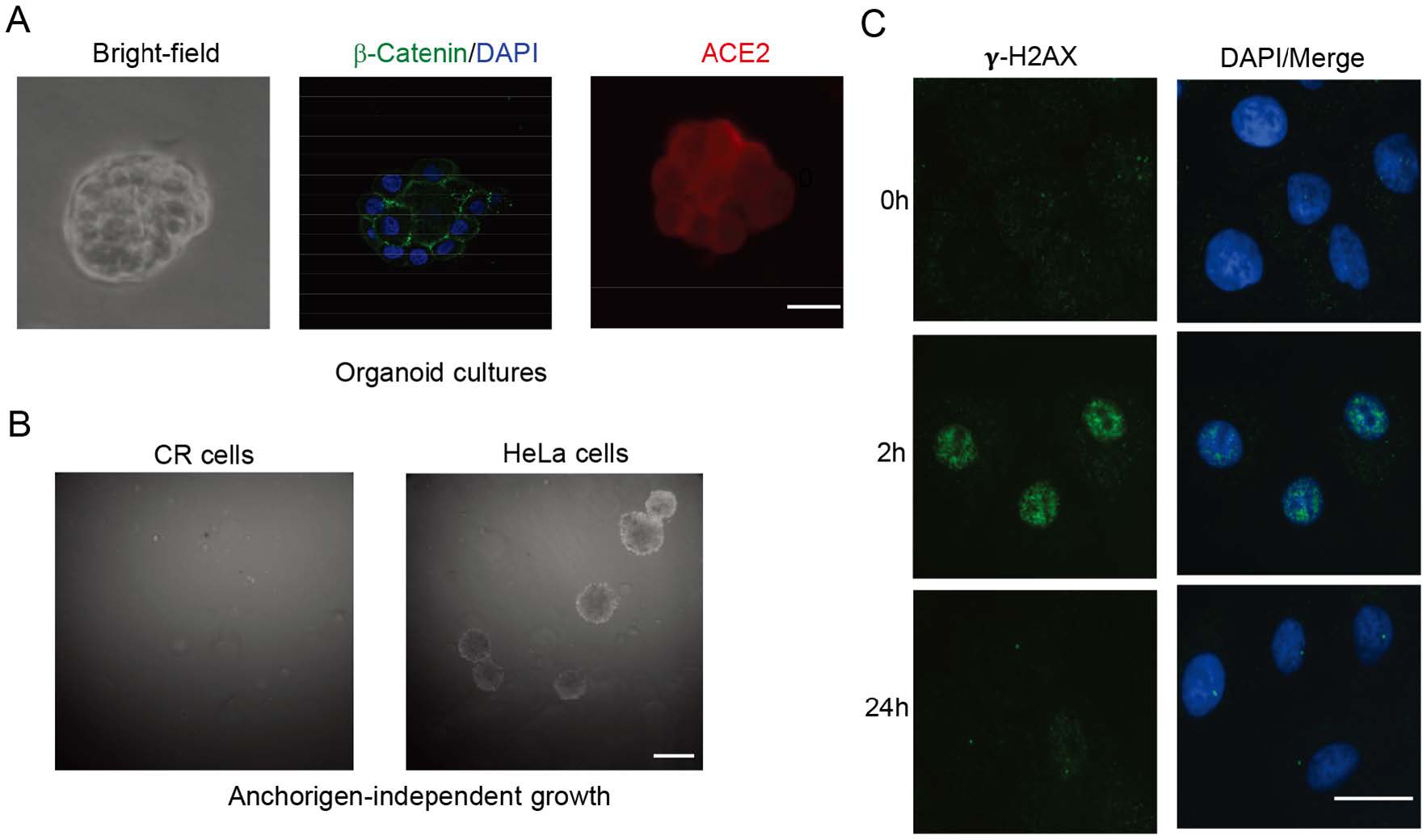
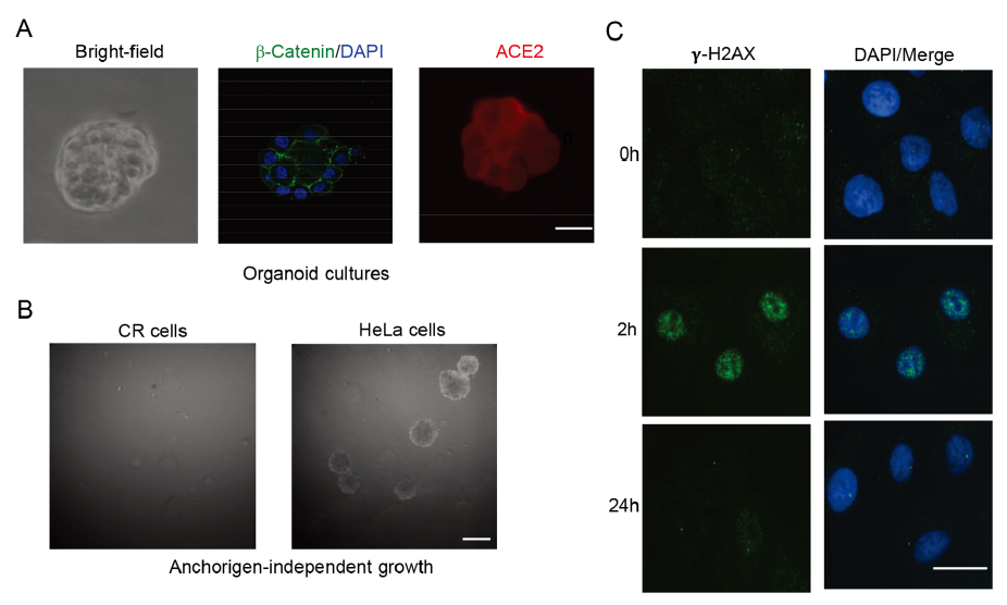
      Figure 2.  Characterization of CR KPTECs. A Organoid cultures of KPTECs. KPTECs were cultured in Matrigel 3D organoid culture as described in Materials and Methods. Left: bright field image of organoids. Middle: beta-catenin and DAPI. Right: Expression of ACE2. Scale bar, 20 μm. B CR KPTECs did not form colonies in soft agar. CR KPTECs did not form colonies in soft agar, but HeLa cells did. Scale bar, 1000 μm. C CR KPTECs maintained ability to repair DNA damage. Phosphorylated H2AX foci and DNA were stained and images were captured using immunofluorensence microscopy. CR KPTECs were able to repair DNA damage induced by irradiation in 24 h. Scale bar, 10 μm.

      Anchorage-independent growth is a hallmark of transformed or malignant cells. The transformed or malignant cells can form colonies when cultured in soft agar, while normal cells are not able to grow under this stringent culture condition. Thus, we tested whether our CR KPTECs were able to form colonies in soft agar. As shown in Fig. 2B, CR KPTECs did not formed colonies in soft agar, indicating that they had no anchorage-independent growth properties as cancer cell line did.

    • It is of special interest to further test if CR KPTECs retain characteristic of original normal tissue at the molecular level. For that purpose, we used immunofluorescence to visualize phosphorylated H2AX foci in CR KPTECs at various times after irradiation (2 h and 24 h). Foci are believed to represent the sites of DNA double-strand break (DSB) and H2AX phosphorylation are the early events in DNA damage response and repair (DDR). After DSBs repair, foci resolution occurs by dephosphorylation of the p-H2AX protein, while persistence of the foci at late time points is thought to represent residual unrepaired DSB. By comparing the formation and resolution of the foci in the cells at the different time points after irradiation, we can determine the DSB repair kinetics and the status of the DDR response. We analyzed the time-dependent change of gamma irradiation induced-H2AX foci in CR KPTECs. The results showed that the DDR response occurred in CR KPTECs at 2 h after irradiation (Fig. 2C). However, almost undetectable fraction of the initial foci persist after 24 h were observed, suggesting that the cells had the ability to repair DNA damage.

    • Since transport functions are unique property of kidney tubule cells, we tested the expression of KPTECs specific sodium-dependent phosphate transporters in CR KPTECs. We performed RT-PCR for detection of SLC34A3 mRNA. Our results demonstrated that CR KPTECs expressed SLC34A3, while HeLa cells did not express SLC34A3 as a negative control (Fig. 3A). Another specific transport system expressed in KPTECs is the megalin/cubilin multiligand receptor, which plays an essential role in endocytotic uptake of filtered proteins by the proximal tubule in kidney. We observed similar pattern for expression of cubilin in CR KPTECs using RT-PCR (Fig. 3A). These results suggested that CR KPTECs maintain lineage function with expression of specific functional genes in the proximal tubules.

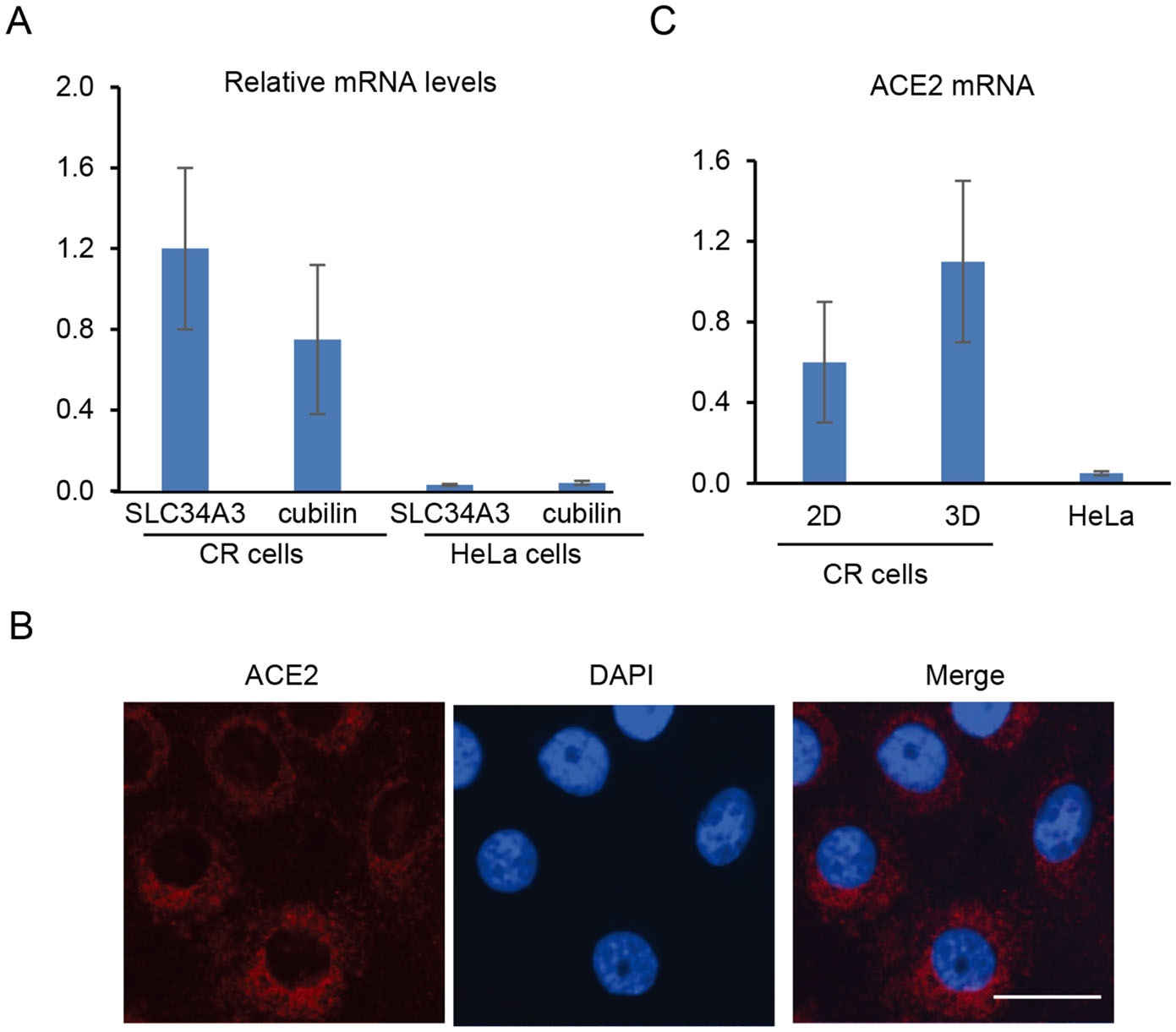
      Figure 3.  Expression of endogenous transporters and ACE2 in KPTECs. A Expression of endogenous SLC34A3 and cubilin. Expression of two transporters was measured using quantitative RT-PCR. β-Actin was used as an internal control. Both SLC34A3 and cubilin were expressed in CR KPTECs, but not in HeLa cells. Expression of endogenous ACE2 in CR KPTECs was measured using IF (B) and quantitative RT-PCR (C). Scale bar, 10 μm.

    • As we described above, some cases with COVID-19 progress to severe pneumonia and multi-organ failure including kidney. To study whether our long term culture system can be used for studies of SARS-CoV-2 and COVID-19, we performed immunofluoresence and RT-PCR to test the expression of ACE2 in 2D and 3D cultures. The ACE2 mRNA and protein were detectable in both 2D and 3D cultures (Figs. 2A, 3B, 3C). Interestingly, our RT-PCR demonstrated expression of endogenous ACE2 in CR KPTECs, not in HeLa cells (Fig. 3C). We also observed a relative higher (around twofold) expression of ACE2 in 3D organoids compared to that in 2D CR conditions (Fig. 3C). Finally, we generated pseudovirions in HEK293T cells transfected with SARS-CoV S plasmid, psPAX, and lenti-Luc. CR KPTECs and HeLa cells were infected by pseudovirions to investigate whether these cells could be infected by SARS-CoV pseudovirions. As shown in Fig. 4, CR KPTECs infected with pseudovirions showed RLUs of 6890 ± 490, while CR KPTECs without pseudovirion infection had RLUs of only 28 ± 8. RLUs were below 100 in HeLa cells with or without SARS-CoV S pseudovirion infection. Thus, CR KPTECs were permissive for SARS-CoV infection. These data suggested that our integrated 2D CR and 3D organoids would be an appropriate physiological human cell system for study of SRAR-CoV and SARS-CoV-2 and their associated kidney injuries, especially for virus entry to KPTECs, innate immune response of host cells, drug screening for anti-SARS-CoV-2 and safety evaluation of kidney functions.

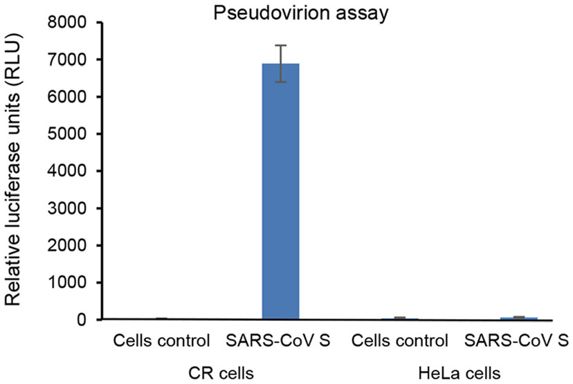
      Figure 4.  Pseudovirion entry experiments of SARS-CoV S in CR KPTECs and HeLa cells. Data are represented as mean ± standard deviation of technical triplicates.

    • In this study, we firstly established long term cultures of human KPTECs using CR technology. These CR cells maintained their differentiation potential and the ability to repair DNA damage induced by irradiation. These cells also expressed lineage functional markers of kidney proximal tubule epithelial cells, including SLC34A3 and megalin/cubilin. These data suggested that CR KPTECs would be used as an ex vivo model for studies of kidney diseases or kidney injury associated with other systemic diseases (e.g., diabetes), and discovery of novel biomarkers and targets.

      As we discussed above, mortality of severe patients with COVID-19 are relative high due to preexisting conditions and multi-organ failure (Wang T et al. 2020; Wang Z et al. 2020; Wu and McGoogan 2020; Xu L et al. 2020; Xu Z et al. 2020; Yang X et al. 2020; Yao et al. 2020; Ye et al. 2020; Zhang H et al. 2020). As SARS-CoV-2 enter host cells via ACE2 protein receptor, it is highly possible that SARS-CoV-2 infect all ACE2 expressing cell types. It was reported that ACE2 is highly expressed in intestine, testis, kidney, heart, etc., and this might help to explain multi-organ injury (Zou X et al. 2020). In particular, a number of studies have reported kidney injury in severe COVID-19 cases (Huang et al. 2020; Li et al. 2020). Furthermore, culturing SARS-CoV-2 virus and SARS-CoV-2 mRNA were detected from urine samples of COVID-19 patients (Ling et al. 2020; Rothe et al. 2020; Xie et al. 2020; Young et al. 2020), indicating the possible replication of SARS-CoV-2 in urological system, especially in kidney and bladder. Thus, it would be important to study the possibility of SARS-CoV-2 infection with urological system.

      In terms of the questions above, we need model systems to study infection of SARS-CoVs in ACE2 expressing cell types, especially in kidney epithelial cells (Hamming et al. 2004; Zou X et al. 2020). Transgenic mice with human ACE2 (Yang et al. 2007; Netland et al. 2008) would be used for modeling disease in vivo, however, it's hard to mimic human diseases due to species-related restriction conditions. Animal models are usually very expensive and need much longer time to complete, thus they cannot be used for any high throughput screening. Another option is human cell lines. Cancer cell lines usually gain the ability to proliferate in very stringent conditions, such as soft agar, and they loss their specific differentiation potential. Thus, those cancer cell lines are not suitable to study "normal" or "physiological" response of host cells to viral infections, although viruses may lead lytic infections in some cancer cell lines. Therefore, much efforts have been made for generating immortalized cell lines (Ryan et al. 1994; Racusen et al. 1997; Kowolik et al. 2004; Orosz et al. 2004; Wieser et al. 2008). As we described above, HPV16 E6/E7, SV40, and hTERT have been widely used for immortalization of human primary cells (Ryan et al. 1994; Racusen et al. 1997; Kowolik et al. 2004; Orosz et al. 2004; Wieser et al. 2008). These cell lines have been widely used in many study fields, including cell biology, production of research reagents and modeling human diseases. However, due to pathways altered by viral or cellular genes or integration of viral/cellular gene to host genome, these immortalized cell lines cannot reflect the physiological status of normal human cells. A related study reported the susceptibilities of a panel of cell lines to SARS-CoV (Kaye 2006). Among the 21 cell lines, including several cell lines from monkey and other species, only three human cell lines (HEK293, Hep G2 and Huh-7) are susceptible (Kaye 2006). Besides, SARS-CoV can achieve high viral load in some cell lines without specific CPE (cytopathic effect) (Kaye 2006). However, none of these cell lines retain physiological functions.

      A recent study demonstrated that active SARS-CoV-2 replication was detected in tissues of the upper respiratory tract, especially virus load and shedding were very high during the first week of symptoms (Wolfel et al. 2020). This suggests an urgent need to establish normal airway cells model from upper (nasal cavity) to lower (lung) respiratory tract to study interactions between SARS-CoV-2 and host epithelial cells. CR has been widely used for generation of various patient-derived cell models that can be used for modeling human disease, drug discovery and tissue regeneration (Borodovsky et al. 2017; Wolf et al. 2017; Yuan et al. 2017; Alkhilaiwi et al. 2018; Brewington et al. 2018; Jin et al. 2018; Moorefield et al. 2018; Peters-Hall et al. 2018; Sayej et al. 2018; Wang et al. 2018; Zhang et al. 2018; Alkhilaiwi et al. 2019; Nicolas et al. 2019; Palechor-Ceron et al. 2019; Wan 2019; Wang et al. 2019; Chai et al. 2020; Krawczyk et al. 2020; Peters-Hall et al. 2020; Wang Y et al. 2020). In the current study, we established long term cell cultures of KPTECs using CR technology. Our CR KPTECs maintain lineage differentiation potential and express cell type-specific function markers and ACE2 protein. These cells can serve as a physiological cell model for the study of SARS-CoVs and kidney functions, innate immune response of kidney cells to viruses and a novel platform for drug discovery and safety evaluation.

    • This work was supported by the National Natural Science Foundation of China (81571396 and 81771528); Science, Technology and Innovation Commission of Shenzhen Municipality (JCYJ20170411090932146, JCYJ20170818110544730).

    • HL: Conceptualization, SX, MW, SC, TZ, LY, JL: Laboratory work and data analysis. SX, MW, HL: Drafting manuscript. All authors have read and approved the submitted version of manuscript.

    • The authors declare no conflict of interest.

    • This article does not contain any studies with human or animal subjects performed by any of the authors.

    Figure (4)  Reference (76) Relative (20)

    目录

    /

    DownLoad:  Full-Size Img  PowerPoint
    Return
    Return