Li Zhao, Ruqin Gao, Roujian Lu, Huijuan Wang, Yao Deng, Peihua Niu, Fachun Jiang, Baoying Huang, Jiwei Liang, Jing Jia, Feng Zhang, Wenling Wang, Guizhen Wu and Wenjie Tan. Profiles of SARS-CoV-2 RNA and Antibodies in Inpatients with COVID-19 not Related with Clinical Manifestation: A Single Centre Study[J]. Virologica Sinica, 2021, 36(5): 1088-1092. doi: 10.1007/s12250-021-00411-w
Citation: Li Zhao, Ruqin Gao, Roujian Lu, Huijuan Wang, Yao Deng, Peihua Niu, Fachun Jiang, Baoying Huang, Jiwei Liang, Jing Jia, Feng Zhang, Wenling Wang, Guizhen Wu, Wenjie Tan. Profiles of SARS-CoV-2 RNA and Antibodies in Inpatients with COVID-19 not Related with Clinical Manifestation: A Single Centre Study .VIROLOGICA SINICA, 2021, 36(5) : 1088-1092.  http://dx.doi.org/10.1007/s12250-021-00411-w

单中心研究结果:COVID-19患者体内SARS-CoV-2 RNA和抗体水平与临床状态无关

  • 通讯作者: 王文玲, wangwl@ivdc.chinacdc.cn, ORCID: http://orcid.org/0000-0002-7360-2913
    ; 武桂珍, wugz@ivdc.chinacdc.cn, ORCID: http://orcid.org/0000-0003-2778-4290
    ; 谭文杰, tanwj@ivdc.chinacdc.cn, ORCID: http://orcid.org/0000-0002-5963-1136
  • 收稿日期: 2020-08-13
    录用日期: 2021-04-30
    出版日期: 2021-07-01
  • 2019冠状病毒病(COVID-19)患者体内严重急性呼吸综合征冠状病毒2(SARS-CoV-2)RNA和抗体水平是否与临床分型相关,该问题需要深入研究。本研究分析了青岛地区53例不同临床分型患者(无症状、轻型、普通型、重症)187份标本中SARS-CoV-2 RNA及抗体水平。结果显示,处于发病第1周的COVID-19患者鼻咽拭子和痰标本中SARS-CoV-2 RNA含量处于峰值状态,处于发病第2周的患者SARS-CoV-2 RNA含量显著下降。不同临床分型患者样本平均病毒载量没有差异,但轻型和普通型COVID-19患者,鼻咽拭子阳性率和病毒载量要高于粪便样本。此外,COVID-19患者血清阳转(IgG和NAb)发生子在第2周,整个病程中IgM的平均滴度均较低。此外,在COVID-19患者配对样本的检测结果表明,RNA和Ab表现出不同的变化趋势。综上所述,呼吸道是SARS-CoV-2排毒的主要途径,SARS-CoV-2 RNA 和抗体水平与临床表现无关。

Profiles of SARS-CoV-2 RNA and Antibodies in Inpatients with COVID-19 not Related with Clinical Manifestation: A Single Centre Study

  • Corresponding author: Wenling Wang, wangwl@ivdc.chinacdc.cn Guizhen Wu, wugz@ivdc.chinacdc.cn Wenjie Tan, tanwj@ivdc.chinacdc.cn
  • ORCID: http://orcid.org/0000-0002-7360-2913; http://orcid.org/0000-0003-2778-4290; http://orcid.org/0000-0002-5963-1136
  • Received Date: 13 August 2020
    Accepted Date: 30 April 2021
    Published Date: 01 July 2021

  • 加载中
    1. Burbelo PD, Riedo FX, Morishima C, Rawlings S, Smith D, Das S, Strich JR, Chertow DS, Davey RT, Cohen JI (2020) Detection of nucleocapsid antibody to SARS-CoV-2 is more sensitive than antibody to Spike protein in COVID-19 patients. China CDC Weekly 9: 469
        doi: 10.1093/infdis/jiaa273

    2. Chinese Center for Disease Control and Prevention (2020) Technical guidelines for COVID-19 laboratory testing. China CDC Weekly. https://doi.org/10.46234/ccdcw2020.085
        doi: 10.46234/ccdcw2020.085

    3. Demey B, Daher N, François C, Lanoix JP, Duverlie G, Castelain S, Brochot E (2020) Dynamic profile for the detection of anti-SARS-CoV-2 antibodies using four immunochromatographic assays. J Infect S0163: 30244–30249
        doi: 10.1016/j.jinf.2020.04.033

    4. Garbati MA, Fagbo SF, Fang VJ, Skakni L, Joseph M, Wani TA, Cowling BJ, Peiris M, Hakawi A (2016) A comparative study of clinical presentation and risk factors for adverse outcome in patients hospitalised with acute respiratory disease due to MERS coronavirus or other causes. PLoS ONE 11: 1–12
        doi: 10.1371/journal.pone.0165978

    5. Han H, Luo Q, Mo F, Long L, Zheng W (2020) SARS-CoV-2 RNA more readily detected in induced sputum than in throat swabs of convalescent COVID-19 patients. Lancet Infect Dis 3099: 30174
        doi: 10.1016/S1473-3099(20)30174-2

    6. Hsueh PR, Huang LM, Chen PJ, Kao CL, Yang PC (2004) Chronological evolution of IgM, IgA, IgG and neutralisation antibodies after infection with SARS-associated coronavirus. Clin Microbiol Infect 10: 1062–1066
        doi: 10.1111/j.1469-0691.2004.01009.x

    7. Imai K, Tabata S, Ikeda M, Noguchi S, Kitagawa Y, Matuoka M, Miyoshi K, Tarumoto N, Sakai J, Ito T, Maesaki S, Tamura K, Maeda T (2020) Clinical evaluation of an immunochromatographic IgM/IgG antibody assay and chest computed tomography for the diagnosis of COVID-19. J Clin Virol 128: 1315
        doi: 10.1016/j.jcv.2020.104393

    8. Lee CYP, Lin RTP, Renia L, Ng LFP (2020) Serological approaches for COVID-19: Epidemiologic perspective on surveillance and control. Front Immunol 11: 1–7
        doi: 10.3389/fimmu.2020.00001

    9. Li Z, Yi Y, Luo X, Xiong N, Liu Y, Li S, Sun R, Wang Y, Hu B, Chen W, Zhang Y, Wang J, Huang B, Lin Y, Yang J, Cai W, Wang X, Cheng J, Chen Z, Sun K, Pan W, Zhan Z, Chen L, Ye F (2020) Development and clinical application of a rapid IgM-IgG combined antibody test for SARS-CoV-2 infection diagnosis. J Med Virol 92: 1518–1524
        doi: 10.1002/jmv.25727

    10. Liu W, Liu L, Kou G, Zheng Y, Ding Y, Ni W, Wang Q, Tan L, Wu W, Tang S, Xiong Z, Zheng S (2020) Evaluation of nucleocapsid and spike protein-based enzyme-linked immunosorbent assays for detecting antibodies against SARS-CoV-2. J Clin Microbiol 58: e00461-e520

    11. Long Q, Deng H, Chen J, Hu J, Liu B, Liao P, Lin Y, Yu L, Mo Z, Xu Y, Gong F, Wu G, Zhang X, Chen Y, Li Z, Wang K, Zhang X, Tian W, Niu C, Yang Q, Xiang J, Du H, Liu H, Lang C, Luo X, Wu S, Cui X, Zhou Z, Wang J, Xue C, Li X, Wang L, Tang X, Zhang Y, Qiu J, Liu X, Li J, Zhang D, Zhang F, Cai X, Wang D, Hu Y, Ren J, Tang N, Liu P, Li Q, Huang A (2020) Antibody responses to SARS-CoV-2 in COVID-19 patients: the perspective application of serological tests in clinical practice. medRxiv doi: 2020.03.18.20038018

    12. Lou B, Li T-D, Zheng S-F, Su Y-Y, Li Z-Y, Liu W, Yu F, Ge S-X, Zou Q-D, Yuan Q, Lin S, Hong C-M, Yao X-Y, Zhang X-J, Wu D-H, Zhou G-L, Hou W-H, Li T-T, Zhang Y-L, Zhang S-Y, Fan J, Zhang J, Xia N-S, Chen Y (2020) Serology characteristics of SARS-CoV-2 infection since exposure and post symptom onset. Eur Respir J 27: 775
        doi: 10.1183/13993003.00763-2020

    13. Lu R, Zou L, Wang Y, Zhao Y, Zhou W, Wu J, Wang W, Wu G, Ke C, Tan W (2015) Sequencing and phylogenetic analyses of structural and accessory proteins of Middle East respiratory syndrome coronavirus from the first imported case in China, 2015. Chinese J Virol 31: 333–340

    14. Nie J, Li Q, Wu J, Zhao C, Hao H, Liu H, Zhang L, Nie L, Qin H, Wang M, Lu Q, Li X, Sun Q, Liu J, Fan C, Huang W, Xu M, Wang Y (2020) Establishment and validation of a pseudovirus neutralization assay for SARS-CoV-2. Emerg Microbes Infect 9: 680–686
        doi: 10.1080/22221751.2020.1743767

    15. Niu P, Lu R, Zhao L, Wang H, Huang B, Ye F, Wang W, Tan W (2020) Three novel real-time RT-PCR assays for detection of COVID-19 virus. China CDC Weekly. https://doi.org/10.46234/ccdcw2020.116
        doi: 10.46234/ccdcw2020.116

    16. Norman M, Gilboa T, Ogata AF, Maley AM, Cohen L, Cai Y, Zhang J, Feldman JE, Hauser BM, Caradonna TM, Chen B, Schmidt AG, Alter G, Charles RC, Ryan ET, Walt DR (2020) Ultra-sensitive high-resolution profiling of anti-SARS-CoV-2 antibodies for detecting early seroconversion in COVID-19 patients. medRxiv doi: 2020.04.28.20083691

    17. Okba N, Muller M, Li W, Wang C, GeurtsvanKessel C, Corman V, Lamers M, Sikkema R, de Bruin E, Chandler F, Yazdanpanah Y, Le Hingrat Q, Descamps D, Houhou-Fidouh N, Reusken C, Bosch B-J, Drosten C, Koopmans M, Haagmans B (2020) Severe acute respiratory syndrome coronavirus 2−specific antibody responses in coronavirus disease patients. Emerg Infect Dis 26: 1478–1488
        doi: 10.3201/eid2607.200841

    18. Ozturk T, Howell C, Benameur K, Ramonell RP, Cashman K, Pirmohammed S, Bassit L, Roback J, Marconi VC, Schinazi RF, Wharton W, Lee FE-H, Hu WT (2020) Cross-sectional IgM and IgG profiles in SARS-CoV-2 infection. medRxiv doi: 2020.05.10.20097535

    19. Pan X, Chen D, Xia Y, Wu X, Li T, Ou X, Zhou L, Liu J (2020a) Asymptomatic cases in a family cluster with SARS-CoV-2 infection. Lancet Infect Dis 20: 410–411
        doi: 10.1016/S1473-3099(20)30114-6

    20. Pan Y, Zhang D, Yang P, Poon LLM, Wang Q (2020b) Viral load of SARS-CoV-2 in clinical samples. Lancet Infect Dis 20: 411–412
        doi: 10.1016/S1473-3099(20)30113-4

    21. Taskforce on C-P, Control CSC (2020) Protocol for the management of asymptomatic persons infected with COVID-19 virus. China CDC Weekly 2: 1–2
        doi: 10.46234/ccdcw2020.001

    22. To KKW, Tsang OTY, Leung WS, Tam AR, Wu TC, Lung DC, Yip CCY, Cai JP, Chan JMC, Chik TSH, Lau DPL, Choi CYC, Chen LL, Chan WM, Chan KH, Ip JD, Ng ACK, Poon RWS, Luo CT, Cheng VCC, Chan JFW, Hung IFN, Chen Z, Chen H, Yuen KY (2020) Temporal profiles of viral load in posterior oropharyngeal saliva samples and serum antibody responses during infection by SARS-CoV-2: an observational cohort study. Lancet Infect Dis 20: 565–574
        doi: 10.1016/S1473-3099(20)30196-1

    23. Wan Y, Li Z, Wang K, Li T, Liao P (2020) Performance verification of detecting COVID-19 specific antibody by using four chemiluminescence immunoassay systems. medRxiv doi: 2020.04.27.20074849

    24. Wang W, Wang H, Deng Y, Song T, Lan J, Wu G, Ke C, Tan W (2016) Characterization of anti-MERS-CoV antibodies against various recombinant structural antigens of MERS-CoV in an imported case in China. Emerg Microbes Infect 5: e113–e116
        doi: 10.1038/emi.2016.114

    25. Wang W, Wang T, Deng Y, Niu P, Ruhan A, Zhao J, PeirisN M, Tang S, Tan W (2019) A novel luciferase immunosorbent assay performs better than a commercial enzyme-linked immunosorbent assay to detect MERS-CoV specific IgG in humans and animals. Biosaf Heal 1: 134–143
        doi: 10.1016/j.bsheal.2019.12.006

    26. Wang W, Xu Y, Gao R, Lu R, Han K, Wu G, Tan W (2020a) Detection of SARS-CoV-2 in different types of clinical specimens. JAMA 323: 1061–1069
        doi: 10.1001/jama.2020.1585

    27. Wang X, Guo X, Xin Q, Pan Y, Hu Y, Li J, Chu Y, Feng Y, Wang Q (2020b) Neutralizing antibodies responses to SARS-CoV-2 in COVID-19 inpatients and convalescent patients. Clin Infect Dis 71: 2688–2694
        doi: 10.1093/cid/ciaa721

    28. Wang Y, Zhang L, Sang L, Ye F, Ruan S, Zhong B, Song T, Alshukairi AN, Chen R, Zhang Z, Gan M, Zhu A, Huang Y, Luo L, Mok CKP, Gethamy MMA, Tan H, Li Z, Huang X, Li F, Sun J, Zhang Y, Wen L, Li Y, Chen Z, Zhuang Z, Zhuo J, Chen C, Kuang L, Wang J, Lv H, Jiang Y, Li M, Lin Y, Deng Y, Tang L, Liang J, Huang J, Perlman S, Zhong N, Zhao J, Peiris M, Li Y, Zhao J (2020c) Kinetics of viral load and antibody response in relation to COVID-19severity. J Clin Invest 130(10): 5235–5244
        doi: 10.1172/JCI138759

    29. Xiang F, Wang X, He X, Peng Z, Yang B, Zhang J, Zhou Q, Ye H, Ma Y, Li H, Wei X, Cai P, Ma WL (2020) Antibody detection and dynamic characteristics in patients with COVID-19. Clin Infect Dis Ciaa. https://doi.org/10.1093/cid/ciaa721
        doi: 10.1093/cid/ciaa721

    30. Xu T, Chen C, Zhu Z, Cui M, Chen C, Dai H, Xue Y (2020) Clinical features and dynamics of viral load in imported and non-imported patients with COVID-19. Int J Infect Dis 94: 68–71
        doi: 10.1016/j.ijid.2020.03.022

    31. Zhao R, Li M, Song H, Chen J, Ren W, Feng Y, Gao GF, Song J, Peng Y, Su B, Guo X, Wang Y, Chen J, Li J, Sun H, Bai Z, Cao WJ, Zhu J, Zhang Q, Sun Y, Sun S, Mao X, Su J, Chen X, He A, Gao W, Jin R, Jiang Y, Sun L (2020) Early detection of SARS-CoV-2 antibodies in COVID-19 patients as a serologic marker of infection. Clin Infect Dis. https://doi.org/10.1093/cid/ciaa523
        doi: 10.1093/cid/ciaa523

    32. Zou L, Ruan F, Huang M, Liang L, Huang H, Hong Z, Yu J, Kang M, Song Y, Xia J, Guo Q, Song T, He J, Yen H-L, Peiris M, Wu J (2020) SARS-CoV-2 viral load in upper respiratory specimens of infected patients. N Engl J Med 382: 1177–1179
        doi: 10.1056/NEJMc2001737

  • 加载中

Figures(1)

Article Metrics

Article views(7175) PDF downloads(3) Cited by(0)

Related
Proportional views
    通讯作者: 陈斌, bchen63@163.com
    • 1. 

      沈阳化工大学材料科学与工程学院 沈阳 110142

    1. 本站搜索
    2. 百度学术搜索
    3. 万方数据库搜索
    4. CNKI搜索

    Profiles of SARS-CoV-2 RNA and Antibodies in Inpatients with COVID-19 not Related with Clinical Manifestation: A Single Centre Study

      Corresponding author: Wenling Wang, wangwl@ivdc.chinacdc.cn
      Corresponding author: Guizhen Wu, wugz@ivdc.chinacdc.cn
      Corresponding author: Wenjie Tan, tanwj@ivdc.chinacdc.cn
    • 1. NHC Key Laboratory of Biosafety, National Institute for Viral Disease Control and Prevention, Chinese Center for Disease Control and Prevention, Beijing, 102206, China
    • 2. Qingdao Center for Disease Control and Prevention, Qingdao, 266033, China
    • 3. Center for Biosafety Mega-Science, Chinese Academy of Sciences, Wuhan, 430071, China

    Abstract: 

    • Dear Editor,

      A novel coronavirus disease (COVID-19) caused by acute respiratory syndrome coronavirus 2 (SARS-CoV-2) first broke out in Wuhan, Hubei Province, China, in 2019. More than 50 million cases of COVID-19 have been confirmed globally with 1,280,868 deaths reported as of November 12, 2020 (https://covid19.who.int/). RNA detection based on real-time reverse transcription polymerase chain reaction (rRT-PCR) assay has been developed and is widely used as the standard of diagnosing COVID-19 (Chinese Center for Disease Control and Prevention 2020). SARS-CoV-2 RNA can be detected from various types of specimens, with respiratory specimens mostly preferred (Pan et al. 2020; Wang et al. 2020; Zou et al. 2020). However, more data are needed to understand the profiles of viral shedding in different types of specimens from COVID-19 patients with different clinical manifestations. Antibody detection plays an important role in identifying the clinical status and outcome of a patient. Although several studies on antibody detection in COVID-19 patients have been published recently (Burbelo et al. 2020; Demey et al. 2020; Lee et al. 2020; Li et al. 2020; Liu et al. 2020; Nie et al. 2020; Okba et al. 2020; Wan et al. 2020; Xiang et al. 2020; Zhao et al. 2020; Wang et al. 2020a, 2020b, 2020c), knowledge gaps regarding the profile, dynamics, and magnitude of the antibody response in COVID-19 patients with different clinical manifestations still exist.

      To understand the profiles of SARS-CoV-2 RNA and antibodies in inpatients with COVID-19, we enrolled 53 COVID-19 inpatients admitted to hospitals in Qingdao between January and March of 2020. The day of symptom onset (fever, cough, or fatigue, etc.) was defined as day 0 for most cases in the subsequent analyses. Specimens were collected in January and February on the basis of easy access by the Qingdao Municipal Center for Disease Control and Prevention according to the Technical Guidelines for COVID-19 Laboratory Testing (China CDC 2020).

      The median age of the patients was 35 years old (range, 5–70 years old), and 43% of the patients were male. Of all patients, 3 (6%), 11 (21%), 33 (62%), and 5 (9%) exhibited asymptomatic, mild, moderate, and severe clinical symptoms, respectively, and clinical information was lacking for one patient. The mean duration between symptom onset and admission for all patients was 3 days (range, 0–22 days). Patients have stayed in hospital for an average of 11 days, and three patients were hospitalised for more than three weeks, with a maximum stay of 49 days. 187 specimens were collected and tested, including 142 tests for viral RNA using six types of samples (nasopharyngeal swabs, sputum, faeces, urine, blood, and conjunctival swabs).

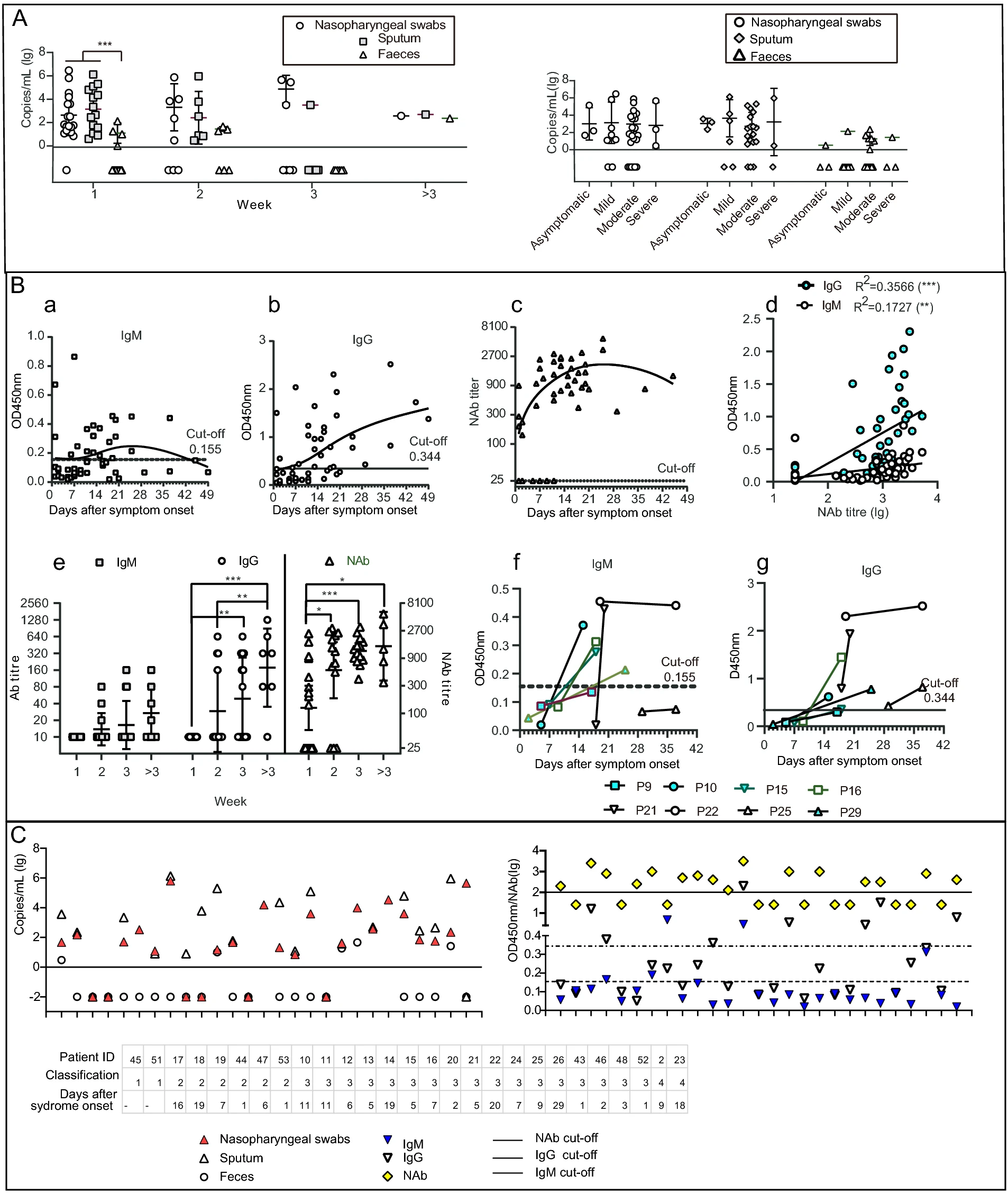
      To determine viral RNA levels in samples, real-time RT-PCR was performed using the nucleocapsid gene as a target (Lu et al. 2015; Niu et al. 2020; Wang et al. 2020). Nasopharyngeal swab and sputum samples had the highest viral loads, up to 2.9 × 10⁶ copies/mL (mean, 1.6 × 10⁶ copies/mL) and 1.3 × 10⁶ copies/mL (mean, 1.1 × 10⁶ copies/mL), respectively, substantially higher than the maximum load of 231 copies/mL (mean, 54 copies/mL) recorded in faecal specimens (Fig. 1A, left). Most nasopharyngeal swabs (95%, 18 of 19 tested) and all sputum samples (15 tested) had detectable viral loads in week 1, compared with 22% of the faecal samples (Fisher's exact test, P < 0.001; Fig. 1A). Positive test rates did not differ among the three types of samples starting in week 2 after symptom onset (Fig. 1A, left). No positive results were obtained from urine (n = 13), blood (n = 17), or conjunctival swab (n = 10) samples.

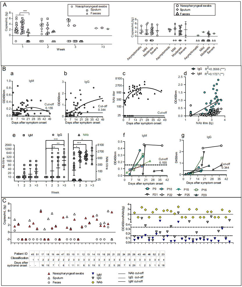
      Figure 1.  Detection of viral RNA and antibodies in samples of COVID-19 patients. A Comparison of viral RNA loads in COVID-19 patients detected by nasopharyngeal swab, sputum, and faecal samples (left) Viral RNA loads in COVID-19 patients with different clinical manifestations. B Serum IgM and IgG profiles in COVID-19 patients as analysed using RBD-based enzyme-linked immunosorbent assay (ELISA). a–c Cross-sectional profiles of absorbance at 450 nm (OD450nm) by IgM (a) and IgG (b) against RBD and by neutralising antibody (NAb) in a pseudovirus particle neutralisation test (c). Each dot represents an individual serum sample. d, Comparison of IgM, IgG, and NAb. e, Chronological changes (weeks after symptom onset) in IgM and IgG titres based on RBD-ELISA, and in NAb titres. f and g Changes in IgM (f) and IgG (g) levels between matched pairs of serum samples from COVID-19 patients. Each line represents an individual patient. C Detection of viral RNA and antibodies in SARS-CoV-2-infected patients. Results of matched-pair analysis to detect RNA (left) and antibody levels (right) in SARS-CoV-2-infected individuals. Classification: 1 indicated as asymptomatic patients, 2 indicated as mild patients, 3 indicated as moderate patients, 4 indicated as severe patients. *P < 0.05, **P < 0.01, ***P < 0.001.

      Viral loads of nasopharyngeal swab did not differ in COVID-19 patients with different illness stage, nor that of sputum, or faecal samples. The positive rate from nasopharyngeal swab samples decreased significantly, to 43%, in week 3 after symptom onset compared to the rate in week 1 (Fisher's exact test, P < 0.05). Similarly, positive rates from sputum samples decreased significantly, from 100% to 14%, from both weeks 1 and 2 to week 3 after symptom onset (Fisher's exact test, P < 0.001 and P < 0.01, respectively). Furthermore, the average viral loads in COVID-19 patients with different clinical manifestations were assessed (Fig. 1A, right). For nasopharyngeal swab, sputum, and faecal samples, the average viral loads did not differ among patients (Fig. 1A, right). However, nasopharyngeal swabs collected from patients with mild disease status produced higher positive test rates compared with faecal samples (Fisher's exact test, P < 0.05). The proportion of positive tests differed more markedly among nasopharyngeal swab, sputum, and faecal samples collected from patients with moderate disease status (Fisher's exact test, P < 0.01).

      To determine the antibody levels in serum samples, an enzyme-linked immunosorbent assay (ELISA) was performed in-house by coating ELISA plates with commercial RBD (Sino Biological Inc., Beijing, China) to detect immunoglobulin (Ig) M and IgG serum samples. The cut-off value was set at three standard deviations above the mean value of the negative cohort. A SARS-CoV-2 pseudovirus particle neutralisation test (ppNT) was performed to detect neutralizing antibody (NAb) as previously described (Nie et al. 2020; Wang et al. 2019, 2016). The median seroconversion times for IgM, IgG, and NAb were 14 (1–37), 16 (1–49), and 14 (1–45) days after symptom onset, respectively. Seroconversion of IgM, IgG, and NAb were observed in 21, 28 and 41 serum samples, respectively, that of both IgM and IgG in 18 serum samples, that of both IgM and NAb in 19 samples, and that of both IgG and NAb in 26 samples (Fig. 1B-a, -b and -c). Both IgM and IgG titres were significantly correlated with NAb (P < 0.01 and P < 0.001, respectively; Fig. 1B-d). Overall, IgM conversion exhibited an increase–decrease pattern (Fig. 1B-a), with 18% of the serum samples (n = 17) testing positive in week 1 and 54% (n = 13), 50% (n = 14) in weeks 2 and 3, respectively. After week 3, seroconversion of IgM decreased to 43% (n = 7). IgG and NAb conversion exhibited a continually increasing trend. IgG had a low conversion rate of 24% (n = 17) in week 1 and 100% (n = 14) at 3 weeks later (Fig. 1B-b). In week 1, 53% (n = 17) of the samples tested positive for NAb, increasing to 77% (n = 13) in week 2, and 100% thereafter. The average IgM titre was low throughout the observation period, whereas the average IgG titre continued to increase at 3 weeks after symptom onset (Fig. 1B-e). IgG titres increased significantly in week 3 compared to week 1 (P < 0.01), and that were significantly higher after week 3 than that in week 1 and week 2 (P < 0.001 and P < 0.01). NAb displayed special pattern with significantly higher levels in week 2 after symptom onset than that in week 1 (P < 0.05), while NAb titers in week 3 and 4 after symptom onset were not significantly higher than that in week 2 (Fig. 1B-e). IgM and IgG levels in the serum of eight patients were compared in a matched-pair analysis (Fig. 1B-f, -g). The dynamic changes of serum antibody titres exhibited diverse trends and occurred in most of the patients at 2 weeks after symptom onset. However, low IgM levels and a slow response was observed in one individual, and IgM levels in another patient decreased at 5 weeks after symptom onset, when a second serum sample was collected. By contrast, IgG levels in the second serum samples were significantly higher than in the first ones. The median seroconversion times were 14 (IgM and NAb) and 16 (IgG) days after symptom onset, similar with the results in other studies (Demey et al. 2020; Imai et al. 2020; Lee et al. 2020; Liu et al. 2020; Lou et al. 2020; Norman et al. 2020; Okba et al. 2020; To et al. 2020).

      In this single centre study, we investigated the profiles of SARS-CoV-2 RNA and antibodies of 53 COVID-19 patients with different clinical manifestation (Garbati et al. 2016; Lou et al. 2020; Xiang et al. 2020). The results proved that SARS-CoV-2 excretion via the respiratory tract was the main viral source, and the profiles of SARS-CoV-2 RNA and antibodies were not related with clinical manifestation. Notably, the dynamics of SARS-CoV-2 RNA were similar in asymptomatic COVID-19 patients with others, and this aspect should be studied in further detail. Our analysis of viral loads confirmed that some asymptomatic cases can host extraordinarily high viral loads, implying that asymptomatic patients who engage in social contact can have high transmission potential (Pan et al. 2020). These cases should be managed early to prevent disease transmission (Joint Taskforce on Control 2020).

      Although IgG and NAb levels increased over time, indicating seroconversion, no significant differences in IgG and NAb levels were found among COVID-19 patients with different clinical manifestations throughout the observation period. These results contradict previous studies (Long et al. 2020; Okba et al. 2020; Ozturk et al. 2020; Wang et al. 2020b, 2020c), considering the difference of longitudinal study and cross-sectional survey.

      In this study, the matched-pair analysis indicated that the occurrences of the virus and antibodies were not mutually exclusive, suggesting that the development of antibodies in serum was not followed by the rapid elimination of viral RNA from the lung. NAb normally includes IgA, which is secreted in respiratory fluids and saliva (Hsueh et al. 2004; Norman et al. 2020; Okba et al. 2020). Further work by our group will focus on IgA detection.

      A major limitation of this study was the limited number of patients enrolled from a single centre. Multicentre, multi-national, prospective studies on the profiles and dynamics of SARS-CoV-2 RNA and antibodies are urgently needed for prediction and diagnosis of the clinical progress and outcomes in COVID-19 patients. In addition, commercial antibody assays with the best performance in clinical settings should be chosen in future studies.

    • This work was supported by the National Key Program for Infectious Disease of China (2018ZX10101002), the National Key Research and Development Program of China (2016YFD0500301, 2020YFC0840900).

    • The authors declare no competing interests.

    • All procedures performed in studies involving human participants were in accordance with the policy on public health investigations of emerging infectious diseases issued by the National Health Commission of China and its later amendments or comparable ethical standards.

    Figure (1)  Reference (32) Relative (20)

    目录

    /

    DownLoad:  Full-Size Img  PowerPoint
    Return
    Return