


-
White spot syndrome virus (WSSV) is a fatal pathogen of shrimp and has a circular, double-stranded DNA genome of about 300 kb (18, 22). Unique aspects of its morphology and genome caused this virus to be classified in a new virus family, Nimaviridae (3, 11). The intact virion is comprised of at least 59 structural proteins, the localization of 44 of which in viral particle have been established; 35 are defined as envelope proteins (including tegument proteins) and nine as nucleocapsid proteins (1, 9, 10, 14, 15, 17, 20, 21).
Like most DNA viruses, WSSV replicates in the nucleus of host cells (13). However, due to the lack of permissive cell lines, the life cycle of WSSV is com-pletely unknown. After entering the cell and passing through the cytosol, entry of the viral genome into the nucleus is a prerequisite for viral genome expression and replication, and is thus essential for initiating the viral life cycle. The available evidence indicates that most nuclear replicating viruses enter the nucleus through nuclear pore complexes (NPCs) (8, 19). Tar-geting and transport of viral genomes to the nucleus depends on nuclear localization sequences (NLSs), exposed on the surface of the capsid particle. This process relies on assistance from cellular import receptors in the form of importins (karyopherins) (8, 19). NLSs are short stretches of residues that mediate the transport of nuclear proteins into the nucleus (2). There are two types of NLS: monopartite NLSs which comprise a short stretch of basic amino acids (7) and bipartite NLSs, consisting of two stretches of basic amino acids separated by a spacer of 10-12 amino acids (12).
A recent report proposed that VP664, VP51C, VP60B and VP15 were the major components of the nucleocapsid. VP664 and VP15 both appear in the nucleocapsid and VP15 is a core protein, involved in the packaging of the WSSV genome in the nucleo-capsid (15, 16, 20, 23). VP15 would therefore be expected to play an important role in transporting the viral genome to the nucleus. Three bipartite NLSs NLS1 (aa 11-27, RRGSKKRSTTAGRISKR), NLS2 (aa 33-49, KKRAGKKSSTVRRRSSK) and NLS3 (aa 44-60, RRRSSKSGKKSGARKSR) (the conserved basic residues are shown in bold letters) were detected in the sequence of VP15, using the computer program ScanProsite (4), but whether or not it is a nuclear transport protein remains unclear. In this study, we characterized the nuclear localization function of VP15 and its three functional bipartite NLSs.
HTML
-
The transient expression vector pIZ/V5-His (In-vitrogen) and pEGFP-N1 (Clontech) were used to express full-and partial-length VP15 in insect cells (Sf9) and baby hamster kidney (BHK) cells, under the control of the O pMNPV immediate-early gene promoter (OpIE2), and the CMV immediate-early gene promoter, respectively.
The full-length DNA encoding VP15 was amplified from the WSSV genome by PCR using forward (5'-GAA AAGCTTAAAATGGTTGCCCGA-3') and reverse (5'-AGGG GTCGACGAACGCTTTGACTT-3') (cor-responding to 164064-164238 of the genome sequence AF369029) primers, containing a Hind Ⅲ and a Sal Ⅰ site (in italics), respectively. The amplified DNA was then ligated into pGEM-T Easy Vector (Promega). After confirming the sequence, the resulting plasmid, pGEM-VP15, was cleaved with Hind Ⅲ and Sal Ⅰ, and cloned into pEGFP-N1. The generated plasmid pEGFP-VP15 was digested with Hind Ⅲ and Not Ⅰ and ligated via the corresponding sites into pIZ/V5-His. As a negative control, the GFP gene was cloned into pIZ/V5-His and expressed.
The two complementary DNA sequences of NLS1, 2, and 3 were synthesized by TaKaRa Bio Inc. (Dalian, China) and all contained a Hind Ⅲ site in the 5'-over hang and a Sal Ⅰ site in the 3'-overhang (in italics). The complementary DNA sequences are listed below: NLS1, 5'- AGCTTATGCGCCGTGGAAGCAAGAA GAGGTCCACCACTGCTGGACGCATCTCCAAGCGG-3' and 5'- TCGACCGCTTGGAGATGCGTCCAGCAGTGGTGGACCTCTTCTTGCTTCCACGGCGCAT A-3'; NLS2, 5'- AGCTTATGAAGAAGCGTGC AGGAAAGAAGAGCTCCACTGTCCGTCGCCGT TCCTCAAAG-3' and 5'- TCGACTTTGAGGAACG GCGACGGACAGTGGAGCTCTTCTTTCCTGCAC GCTTCTTCAT A-3'; NLS3, 5'- AGCTTATGCGTCG CCGTTCCTCAAAGAGCGGAAAGAAGTCTGGAGCCCGCAAGTCAAGG-3' and 5'- TCGACCTTGA CTTGCGGGCTCCAGACTTCTTTCCGCTCTTTGAGGAACGGCGACGCAT A-3'. The doublestranded DNA was inserted into the corresponding sites at the 5'-end of the GFP gene in plasmid pEGFP-N1. The resulting plasmids, pEGFP-NLS1/2/3, were then digested with Hind Ⅲ and Not Ⅰ and cloned into pIZ/ V5-His. The full-length DNA fragment of VP15 with a deletion of NLS1 was amplified by modifying the forward primer to 5'-GTC AAGCTT ATGAGGAGCC CATCA-3', con-taining a Hind Ⅲ site. The full-length DNA fragment of VP15 with a mutation of two basic amino acids (11RR12), which were replaced by two alanines (11AA12), was amplified by modifying the forward primer to 5'-ACA AAGCTTATGGTTGCCCG AAGCTCCAAGACCAAATCCGCTGCTGGAAGC-3'. The amplified DNA was cloned into pEGFP-N1 and pIZ/V5-His, as described above, generating the plas-mids pEGFP-VP15ΔNLS1, pEGFP-VP15(11AA12), pIZ/ V5-GFP-VP15 ΔNLS1 and pIZ/V5-GFP-VP15M(11AA12).
-
Sf9 cells were maintained at 27 ℃ in Grace's medium (GIBCO/BRL) supplemented with 10% bovine calf serum. BHK cells were cultured at 37 ℃ in Dulbecco's Modified Eagle's Medium (DMEM) (GIBCO/BRL) supplemented with 10% bovine calf serum. For transfection, cells (about 105) were plated onto a glass cover slip in a cell culture dish. After overnight incubation, about 1 μg of plasmid DNA was mixed with Lipofectin Reagent (Invitrogen) and laid over the cultured cells, according to the manufacturer's instructions.
-
Twenty-four hours after transfection, cells on the glass coverslips were fixed in 4% paraformaldehyde (Sigma) for 30 min at 4 ℃, then washed with PBS, and the nuclei were stained with Hoechst 33258 (Sigma). Samples were observed using a confocal microscope (LEICA/TCS-SP2).
Plasmid construction
Cell culture and transient transfection
Microscopy analysis
-
When the GFP-containing plasmids were trans-fected into Sf9 and BHK cells, GFP diffused throug-hout the cells (in both the cytoplasm and the nucleus), whereas the VP15-GFP fusion protein accumulated exclusively in the nucleus (Fig. 1. A, B and C), in both the insect and the mammalian cells. The NLS1-GFP fusion protein was also directed to the nucleus, but not efficiently, whereas NLS2-GFP and NLS3-GFP diffused throughout the cell, in both Sf9 (Fig. 1. D, E, F) and BHK cells (data not shown). However, if NLS1 was deleted from VP15, the remaining sequence (containing NLS2 and NLS3) could still direct GFP from the cytoplasm to the nucleus, though a small amount of fluorescence still remained in the cytoplasm (Fig. 1. G). The mutated VP15, in which the two basic amino acids (11RR12) of NLS1 were modified to two alanines (11AA12), localized exclusively in the cytoplasm of both Sf9 (Fig. 1 H) and BHK cells (data not shown). These results demonstrated that VP15 is a nuclear locali-zation protein and needs the cooperation of three NLSs. The two residues (11RR12) of NLS1 play a key role in the nuclear localization function of the full-length VP15 protein. Moreover, we observed that VP15-GFP, VP15ΔNLS1-GFP and NLS1 all formed speckles in the nucleus. We speculate that VP15 localized in some specific region of the nucleus and formed speckle structures with nuclear factors, probably cellular chromatin.
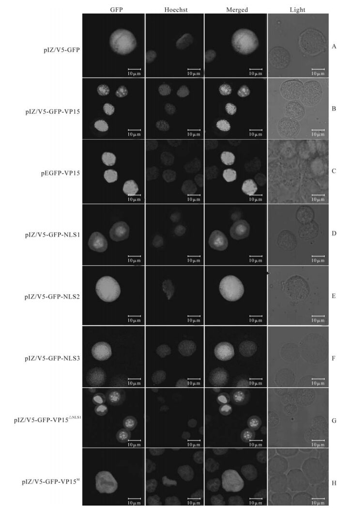
Figure 1. Functional analysis of the NLSs of VP15 in cells, demonstrated by confocal microscopy. The plasmids used for transfection are labeled in the left of the picture. The first column indicates the green fluorescence of GFP; the second column indicates the blue fluorescence of the nucleus stained by Hoechst 33258; the third column indicates the merged image of the first and second columns; and the fourth column indicates the cells observed with transmitted light. Bar=10μm.
In eukaryotic cells, molecular transport between the nucleus and cytoplasm occurs through nuclear pore complexes (NPCs). Small molecules ( < 9 nm in size) can diffuse through NPCs, but larger molecules must be actively transported (6). Active transport through NPCs is mediated by receptor proteins (also known as importins, exportins and transportins, or karyopherins), which interact with localization signals on cargo molecules, RanGTP, and proteins of the NPC (nucleoporins or Nups) (6). VP15 of WSSV is a major nucleocapsid protein of 14 kDa, and its theoretical molecular size as a monomer is below the NPC restriction, allowing it to diffuse passively through the NPC. In this study, however, we demonstrated that VP15 entered the nucleus through signal-mediated transport, implying that VP15 is not present in cells as a monomer. Our result is in agreement with a previous report showing that VP15 can form homomultimers when expressed in bacteria (20).
VP15 needs three NLSs to function efficiently as a nuclear localization protein. Functional cooperation between multiple NLSs in one, or even two proteins, occurs in some nuclear localization proteins. For example, a basic leucine zipper protein, HBZ, of the human T-cell leukemia virus type Ⅰ (HTLV-Ⅰ), is a nuclear localization protein whose activity is mediated by three distinct nuclear localization signals. Tran-siently expressed HBZ also forms nuclear speckles which have been proven to co-localize with cellular heterochromatin (5). In our study, the full-length VP15, as well as the truncated VP15, VP15ΔNLS1 and NLS1, all contained basic residues but in decreasing numbers. The efficiency of nuclear translocation and the density of nuclear speckles formed by these three sequences corresponded with their number of basic residues. Considering that VP15 is a core nucleo-capsid protein and WSSV replicates in the nucleus, the multiple NLSs of VP15 are necessary not only for its function as a nuclear transport protein, but also for DNA-binding in the nucleus.
The process of virus entry into the nucleus varies between different viruses (19). Influenza and adeno-viruses undergo extensive disassembly prior to genome import; herpes viruses release their genome into the nucleus without immediate capsid disassembly. Polyoma virus, parvovirus, and lentivirus preintegration com-plexes are thought to enter in intact form, whereas the corresponding complexes of onco-retroviruses have to wait for mitosis, because they cannot infect inter-phase nuclei. The nucleocapsid of WSSV measures about 300 × 70 nm and cannot pass the NPCs even in its longitudinal orientation. VP664 and VP15 are two major nucleocapsid proteins that maintain WSSV nucleocapsid structure and should play key roles in transporting the WSSV genome to the nucleus, espe-cially during its binding to nuclear receptors. We have now proved that VP15 has the ability to localize itself to the nucleus, through NLS-mediated transport in vitro. VP664 is the largest viral protein found to date (6077 aa). Whether the NLSs of VP664 are functional signals, and whether VP664 cooperates with VP15 during entry of the WSSV genome to the nucleus, are interesting questions that require further investigation.


 DownLoad: