


-
Foot-and-mouth (FMD) is an acute, highly contagious infection and febrile disease of ungulates induced by the Foot-and-mouth virus (FMDV). The highly contagious nature of the disease and severe mortality in young stock, the loss of yield and the significant suffering coupled with the major constraint to international trade in livestock products are the ultimate reasons for controlling FMDV[10] FMD is endemic and at a high prevalence in many countries in Africa, the Middle East and Asia and is also present in parts of South America. Animal vaccination strategies are commonly applied in these regions to control the disease. Vaccination is carried out after swift assessment of the serotype involved and advice is given on the suitability of available vaccines for disease control and any delay in laboratory testing can hinder the effectiveness of disease control measures[10]. In China, outbreaks of serotype A FMDV occurred in early 2009 and a 24-h stamping-out policy, in which farm livestock showing clinical signs of FMD are condemned, has been employed during outbreaks. A fast and sensitive diagnosis that can clearly distinguish different serotypes of FMDV from other disease that provoke some or all symptoms similar to FMDV is critical in the case of a suspected outbreak. The quick identification of the serotype of FMEV is essential for an emergency vaccination and for tracking the origin of an outbreak. Additionally, a fast and sensitive diagnostic method also has economic benefits for the detection of FMDV free live-stock and meat and for international animal trade.
In China, routine diagnosis of FMD is carried out at the National Foot-and-mouth Disease Reference Laboratory of China (NFMDRLC) at Lanzhou Veterinary Research Institute (LVRI). The laboratory routinely receives tissue samples, mainly epithelial material, for FMDV diagnosis and serotyping. Reverse-hemagglutination-inhibition (RHAI) has been success fully used for many years in detecting and typing FMDV in epithelial tissue samples. Recently, indirect-sandwich ELISA[3] and RT-PCR[17, 18] have been widely employed, conferring higher sensitivity and specificity. However, these diagnostic methods can only be done in centralized laboratories and have limited applications for on-site detection of viruses because they require highly trained laboratory personnel, stable reagents, expensive equipment and multi-step sample handling and preparation procedures. Immuno-chromatographic assays, also known as lateral flow assays or strip assays, has been available for some time. Currently, it has been widely applied as one of the new and emerging lab-on-site technologies to improve laboratory and on-site detection of viruses in animals and animal products. This technique is based on an immunochromatographic procedure that utilizes antigen-antibody properties in a novel manner and provides rapid detection of analyte[7, 11, 19]. It can be easily performed by unskilled personnel and the result could be obtained within 15-20 min. The easy appli-cation of test strip for on-site diagnosis in the case of a suspected disease outbreak would circumvent problems associated with the transportation of samples to the laboratory and would speed up diagnosis in emergency situations. Several rapid chromatographic strip tests or lateral flow devices (LFD) for the pen-side diagnosis of FMD have previously been reported [4, 5, 15, 16]. However, a specific immunochromatographic strip test for typing FMDV serotypes A has not yet been developed. In present report, we describe the selection of a pair of high-specificity anti-serotype A FMDV polyclonal antibodies to facilitate pen-side diagnosis of serotype-specific FMDV antigens and the development and validation of a rapid and sensitive sandwich immuno-chromatographic assay for FMDV serotype A detection.
HTML
-
Agar gel immunodiffusion (AGID) antigens of FMDV type A, the cell culture-adapted FMDV strain A/CHA/09 and guinea-pig antisera (the source of the capture antibody) to A/AV88(L) (originating from Russia in 1958) were prepared at the National Foot-and-mouth Disease Reference Laboratory of China (NFMDRLC) at Lanzhou Veterinary Research Institute (LVRI). Gold chloride (HAuCl4), bovine serum albumin (BSA) (fraction V), and goat Anti-guinea-pig antibody were obtained from Sigma (St. Louis, MO, USA). The sample pad, the conjugate release pad, the nitrocellulose membrane, and the absorbent pad were obtained from Millipore (Bedford, MA. USA). Protein-A Sepharose colum was obtained from GE(.USA).
-
680 vesicular epithelia and vesicular fluid samples that were collected clinically from suspected FMD cases around the Chinese endemic regions and submitted to the NFMDRLC were used to evaluate the diagnostic sensitivity and specificity of the LFI. These samples were collected at different geographical origins and showed antigenic and molecular variations within each of the FMDV serotypes and had previously been identified through virus isolate, RIHA, ELISA and RT-PCR/nucleotide sequencing. No FMDV was detected in 285 of the samples and these were classified as the no virus detected (NVD) group. 167 negative control samples were collected from healthy and FMDV free animals. Samples from FMDV type A(n=62) O (n=33), Asia 1(n=127), C (n=3) and swine vesicular disease virus (SVDV, n=1), Vesicular stomatiti svirus(VSV, n=1) and vesicular exanthema of swine virus(VESV, n=1) which were stored at the NFMDRLC were used.
-
As previously described, the cell culture-adapted strain of FMDV type A/CHA/09 was propagated in a BHK-21 cell line and inactivated using binaryethy-leneimine (BEI), the 146s antigen was then purified through CsCl density gradient centrifugation[1]. To prepare the antiserum, 2 week old Chinese white rabbits were intramuscularly inoculated with inactivated 146s antigen of virus A/CHA/09 emulsified in Freund's complete adjuvant. Rabbits were subsequently injected three times with the 146s antigen mixed with Freund's incomplete adjuvant at two-week intervals. One week after the last injection, specific anti FMDV-A antibody titres in the immunized rabbits were evaluated with an AGID test using commercial-antigens as recommended by the manufacturer[14]. Rabbits with high titer of specific antibodies were sacrificed and sera were pooled. Serum immuno-globulin was precipitated by adding 33% ammonium sulfate and centrifuging at 3 000r/min for 20minutes after 2 h incubation at 4℃. The total IgGs were purified through a Protein-A Sepharose chromato-graphy column (Amicon, Beverly, MA) according to the manufacture's instructions. The flow through antibody solution was subsequently concentrated by ultrafiltration (Amicon, Beverly, MA), quantified by the Bradford method, and stored at -20℃. Anti-FMDV guinea pig antisera were prepared by immunizing (3-5months old, weight 500g) by injection of suckling-mouse adapted strains A/AV88(L) which had been prepared by LVRI laboratory. The preparation, purification and quantification of guinea pig IgGs were performed with the methods described above.
-
Commercial colloidal gold beads were first treated prior to conjugation according to the following procedures[6]. 0.01% (w/v) HAuCl4 was heated to boiling and Trisodium citrate (2mL of a 1% solution) was rapidly added to the mixture, which was boiled for an additional 5min.The suspension color was turned from black to red. The colloidal gold was passed through a 0.45µm micropore filter and was observed by a CM100 transmission electron microscope (TEM) (Philips Co., The Netherlands).
Polyclonal antibody required for stabilizing colloidal gold particles was prepared as described before[6]. The UV absorptions of the colloidal gold and antibody–colloidal gold solutions were monitored immediately after preparation at λ200–750 nm. The pH of the colloidal gold solution was adjusted to 8 by addition of 0.02 mol/L K2CO3 before the polyclonal antibody was added. Capture antibodies were added drop-wise to the colloidal gold solution The solution was centrifuged to remove unconjugated antibody at 12 000×g for 30 minutes at 4℃. The pellet was then re-dispersed in 0.01mol/L phosphate-buffered saline (PBS, pH adjusted to 7.4) containing 1% bovine serum albumin (BSA) and stored at 4℃.
-
The immunochromatographic test strip was prepared as follows: goat anti-guinea pig IgG (0.5mg/mL, Sigma) and FMDV A serotype IgG (1mg/mL in 0.01mol/L PBS, pH7.4) were separately sprayed onto a NC membrane to form a control line and a test line; the membrane was then dried for 1h at 37℃ and blocked by incubation with 2% (w/v) BSA (in 0.01mol/L PBS, pH7.4) for 30min at RT. The membrane was washed with water. The FMDV A serotype IgG coated colloidal gold conjugation was deposited onto a glass fiber pad at 10 µL/cm, using air-brush equipment (Bio-dot XYZ 3050, USA) overlapping with the nitrocellulose membrane. Finally, the absorption pad and the sample pad were adhered onto the plate to complete the fabrication of the device. The plate was cut into 2.5mm wide strips (BiodotCM4000, USA) and then the test strip for detecting FMDV serotypes A was sealed in a plastic bag and stored at 4℃ with desiccant gel until use.
-
To detect the lowest detection limit of the strip test, add 10µL of the standard 146s FMDV A antigen from a culture solution (150ng/mL) to 190 mL sample buffer (0.01mol/L PBS, pH 7.4), then do 2-fold serial dilution of this soulution with sample buffer up to eight dilutions. 100µL aliquot of each dilution (from 1:20 to 1:1 280) was subjected to the immuno-chromatographic test strip for the test.
-
To determine the cross-reactivity of the test strip, FMDV serotypes O, A, Asia 1 and C and swine vesicular disease (SVD), vesicular stomatitis (VS) and vesicular exanthema of swine (VES) strongly positive clinical samples were tested with the test strip. 20 µL of 10 times diluted epithelial sample suspension was added into the serotype A-specific test strip and the results were observed within 20 min after the sample being applied.
-
Each epithelial sample was suspended in 0.04 mol/L phosphate buffer (pH 7.2-7.6) containing antibiotics and stored at –80℃[3]. The virus serotype of each clinical sample and the suckling-mouse-adapted sample had been determined by an indirect sandwich ELISA from NFMDRLC. Furthermore, the serotype of each sample had been verified by the virus isolation and RT-PCR tests (data not shown). The results of the indirect sandwich ELISA and the type A test strip were compared.
Materials
Test samples
Preparation and affinity purification of antibodies
Preparation of the colloidal gold conjugated probe
Development of the immunochromatographic test strip
Sensitivity test of the test strip
Specificity test of the test strip
Field test of the test strip
-
The titer of the rabbit antisera to homologous FMDV type A was 1:16 and to heterologous FMDV types (include O, and Asia 1) was < 1:2 as determined by AGID, indicating no cross-reactivity with other serotypes of FMV. The specificity and sensitivity of the guinea pig antisera was verified in a complement fixation (CF) test (data not shown) by NFMDRLC. The final concentration of the purified rabbit antibodies (detection antibody) to the FMDV type A/CHA/09 vaccine strains was 1.8 mg/mL and the concentration of the purified guinea pig antibodies (capture antibody) is 2.0 mg/mL.
The infective FMDV-particle has a sedimentation constant of 146S, and comprises one molecule of ssRNA and 60 copies of each of the four polypeptides VP1, VP2, VP 3 and VP4, and is considered responsible for inducing resistance against the infection. the I46S particles of the seven serotypes of produce specific neutralizing antibody against FMDV[2].Thereforce, 146s was used as immunogen in the test.
In a traditional sandwich method assay, antibodies are usually prepared by immunizing different animal species with the same antigen. However, in this study, the test strip adopts two FMDV virus strains of polyclonal antibodies as the capture and test antibodies; these are most representative of the epidemic FMDV strains in China and the antibodies generated by them are expected to react well with a wide range of clinical samples. Thus, tt will increase the dominant epitope recognized, extend the screening range of the spectrotype of homotype strains, improve the accuracy and sensitivity of this method and reduce the omission factor.
In common with the antigen diagnostic, MAbs show higher specificity and sensitivity in an immunoassay than. PAbs. However, very few highly specific MAbs that recognize wide range of FMDV serotype A have been generated and cocktails of FMDV serotype specific MAbs may require the development of a highly serotype specific immunoassay[8, 12, 13, 15-17]Thus, the coupling of PAbs produced with two different FMDV serotype A stains in rabbits and guinea pigs could produce a considerably highly specific assay for FMDV detection as being demonstrated in this study.
-
Transmission electron microscopy (TEM) images show that colloidal gold particles were well-dispersed in water and form a clear colloidal solution (Fig. 1). The average diameter of the colloidal gold particles was 40 ±1.5 nm. The colloidal gold particles were in a monodispersional state with a narrow diameter distri-bution, which provided the best basis for preparation of the test strip. Colloidal gold particles showed the largest optical absorption with a peak at 525 nm the largest optical absorption in UV-Vis spectroscopy (Fig. 2). The visible spectrum of the antibody-colloidal gold conjugate showed a maximum absorbance at 530 nm. By comparing the absorption at 530 nm, the optimal pH for coating antibody to colloidal gold particles was found to be 8.0. The visible spectrum of the antibody– colloidal gold conjugate showed a maximum absorbance at 530 nm. As the absorbance from the tyrosine and tryptophane residues in the IgG, it was observed a peak at approximately 280 nm. However, as centrifugation and removal of the unconjugated antibody molecules from the solution, the intensity of the surface resonance band and the protein absorbance band at 280 nm was decreased abruptly.
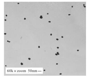
Figure 1. TEM images showing the size distribution of particles.
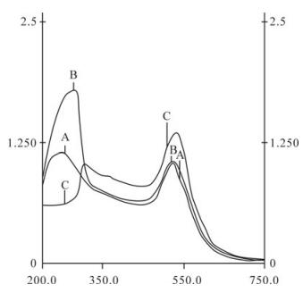
Figure 2. UV-vis spectra of colloidal gold and antibody-colloidal gold conjugates. Curve A: Colloidal gold solution. Curve B: Anti-FMDV type A immediately after addition of antibody to gold solution. Curve C: Anti-FMDV type A after centrifugation to remove the unbound antibodies.
-
In the experiment 0.05mg/mL of FMDV A serotype antibodies were confirmed to be the minimum amount required for stabilizing colloidal gold solution. However, the 0.06 mg/mL(20% greater) of PAbs for the con-jugation was considered as the selected PAbs concentration because this greater concentration resulted in better stabilization and more extensive of colloidal gold.
The optimum nitrocellulose membrane blocking agent was one that produced a short reaction time and a light-pink back ground color. Based on the speed at which antibody-colloidal gold conjugates moved and the background color of nitrocellulose membrane after reaction, 2.0% (w/v) BSA was found to be the optimal blocking buffer.
Conjugated with colloidal gold sprayed at 10 μL/cm on the gold pad, goat anti-guinea pig IgG and FMDV A serotype IgG at 1.0 μL/cm on nitrocellulose membrane at the control and test line gave the best assay result, yielding the strongest band and not obtaining false-positive results.
-
To test a sample, the test sample was mixed with an equal volume of sample buffer and loaded on the serotype A-specific test strip. After 15 min sample application, the result was observed and interpreted. As shown in Fig. 3, the appearance of one or two red lines were used to judge the negative and positive results of a single test strip. A positive result (+) is obtained if two red lines appear in both control and test lines and a negative result (–) is recorded if only a single red line appeared in the place of control line. The result will be inconclusive (+/–) if a weak red color line is seen in the test regions[9].
Figure 3. The schematic of LFI test results. The results of single immunochromatography test strip. The positive result was evaluated by two red lines in the control and test region, negative results is judged by the appearance of only a single color line in the control line. An inconclusive result is obtained by the appearance of a weak line in the test region and a red color line in the control region. The test is invalid if no line is present in the control region.
-
The sensitivity of this test strip was determined by assaying serial samples of various concentrations of FMDV A 146s antigen (750, 375, 186, 94, 47, 23, 11 ng/mL). It was determined that the lower detection limit of the test strip for FMDV A 146s antigen was 15µg/mL when the results was read at 10 min after sample loading (Fig. 4). It was also observed that the red line of the test tape will develop lighter when a positive specimen was diluted down with a dilution buffer and tested in a test strip, indicating that the specific detection of this test strip is virus concentration dependent.
Figure 4. Immunochromatographic assay of FMDV serotype A. A series of dilutions (1:20-1:1280) mL) of type A FMDV 146s antigen (150ng/mL) was prepared in sample buffer. The lowest detection limit of A 146s antigen was the titer of 1:1280 with a concentration of 0.11ng/mL of viral particles.
-
As shown in Fig 5, only FMDV serotypes A samples showed positive at 1:20–1:80 dilutions on the test strip. Although a cross-reaction was seen with three Asia Ⅰ FMDV positive samples at 1:40 dilution, other clinical samples containing FMDV serotype O, Asia 1, C, SVDV, VES and VS show clear negative results at less than 1:20 dilutions. Therefore, the A test strip has high specificity and will not show crossreaction with other type of FMDV at a 1:80 or higher dilutions.
Figure 5. Cross-reactivity assay of type A test strip. Samples containing FMDV serotype A, O, Asia 1, C and SVDV, VS, VES were applied to type A test strip, while only FMDV A sample produced a red band in the test line. Non-specific binding was not visualized in the absence of A type of FMDV antigen.
These results suggest that it is important to choose an optimal dilution for testing each sample before a final conclusion is made. The positive results can be determined with 1:20 dilution of samples for the majority of FMDV serotype A clinical samples collected at one animal site in the field. However, a few positive samples were only determined as positive at a dilution of 1:80 (data not shown). A weak cross reactivity was observed in some samples containing high concen-tration of Asia Ⅰ viruses at a dilution less than, 1:80.This kind of cross reactivity may be due to the close phylogenetic relationship between these virus types.
-
A total of 680 clinical samples of different suspected FMDV animals collected on-site from various regions in China have been tested with the type A strip test and the indirect-sandwich ELISA in parallel in this study. As summarized in Table. 1-2, the sensitivity of the test strip (88.7%) is as sensitive as indirect-sandwich ELISA (88.7%). Although the cross-reactivity of this LFI for SAT 1 and 3 were not tested due to the restriction of governmental policy in which SAT 1 and 3 type FMD viruses ware strictly forbidden to be used in the Chinese laboratories, this strip test has detected no other FMDV serotypes but Serotype A. All the samples of type O, Asia 1 and C of FMDV, SVDV, VSV, VESV and NVD produced negative results on the test strip. None of negative samples (n=167) obtained from healthy animals were positive in both the ELISA and the test strip. Positives were observed in only a few type O and Asia 1 samples with the type A test strip. In comparison to the specificity of 98.7% in the indirect-sandwich ELISA, the specificity of 98.2% was obtained with these samples in the type A strip test, suggesting the specificity of FMDV serotype A immunochromatographic test strip are slight lower than those of ELISA for the detection of serotype A FMDV clinical samples.

Table 1. Sensitivity of Test strip in comparison with FMDV Indirect ELISA

Table 2. Specificity of Test strip in comparison with FMDV Indirect ELISA
In this study, the test trip-A could detect most of serotype A FMDV strains isolated in China, and only 11 false positive from Chinese isolates of FMDV serotype O, Asia 1, C were recorded; no FMDV isolates from other sources have been evaluated. Further work for evaluating the performance of the test strip with clinical samples from other regions where other FMDV serotypes are prevalent in the world are the logical next step. This type of colloidal gold assay is commonly used to provide qualitative or semi-quantitative data. The identification and successful application of serotype A test strip for rapid diagnosis of Serotyp A FMDV on-site provides invaluble information for the development of a highly sensitive, specific and quantitative immnochromatographic assay using special tracer molecules such as fluorescent or magnetic particles with specialized reader devices in the future.
Characterization of the rabbit and guinea-pig antibodies
Characterization of the colloidal gold probe
Optimization of the immunochromatographic test strip
Analysis of the results of the test strip
Sensitivity of the test strip
Specificity test of the test strip
Field testing
-
In this study, the development of a simple and rapid immunochromatographic strip test for detection of FMDV of A in specimens from animal vesicular and epithelial fluids has been described. High specificity and ood correlation with ELISA results for detecting A serotype of FMDV have been demonstrated with this test with various clinical samples from the fields. The results can be obtained within 15-20 min after sample received and this test is much faster than current ELISA or RT-PCR tests in which minimum of 4h is required. Recently, several devastating outbreaks of A of FMD have been reported in a few provinces in China. This fast assay will be an invaluable tool for FMDV vaccine selection and in epidemiological studies. This test will also become a very useful test for FMD control programmers who work in FMD endemic regions of undeveloped countries where diagnostic facilities are limited.


 DownLoad: