2012 Vol.27(3)
|
Developments of Subunit and VLP Vaccines Against Influenza A Virus*
2012, 27(3): 145 doi: 10.1007/s12250-012-3241-1
Received: 17 January 2012
Accepted: 11 May 2012

Influenza virus is a continuous and severe global threat to mankind. The continuously re-emerging disease gives rise to thousands of deaths and enormous economic losses each year, which emphasizes the urgency and necessity to develop high-quality influenza vaccines in a safer, more efficient and economic way. The influenza subunit and VLP vaccines, taking the advantage of recombinant DNA technologies and expression system platforms, can be produced in such an ideal way. This review summarized the recent advancements in the research and development of influenza subunit and VLP vaccines based on the recombinant expression of hemagglutinin antigen (HA), neuraminidase antigen (NA), Matrix 2 protein (M2) and nucleocapsid protein (NP). It would help to get insight into the current stage of influenza vaccines, and suggest the future design and development of novel influenza vaccines.

Complete Genome Sequence Analysis of Duck Circovirus Strains from Cherry Valley Duck*
2012, 27(3): 154 doi: 10.1007/s12250-012-3214-4
Received: 25 July 2012
Accepted: 02 May 2012
To investigate molecular epidemiology of DuCV in Cherry Valley ducks in China, the complete genomes of six DuCV strains, which were detected from Cherry Valley ducks in China between 2007 and 2008, were sequenced. Sequence and phylogenetic analysis were carried out to compare these six strains with another 27 DuCV strains from Mulard duck, Muscovy duck, Pekin ducks and Mule duck. The analysis showed that the six DuCV strains exhibited typical genetic features of the family of DuCV, such as a stem-loop structure, three major open reading frames (Rep, Cap and ORF3), four intergenic repeats and the conserved motifs for rolling circle replication and for the dNTP binding domain located in the Rep protein. Phylogenetic analysis of the nucleotide sequences of the complete genome and Cap gene of these strains together with those that have been previously published demonstrated two distinct DuCV genotypes. The DuCV strains with complete genomes containing 1988 and 1989 nucleotides clustered in genotype A, whereas the strains with complete genomes containing 1991, 1992, 1995 and 1996 nucleotides lay in genotype B. The six DuCV strains from Cherry Valley ducks were divided into the two groups. The results of the study provides some insight into the variation of DuCVs in Cherry Valley ducks.
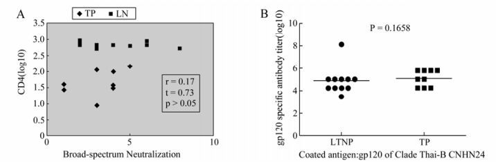
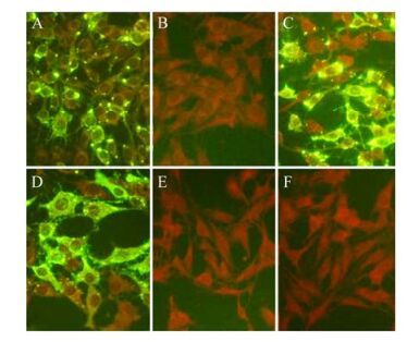
Similar Neutralizing Activity in the HIV-1 Infected Long Term Non-progressors(LTNPs) and Typical Progressors(TPs)*
2012, 27(3): 165 doi: 10.1007/s12250-012-3239-8
Received: 31 December 2011
Accepted: 08 May 2012
Neutralizing antibodies are considered to be an important protective parameter used in HIV-1 vaccine evaluation. However, the exact role that neutralizing antibodies plays in controlling the disease progression of HIV-1 infected peoples is still undetermined. In this paper, we compared the protective function of the neutralizing antibody response in the plasma from LTNP and TP against clade B and clade C pseudoviruses. No difference in the neutralizing activities between the plasma from LTNP and TP was found, which was consistent with the most recent reports. In addition, no correlations between the titer or breadth and CD4+ or viral load in HIV-1 infected individuals were found. The protective roles played by neutralizing antibodies in controlling disease progression of HIV-1 infected people need to be considered in a new viewpoint.
Preparation and Identification of Anti-rabies Virus Monoclonal Antibodies*
2012, 27(3): 172 doi: 10.1007/s12250-012-3242-0
Received: 08 February 2012
Accepted: 28 March 2012
To provide a foundation for the development of rapid and specific methods for the diagnosis of rabies virus infection, anti-rabies virus monoclonal antibodies were prepared and rabies virus nucleoprotein and human rabies virus vaccine strain (PV strain) were used as immunogens to immunize 6-8 week old female BALB/c mice. Spleen cells and SP2/0 myeloma cells were fused according to conventional methods: the monoclonal cell strains obtained were selected using the indirect immunofluorescence test; this was followed by preparation of monoclonal antibody ascitic fluid; and finally, systematic identification of subclass, specificity and sensitivity was carried out. Two high potency and specific monoclonal antibodies against rabies virus were obtained and named 3B12 and 4A12, with ascitic fluid titers of 1:8000 and 1:10000, respectively. Both belonged to the IgG2a subclass. These strains secrete potent, stable and specific anti-rabies virus monoclonal antibodies, which makes them well suited for the development of rabies diagnosis reagents.
Simultaneous Detection of Three Arboviruses Using a Triplex RT-PCR Enzyme Hybridization Assay *
2012, 27(3): 179 doi: 10.1007/s12250-012-3246-9
Received: 18 February 2012
Accepted: 07 May 2012
Arboviruses represent a serious problem to public health and agriculture worldwide. Fast, accurate identification of the viral agents of arbovirus-associated disease is essential for epidemiological surveillance and laboratory investigation. We developed a cost-effective, rapid, and highly sensitive one-step “triplex RT-PCR enzyme hybridization” assay for simultaneous detections of Japanese Encephallitis virus (JEV, Flaviviridae), Getah virus (GETV, Togaviridae), and Tahyna virus (TAHV, Bunyaviridae) using three pairs of primers to amplify three target sequences in one RT-PCR reaction. The analytical sensitivity of this assay was 1 PFU/mL for JEV, 10 PFU/mL for GETV, and 10 PFU/mL for TAHV. This assay is significantly more rapid and less expensive than the traditional serological detection and single RT-PCR reaction methods. When “triplex RT-PCR enzyme hybridization” was applied to 29 cerebrospinal fluid (CSF) samples that were JEV-positive by normal RT-PCR assay, all samples were strongly positive for JEV, but negative for GETV and TAHV, demonstrating a good sensitivity, specificity, and performance at CSF specimen detection.

Comparison of RFFIT Tests with Different Standard Sera and Testing Procedures*
2012, 27(3): 187 doi: 10.1007/s12250-012-3247-8
Received: 27 February 2012
Accepted: 07 May 2012
The World Health Organization (WHO) standard assay for determining antibody level is the rapid fluorescent focus inhibition test (RFFIT) and is used to determine the degree of immunity after vaccination against rabies. To compare the difference in RFFIT results between the laboratories of The National Institute of Infectious Disease in Japan (NIID) and the Chinese Centre for Disease Control (CCDC) as well the influence of the choice of standard serum (STD) for the detection, the two laboratories detection methods were simultaneously manipulated by RFFIT. The reference serums used in NIID and the WHO standard serum used in CCDC were compared in the same RFFIT detection to determine the titer of four sera samples C1, S1, S2 and S4 in parallel, and the titers of the detected sera samples were calculated using the standard formula for neutralizing antibody titer. No significant difference was found in RFFIT methods from the two laboratories and the RFFIT testing procedures of the two laboratories have good consistency. However, different titers were obtained with the tentative internal standard serum (TI-STD) produced by adjusting to 2.0 IU of WHO standard serum in NIID and the WHO STD. The titer determined with the TI-STD was higher than that determined with WHO STD, This difference appears to be significant and requires further investigation.
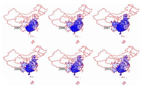
Molecular Characterization of Viral G Gene in Emerging and Re-emerging Areas of Rabies in China, 2007 to 2011 *
2012, 27(3): 194 doi: 10.1007/s12250-012-3248-7
Received: 27 February 2012
Accepted: 23 April 2012
In recent years (2007 to 2011), although the overall number of rabies cases in China has decreased, there is evidence of emerging or re-emerging cases in regions without previous rabies cases or with low incidence of rabies. To investigate the origin and the factors affecting the spread of rabies in China, specimens were collected from 2007 to 2011 from provinces with emerging and re-emerging cases and tested for the presence of the rabies virus. Positive specimens were combined with sequences from GenBank to perform comparisons of homology and functional sites, and to carry out phylogenetic analyses. Out of these regions, five provinces had 9 positive specimens from canine and cattle, and 34 canine or human specimens were obtained from previously high-incidence provinces. Complete sequences of G gene were obtained for these samples. Homology of the sequences of these 43 specimens was 87%-100% at the nucleotide level and 93.7% -100% at the amino acid level. These G gene sequences were combined with reference sequence from GenBank and used to construct a phylogenetic tree. The results showed that 43 specimens were all assigned to China clade I and clade II, with all specimens from emerging and re-emerging areas placed within clade I. Specimens isolated from Shanxi and Inner Mongolia in 2011 were distinct from previously-isolated local strains and had closer homology to strains from Hebei, Beijing and Tianjin whereas new isolates from Shanghai were tightly clustered with strains isolated in the 1990s. Finally, Shaanxi isolates were clustered with strains from adjacent Sichuan. Our results suggest that the rabies cases in emerging and re-emerging areas in China in the last 5 years are a consequence of the epidemic spreading from of neighboring provinces and regions experiencing a serious epidemic of rabies.

The Full-length Genome Analysis of a Street Rabies Virus Strain Isolated in Yunnan Province of China *
2012, 27(3): 204 doi: 10.1007/s12250-012-3251-z
Received: 13 March 2012
Accepted: 03 May 2012
The epidemic of rabies has rapidly increased and expanded in Yunnan province in recent years. In order to further analyze and understand the etiological reasons for the rapid expansion of rabies in Yunnan, a strain of rabies virus CYN1009D in Yunnan was isolated, and the complete genomic sequencing was carried out, and the bioimfomative analysis on genes/encoded proteins and phylogeny with reference to sequences in GenBank was performed. The complete genome of CYN1009D was 11923 nt in length and belonged to genotype I. The genes encoding different structural proteins were all conserved in their lengths, in comparison to other strains in China. The amino acid sequence was conserved at different antigen sites of NP, but the variation was detected at the secondary phosphorylation site of position 375; variations were also detected in the phosphorylation sites at positions 63-63 and 162 of PP; the sites playing important roles in virus synthesis, budding and viral morphology in MP were conserved; two glycosylation sites were detected at Asn37 and Asn319 in GP, the neutralizing antigen sites in GP were conserved; the initial amino acid of LP (ML) was different from that of most of the strains in China (MM); the variations in G-L region in the intergenic region were significant. The phylogenic tree showed that CYN1009D has a closer genetic relationship to the strains in Southeast Asia, indicating that prevention and control on rabies in borderland areas should be reinforced meanwhile efforts are made to control rabies in China.
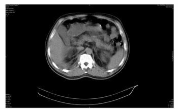
Hemorrhagic Fever with Renal Syndrome Associated with Acute Pancreatitis
2012, 27(3): 214 doi: 10.1007/s12250-012-3231-3
Received: 13 November 2011
Accepted: 16 April 2012
Hemorrhagic fever with renal syndrome (HFRS) is a systemic infectious disease caused by Hantaviruses and characterized by fevers, bleeding tendencies, gastrointestinal symptoms and renal failure. It encompasses a broad spectrum of clinical presentations, ranging from unapparent or mild illnesses to fulminant hemorrhagic processes. Among the various complications of HFRS, acute pancreatitis is a rare find. In this report, based on clinical data, laboratory and radiologic examination findings, we describe a clinical case, with HFRS from Dobrava virus, associated with acute pancreatitis. The patient was successfully treated by supportive management. Clinicians should be alert to the possibility of HFRS when examining patients with epidemiological data and symptoms of acute pancreatitis.