2010 Vol.25(6)
Detection of Bluetongue Virus Group-specific Antigen Using Monoclonal Antibody Based Sandwich ELISA
2010, 25(6): 390 doi: 10.1007/s12250-010-3160-y
Received: 21 June 2010
Accepted: 29 September 2010

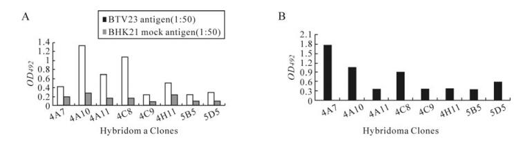
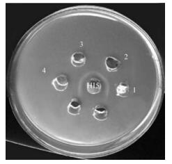
A monoclonal antibody (MAb) specific for the bluetongue virus (BTV) group specific antigen (VP7) was characterized for its reactivity with purified virus and recombinant BTV VP7 (rVP7) protein and its suitability for use in the sandwich ELISA. The MAb, designated as 5B5 was specific to VP7 and belongs to IgG2a subclass and was selected for the development of the sELISA in this study. The MAb had a titer of 1:25 with BTV and 1:2 with the rVP7 protein. The sELISA is based on capturing of BTV antigen with VP7 specific MAb followed by detection using BTV polyclonal antiserum raised in rabbits. The assay was evaluated with six cell culture adapted serotypes of BTV that have been isolated from India, 1, 2, 15, 17, 18 and 23. The assay could detect BTV antigen as early as day 8 in blood. It was also successfully applied for the detection of BTV group specific antigen in clinical samples of blood, washed RBCs, buffy coat and plasma. A total of 102 field samples from animals, suspected of being infected with BTV, were tested and 29.42% were positive. The blood samples were also amplified in cell culture which improved the sensitivity of the assay. Results confirmed that the sELISA is rapid and specific.
Expression of Rice Gall Dwarf Virus Outer Coat Protein Gene (S8) in Insect Cells
2010, 25(6): 401 doi: 10.1007/s12250-010-3152-y
Received: 04 June 2010
Accepted: 09 October 2010
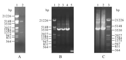
To obtain the P8 protein of Rice gall dwarf virus (RGDV) with biological activity, its outer coat protein gene S8 was expressed in Spodoptera frugiperda (Sf9) insect cells using the baculovirus expression system. The S8 gene was subcloned into the pFastBacTM1 vector, to produce the recombinant baculovirus transfer vector pFB-S8. After transformation, pFB-S8 was introduced into the competent cells (E. coli DH10Bac) containing a shuttle vector, Bacmid, generating the recombinant bacmid rbpFB-S8. After being infected by recombinant baculovirus rvpFB-S8 at different multiplicities of infection, Sf9 cells were collected at different times and analyzed by SDS-PAGE, Western blotting and immunofluorescence microscopy. The expression level of the P8 protein was highest between 48-72 h after transfection of Sf9 cells. Immunofluorescence microscopy showed that P8 protein of RGDV formed punctate structures in the cytoplasm of Sf9 cells.
Sequence Analysis of Attachment Gene of Lumpy Skin Disease and Sheep Poxviruses
2010, 25(6): 409 doi: 10.1007/s12250-010-3150-0
Received: 21 May 2010
Accepted: 29 September 2010
Egypt, protection of cattle against lumpy skin disease (LSD) was carried out using a sheep poxvirus (Kenyan strain) vaccination strategy. In the present study 15 skin nodules from LSD suspected cows and 5 scab samples from sheep pox (SP) suspected sheep were collected. Hyperimmune rabbit sera to Lumpy skin disease virus (LSDV)/Ismailyia88 strain and sheep pox virus (SPV)/ Kenyan vaccinal strain were prepared. The causative agent in the collected samples was identified using immunoflourescence (IF) and immunoperoxidase techniques. Of the 15 skin nodules suspected of LSD, 10 showed a positive reaction and 3 out of 5 skin scabs suspected of sheeppox were found to be positive. An antigenic correlation between field skin isolate of LSDV, tissue culture adapted LSDV/Ismailyia88 strain, field skin isolate of SPV and SPV/Kenyan vaccinal strain was studied using prepared hyperimmune sera. Also, nucleotide sequence of the PCR amplified attachment gene fragments of field skin isolate of LSDV, tissue culture adapted LSDV/Ismailyia88 strain, field skin isolate of SPV and SPV /Kenyan vaccinal strain were compared. The results revealed that the four used viruses were antigenically identical. Sequence analysis indicated that field skin LSDV isolate is more related to tissue culture adapted LSDV/Ismailyia88 strain than to vaccinal SPV/ Kenyan strain and the skin isolate of SPV is more closely related to field skin isolate of LSDV than to SPV/Kenyan vaccinal strain. Thus, further study should be applied on the advantage of a LSD vaccine prepared from LSDV in protection of cattle against LSD compared to the commonly used sheep pox vaccine.
Transcriptional Regulation by HSV-1 Induced HTRP via Acetylation System
2010, 25(6): 417 doi: 10.1007/s12250-010-3147-8
Received: 07 May 2010
Accepted: 28 July 2010
The protein HTRP (human transcription regulator protein) is encoded by the differential gene htrp and induced by Herpes simplex virus type 1 (HSV-1) infection in KMB-17 cells. HTRP was found to interact with SAP30 (mSin3A Association Protein), one of the components of co-repressor complex mSin3A, which is part of the deacetylation transfer enzyme HDAC. To reveal the biological significance of the interaction between HTRP and SAP30, real- time PCR and a dual-luciferase detecting system was used. The results indicate that HTRP could inhibit the transcription of a viral promoter, whose interaction with SAP30 synergistically affects transcriptional inhibition of the viral genes, and is related to HDAC enzyme activity. ChIP experiments demonstrate that HTRP could promote HDAC activity by increasing the deacetylation level of lysine 14 and lysine 9 in histone H3.
Reference Gene Selection for Normalization of PCR Analysis in Chicken Embryo Fibroblast Infected with H5N1 AIV
2010, 25(6): 425 doi: 10.1007/s12250-010-3114-4
Received: 08 November 2009
Accepted: 28 July 2010
Chicken embryo fibroblasts (CEFs) are among the most commonly used cells for the study of interactions between chicken hosts and H5N1 avian influenza virus (AIV). In this study, the expression of eleven housekeeping genes typically used for the normalization of quantitative real-time PCR (QPCR) analysis in mammals were compared in CEFs infected with H5N1 AIV to determine the most reliable reference genes in this system. CEFs cultured from 10-day-old SPF chicken embryos were infected with 100 TCID50 of H5N1 AIV and harvested at 3, 12, 24 and 30 hours post-infection. The expression levels of the eleven reference genes in infected and uninfected CEFs were determined by real-time PCR. Based on expression stability and expression levels, our data suggest that the ribosomal protein L4 (RPL4) and tyrosine 3-monooxygenase tryptophan 5-monooxygenase activation protein zeta polypeptide (YWHAZ) are the best reference genes to use in the study of host cell response to H5N1 AIV infection. However, for the study of replication levels of H5N1 AIV in CEFs, the β-actin gene (ACTB) and the ribosomal protein L4 (RPL4) gene are the best references.
Antiviral Activity of Recombinant Cyanovirin-N against HSV-1
2010, 25(6): 432 doi: 10.1007/s12250-010-3131-3
Received: 01 May 2010
Accepted: 30 July 2010
In this study, a standard strain of HSV-1 (strain SM44) was used to investigate the antiviral activity of the recombinant Cyanovirin-N (CV-N) against Herpes simplex virus type 1 (HSV-1) in vitro and in vivo. Cytopathic effect (CPE) and MTT assays were used to evaluate the effect of CV-N on HSV-1 in Vero cells. The number of copies of HSV-DNA was detected by real-time fluorescence quantitative PCR (FQ-PCR). The results showed that CV-N had a low cytotoxicity on Vero cells with a CC50 of 359.03±0.56 μg/mL, and that it could not directly inactivate HSV-1 infectivity. CV-N not only reduced the CPE of HSV-1 when added before or after viral infection, with a 50% inhibitory concentration (IC50) with 2.26 and 30.16μg/mL respectively, but it also decreased the copies of HSV-1 DNA in infected host cells. The encephalitis model for HSV-1 infection was conducted in Kunming mice, and treated with three dosages of CV-N ( 0.5, 5 & 10 mg/kg) which was administered intraperitoneally at 2h, 3d, 5d, 7d post infection. The duration for the appearance of symptoms of encephalitis and the survival days were recorded and brain tissue samples were obtained for pathological examination (HE staining). Compared with the untreated control group, in the 5mg/kg CV-N and 10mg/kg CV-N treated groups, the mice suffered light symptoms and the number of survival days were more than 9d and 14d respectively. HE staining also showed that in 5mg/kg CV-N and 10mg/kg CV-N treated groups, the brain cells did not show visible changes, except for a slight inflammation. Our results demonstrated that CV-N has pronounced antiviral activity against HSV-1 both in vitro and in vivo, and it would be a promising new candidate for anti-HSV therapeutics.
PrP 106-126 Altered PrP mRNA Gene Expression in Mouse Microglia BV-2 Cells
2010, 25(6): 440 doi: 10.1007/s12250-010-3134-z
Received: 09 April 2010
Accepted: 28 July 2010
Prion diseases are infectious and fatal neurodegenerative diseases. The pathogenic agent is an abnormal prion protein aggregate. Microglial activation in the centre nervous system is a characteristic feature of prion disease. In this study, we examined the effect of PrP 106-126 on PrP mRNA gene expression in Mouse microglia cells BV-2 by real-time quantitative PCR. PrP mRNA expression level was found to be significantly increased after 18 h exposure of BV-2 cells to PrP 106-126, with 3-fold increase after 18 h and 4.5-fold increase after 24 h and BV-2 cells proliferating occurred correspondingly. Our results provide the first in vitro evidence of the increase of PrP mRNA levels in microglial cells exposed to PrP 106-126, and indicate that microglial cells might play a critical role in prion pathogenesis.
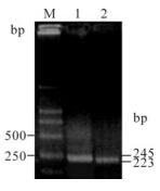
Antiviral Activity of the Effective Monomers from Folium Isatidis Against Influenza Virus in Vivo
2010, 25(6): 445 doi: 10.1007/s12250-010-3142-0
Received: 06 April 2010
Accepted: 15 September 2010
In order to evaluate the anti-influenza virus activity of the effective monomer from Folium Isatidis (FI) in vivo, we established mice model with viral pneumonia and divided them into 3 different dose groups, then observed their lung indexes, pulmonary pathological changes, pulmonary virus hemagglitination titers, living time and death rates. The results showed that the monomer could reduce the pulmonary index from 2.64 to 1.93, 1.63 and 1.40 (P0.01) and decrease the hemagglitination titer from 1.15 to 0.84, 0.70 and 0.59 (P0.01). In addition, different groups of FI could significantly lessen the mortality rate from 100% to 30%, 25% and 15%, and prolong the living time from 5.1d to 6.5d, 8.4d and 8.9d respectively(P0.01). The high dose (75 mg/kg/d) has the similar effect with 100 mg/kg/d dose of virazole(P0.05), and more effective than 200 mg/kg/d dose of antiviral liquor (P0.05).