


2014年29卷4期
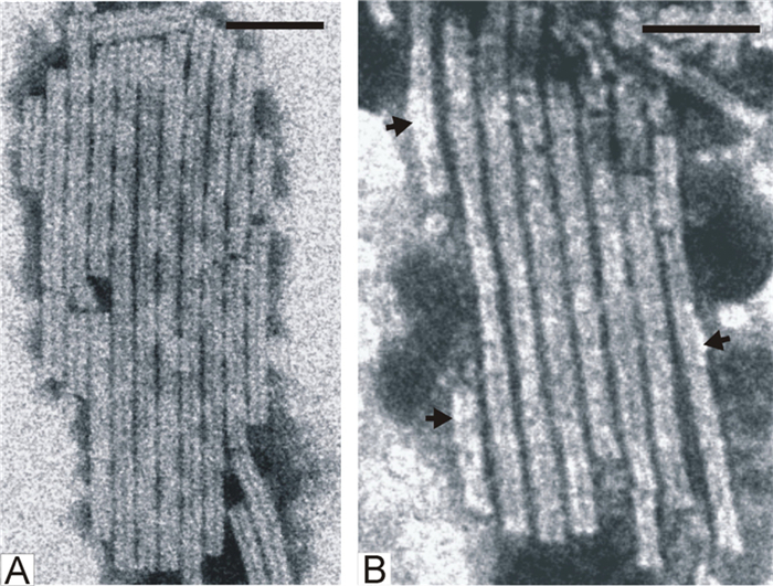
Labeling of virus particles facilitates the study of early events in virus infection.In this issue, Ilyushina and collogues have provided a new and effective approach to label infl uenza viruses. The Function-Spacer-Lipid (FSL) constructs labeling with fluorescent (fluo-Ad-DOPE) or biotin-labeled (biot-CMG2-DOPE) probes were gently inserted into the membrane of seven human/avian Infl uenza viruses, and each virion contained several thousand copies of lipid probes. Virus infectivity and viral affi nity to sialyl receptors were tested and found not much affected in vitro. This lipid labeling approach provides a useful tool in the studies of virus attachment, virus endocytosis, intracellular traffi cking, and more. The cover image shows the transmission electron microscope (TEM) image of influenza viruses (pseudo-colored in indigo), and one virion in the middle labeled with the lipid construct is illustrated (pseudo-colored in fluorescent green). (The original TEM image was provided by the courtesy of Dr. Ding Gao, from the Core Facility Center of Wuhan Institute of Virology.)
|
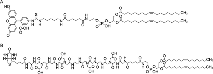
Labeling of influenza viruses with synthetic fluorescent and biotin-labeled lipids
2014, 29(4): 199 doi: 10.1007/s12250-014-3475-1

Direct labeling of virus particles is a powerful tool for the visualization of virus-cell interaction events. However, this technique involves the chemical modification of viral proteins that affects viral biological properties. Here we describe an alternative approach of influenza virus labeling that utilizes Function-Spacer-Lipid (FSL) constructs that can be gently inserted into the virus membrane. We assessed whether labeling with fluorescent (fluo-Ad-DOPE) or biotin-labeled (biot-CMG2-DOPE) probes has any deleterious effect on influenza virus hemagglutinin (HA) receptor specificity, neuraminidase (NA) activity, or replicative ability in vitro. Our data clearly show that neither construct significantly affected influenza virus infectivity or viral affinity to sialyl receptors. Neither construct influenced the NA activities of the influenza viruses tested, except the A/Puerto Rico/8/34 (H1N1) strain. Our data indicate that lipid labeling provides a powerful tool to analyze influenza virus infection in vitro.
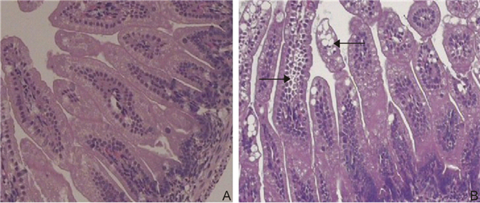
Involvement of aquaporins in a mouse model of rotavirus diarrhea
2014, 29(4): 211 doi: 10.1007/s12250-014-3469-z
Rotavirus diarrhea is a major worldwide cause of infantile gastroenteritis; however, the mechanism responsible for intestinal fluid loss remains unclear. Water transfer across the intestinal epithelial membrane seems to occur because of aquaporins (AQPs). Accumulating evidence indicates that alterations in AQPs may play an important role in pathogenesis. Here, we focus on changes in AQPs in a mouse model of rotavirus diarrhea. In the present study, 32 of 35 mice developed diarrhea and mild dehydration within 24 hours after infection with rotavirus strain SA11. Intestinal epithelial cells demonstrated cytoplasmic vacuolation, malaligned villi, and atrophy. AQP1 expression was significantly attenuated in the ileum and colon in comparison with controls; likewise, AQP4 and-8 protein expression were significantly decreased in the colon of rotavirus diarrhea-infected mice. In contrast, AQP3 protein expression was significantly increased in the colon of rotavirus-infected mice in comparison with controls. These results indicate that rotavirus diarrhea is associated with the downregulation of AQP1,-4, and-8 expression. Therefore, AQPs play an important role in rotavirus diarrhea.

Immunogenicity and protective efficacy of recombinant M2e. Hsp70c (Hsp70359-610) fusion protein against influenza virus infection in mice
2014, 29(4): 218 doi: 10.1007/s12250-014-3428-8
New strategies in vaccine development are urgently needed to combat emerging influenza viruses and to reduce the risk of pandemic disease surfacing. Being conserved, the M2e protein, is a potential candidate for universal vaccine development against influenza A viruses. Mycobacterium tuberculosis Hsp70 (mHsp70) is known to cultivate the function of immunogenic antigenpresenting cells, stimulate a strong cytotoxic T lymphocyte (CTL) response, and stop the induction of tolerance. Thus, in this study, a recombinant protein from the extracellular domain of influenza A virus matrix protein 2 (M2e), was fused to the C-terminus of Mycobacterium tuberculosis Hsp70 (Hsp70c), to generate a vaccine candidate. Humoral immune responses, IFN-γ-producing lymphocyte, and strong CTL activity were all induced to confirm the immunogenicity of M2e.Hsp70c (Hsp70359-610). And challenge tests showed protection against H1N1 and H9N2 strains in vaccinated groups. Finally these results demonstrates M2e.Hsp70c fusion protein can be a candidate for a universal influenza A vaccine.
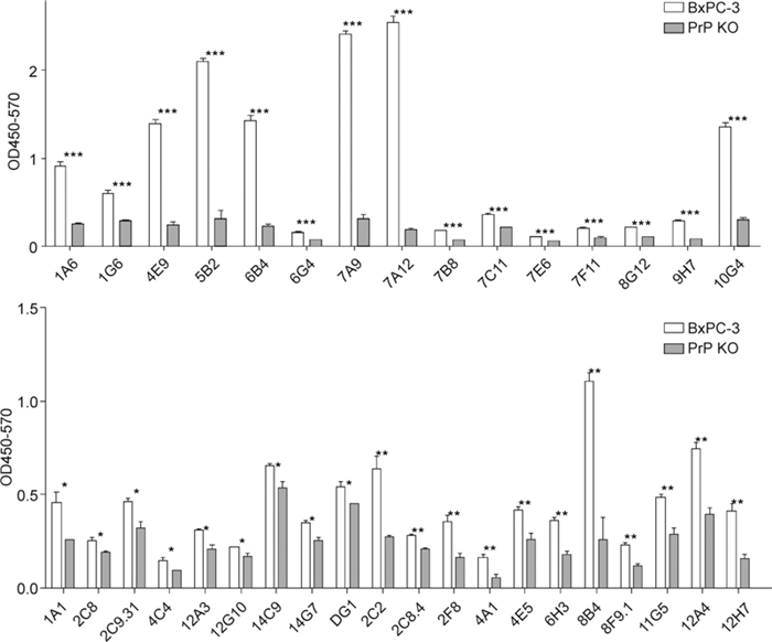
A panel of monoclonal antibodies against the prion protein proves that there is no prion protein in human pancreatic ductal epithelial cells
2014, 29(4): 228 doi: 10.1007/s12250-014-3480-4
Prion diseases are a group of neurodegenerative diseases that are fatal. The study of these unique diseases in China is hampered by a lack of resources. Amongst the most important resources for biological study are monoclonal antibodies. Here, we characterize a panel of monoclonal antibodies specific for cellular prion protein by enzyme-linked immunosorbent assay (ELISA), immunofluorescent staining, flow cytometry, and western blotting. We identify several antibodies that can be used for specific applications and we demonstrate that there is no prion protein expression in human pancreatic ductal epithelial cells (HPDC).

Prevalence and patterns of drug-resistance mutations among HIV-1 patients infected with CRF07_BC strains in Sichuan province, China
2014, 29(4): 237 doi: 10.1007/s12250-014-3487-x
Little information is available on the prevalence of drug-resistance mutations in patients harboring the human immunodeficiency virus type 1 (HIV-1) circulating recombinant form (CRF)07_BC variant in Sichuan, China. This study examined 375 plasma samples from patients with HIV-1 who were infected with the CRF07_BC strain, including 104 drug-naive participants and 271 in whom antiretroviral therapy (ART) had failed. Only one participant in the drug-naive group had a drug-resistance mutation (M46L), compared with 31.73% of those in whom ART had failed. Further analysis showed that 19.56% of strains contained mutations conferring resistance to nonnucleoside reverse transcriptase inhibitors (NNRTIs) alone, 0.74% were resistant to nucleoside reverse transcriptase inhibitors (NRTIs) alone, and 11.44% were dual-resistant to both NRTIs and NNRTIs. The most common mutation in the ART-failure group was M184V (35.88%), K103N (45.01%), Y181C (17.33%), and G190S/A (15.88%). The percentages of HIV-1 strains resistant to lamivudine, emtricitabine, efavirenz, etravirine, and nevirapine were 10.70%, 10.70%, 28.04%, 7.75%, and 26.20%, respectively. To explore site variants possibly related to drug resistance, variations in the ancestor/consensus CRF07_BC sequences from the therapy-naive and ART-failure groups were compared, and seven mutations at six positions were identified as being significantly differently distributed between the two groups (p<0.05). Detailed sequence data will provide information on CRF07_BC genetic characterizations, and improve our understanding of antiretroviral susceptibility and the evolution of drug-resistance mutations. This will be valuable in developing and implementing local public-health approaches for HIV drug-resistance prevention and treatment.

Association of swine influenza H1N1 pandemic virus (SIVH1N1p) with porcine respiratory disease complex in sows from commercial pig farms in Colombia
2014, 29(4): 242 doi: 10.1007/s12250-014-3471-5
Porcine respiratory disease complex (PRDC) is a serious health problem that mainly affects growing and finishing pigs. PRDC is caused by a combination of viral and bacterial agents, such as porcine reproductive and respiratory syndrome virus (PRRSV), swine influenza virus (SIV), Mycoplasma hyopneumoniae (Myh), Actinobacillus pleuropneumoniae (APP), Pasteurella multocida and Porcine circovirus 2 (PCV2). To characterize the specific role of swine influenza virus in PRDC presentation in Colombia, 11 farms from three major production regions in Colombia were examined in this study. Nasal swabs, bronchial lavage and lung tissue samples were obtained from animals displaying symptoms compatible with SIV. Isolation of SIV was performed in 9-day embryonated chicken eggs or Madin-Darby Canine Kidney (MDCK) cells. Positive isolates, identified via the hemagglutination inhibition test, were further analyzed using PCR. Overall, 7 of the 11 farms were positive for SIV. Notably, sequencing of the gene encoding the hemagglutinin (HA) protein led to grouping of strains into circulating viruses identified during the human outbreak of 2009, classified as pandemic H1N1-2009. Serum samples from 198 gilts and multiparous sows between 2008 and 2009 were obtained to determine antibody presence of APP, Myh, PCV2 and PRRSV in both SIV-H1N1p-negative and-positive farms, but higher levels were recorded for SIVH1N1p-positive farms. Odds ratio (OR) and P values revealed statistically significant differences (p<0.05) in PRDC presentation in gilts and multiparous sows of farms positive for SIV-H1N1p. Our findings indicate that positive farms have increased risk of PRDC presentation, in particular, PCV2, APP and Myh.
Effect of chitosan on tobacco mosaic virus (TMV) accumulation, hydrolase activity, and morphological abnormalities of the viral particles in leaves of N. tabacum L. cv. Samsun
2014, 29(4): 250 doi: 10.1007/s12250-014-3452-8
The effect of chitosan on the development of infection caused by Tobacco mosaic virus (TMV) in leaves of Nicotiana tabacum L. cv. Samsun has been studied. It was shown that the infectivity and viral coat protein content in leaves inoculated with a mixture of TMV (2 μg/mL) and chitosan (1 mg/mL) were lower in the early period of infection (3 days after inoculation), by 63% and 66% respectively, than in leaves inoculated with TMV only. Treatment of leaves with chitosan 24 h before inoculation with TMV also caused the antiviral effects, but these were less apparent than when the virus and polysaccharide were applied simultaneously. The inhibitory effects of the agent decreased as the infection progressed. Inoculation of leaves with TMV together with chitosan considerably enhanced the activity of hydrolases (proteases, RNases) in the leaves, in comparison with leaves inoculated with TMV alone. Electron microscope assays of phosphotungstic acid (PTA)-stained suspensions from infected tobacco leaves showed that, in addition to the normal TMV particles (18 nm in diameter, 300 nm long), these suspensions contained abnormal (swollen, "thin" and "short") virions. The highest number of abnormal virions was found in suspensions from leaves inoculated with a mixture of TMV and chitosan. Immuno-electron microscopy showed that "thin" virus particles, in contrast to the particles of normal diameter, lost the ability to bind to specific antiserum. It seems that the chitosan-induced activation of hydrolases stimulates the intracellular degradation of TMV particles and hence hydrolase activation may be considered to be one of the polysaccharide-mediated cellular defense mechanisms that limit virus accumulation in cells.

A novel mitovirus from Buergenerula spartinae infecting the invasive species Spartina alterniflora
2014, 29(4): 257 doi: 10.1007/s12250-014-3470-6
ISSN 1674-0769
EISSN 1995-820X
CN 42-1760/Q
主编: 石正丽
影响因子: 5.5*
*源于2022年JCR


- [01/11]《中国病毒学(英文)》期刊编辑部招聘启事
- [05/07]Q1区!VS最新影响因子5.5!
- [22/02]2022年VS高被引论文奖发布
- [21/10]第十届新生病毒性疾病控制学术研讨会 | 第一轮通知
- [09/09]肝癌细胞中CK1α上调IFNAR1的表达,从而促进I型IFN抑制HBV复制
- [09/09]一种新的干扰素诱导的长非编码RNA ZAP-IT1阻断寨卡病毒在A549细胞中的复制
- [09/09]首发精神分裂症中,驯化的人内源性逆转录病毒W家族包膜蛋白通过降低5-HT4受体的水平激活SK2
- [09/09]发热伴血小板减少综合征病毒L蛋白功能域和保守残基研究为理解病毒RNA转录/复制机制提供新思路
- [09/09]亲环素A结合AKT1并通过介导AKT/mTOR/NF-κB正反馈环路的激活促进EB病毒的致瘤作用 | VS推荐
- [09/09]转录组分析显示克里米亚刚果出血热病毒调控的关键细胞过程及III型干扰素的抗病毒作用 | VS推荐

