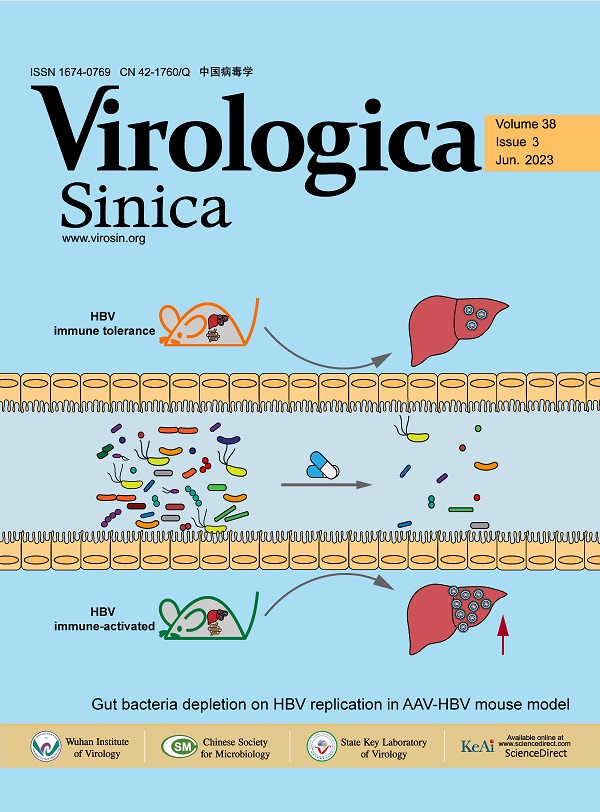
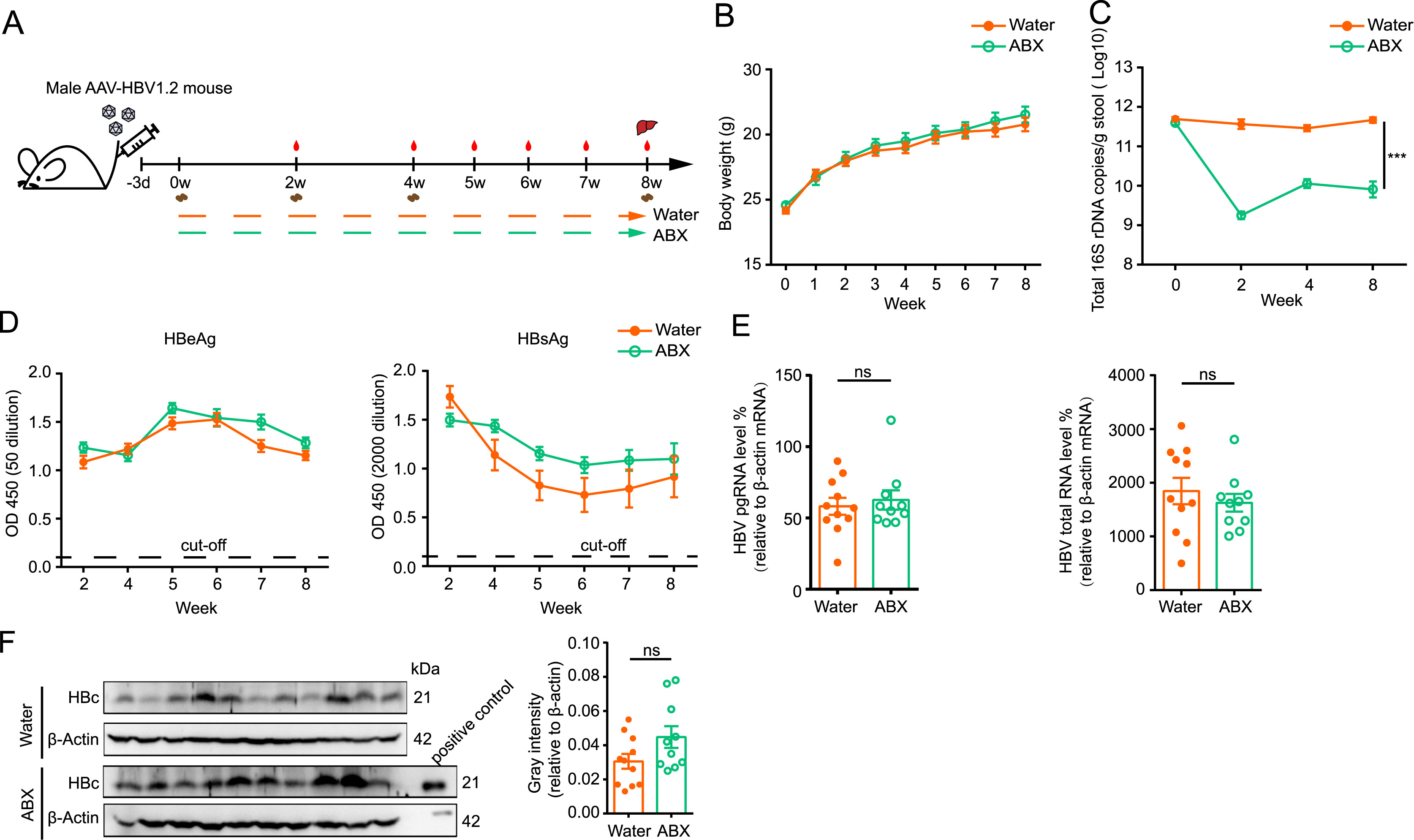
Commensal microbiota is closely related to hepatitis B virus (HBV) infection. However, existing studies have not elucidated the effect of gut bacteria on HBV immune tolerant phase yet. In this issue, Bu et al. altered gut bacteria richness and diversity using broad spectrum antibiotic mixtures and generated HBV immune tolerance mouse model via recombinant adeno-associated virus-HBV transduction to investigate the effects of gut bacteria on HBV replication. Their study provides new thoughts for elucidating the correlation between gut bacteria dysbiosis caused by antibiotic abuse and clinical chronic HBV infection. The cover image describes that antibiotic-induced gut bacteria depletion fails to affect HBV replication in HBV immune tolerance mouse model but contributes to increase HBV replication after immune activation (kindly designed and provided by Yuchen Xia). See page 335–343 for details.
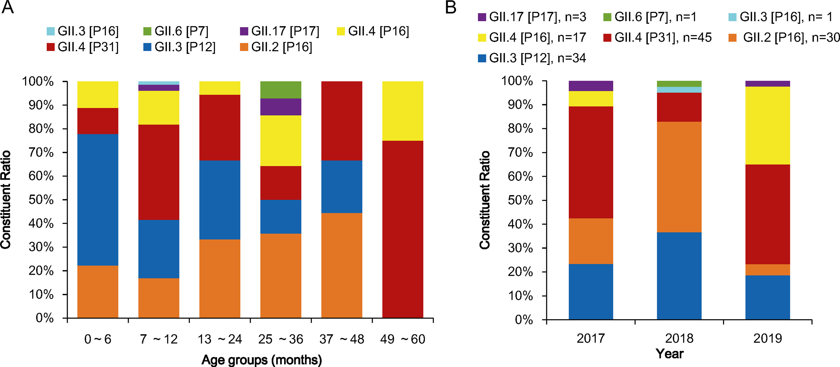
诺如病毒(NoV)是病毒性急性胃肠炎(AGE)的重要病因。为了解湖北省儿童NoV的流行病学特征和遗传多样性,对2017年1月至2019年12月在AGE监测下采集的1216份≤5岁儿童粪便样本进行了分析。结果显示,NoV占AGE病例的14.64%,其中7-12月龄儿童检出率最高(19.76%)。男女感染率差异有统计学意义(χ2=8.108,P=0.004)。RdRp和VP1序列遗传分析显示,NoV GII基因型为GII.4 Sydney[P31](34.35%)、GII.3[P12](25.95%)、GII.2[P16](22.90%)、GII.4 Sydney[P16](12.98%)、GII.17[P17](2.29%)、GII.6[P7]和GII.3[P16](各为0.76%)。湖北GII.17[P17]毒株主要分为Kawasaki323-like世系和Kawasaki308-like世系。在GII.4 Sydney 2012株和GII.4 Sydney 2016株之间检测到独特的重组事件。值得注意的是,所有GII.4/GII.2毒株相关的GII.P16序列与2016年德国重新出现的GII.2[P16]相关。对来自湖北所有GII.4变体的VP1完整序列进行抗原位点分析,发现与抗体结合相关的氨基酸位点存在差异。连续的AGE监测诺如病毒基因型别和VP1抗原位点的观察有助于NoV的监测。
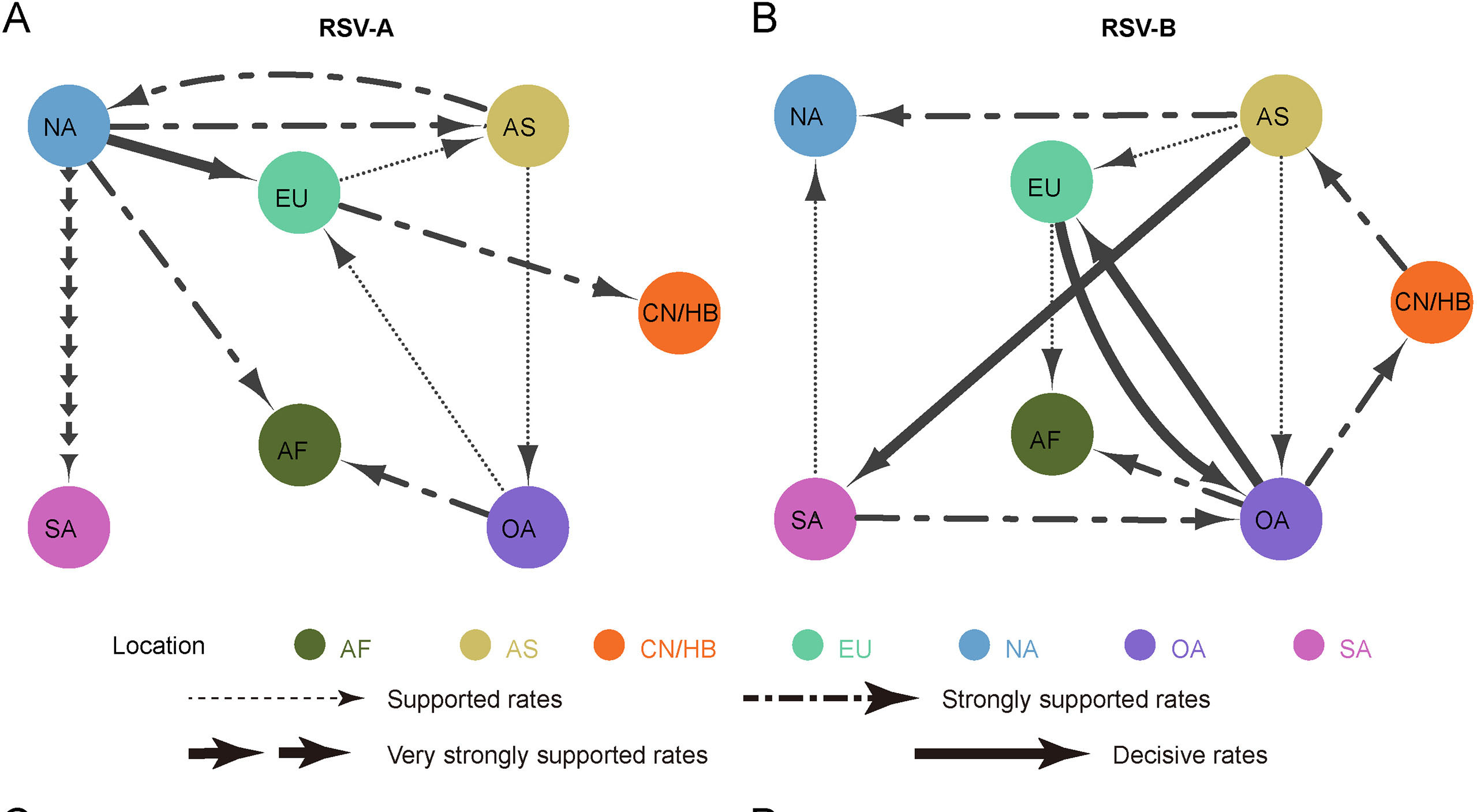
人呼吸道合胞病毒(Respiratory syncytial virus,RSV)是儿童急性下呼吸道感染的主要病原,对儿童健康带来严重威胁。然而目前人们对RSV的宿主内进化和区域间扩散知之甚少。因此在本研究中,我们对2020-2021年湖北地区住院儿童进行了系统监测,通过临床和宏基因组下一代测序(Metagenomic next generation sequencing,mNGS)共检测到106份RSV阳性样本。结果显示在监测期间RSV-A和RSV-B共同流行,但RSV-B占优势。其中有46个高质量的基因组被用于进一步的分析。分析结果显示在34个样本中共检测到163个宿主内单核苷酸变异(intra-host single nucleotide variation,iSNV)位点,G基因是iSNV最丰富的基因,其非同义替换多于同义替换。进化动力学分析表明本研究中G和NS2基因的进化速率较高,RSV病毒群体的种群大小随时间的推移而变化。此外我们还发现了RSV-A和RSV-B分别从欧洲和大洋洲向湖北地区扩散的证据。本研究重点研究了RSV在宿主内和宿主间的进化过程,并为了解RSV的进化过程提供了一些证据。
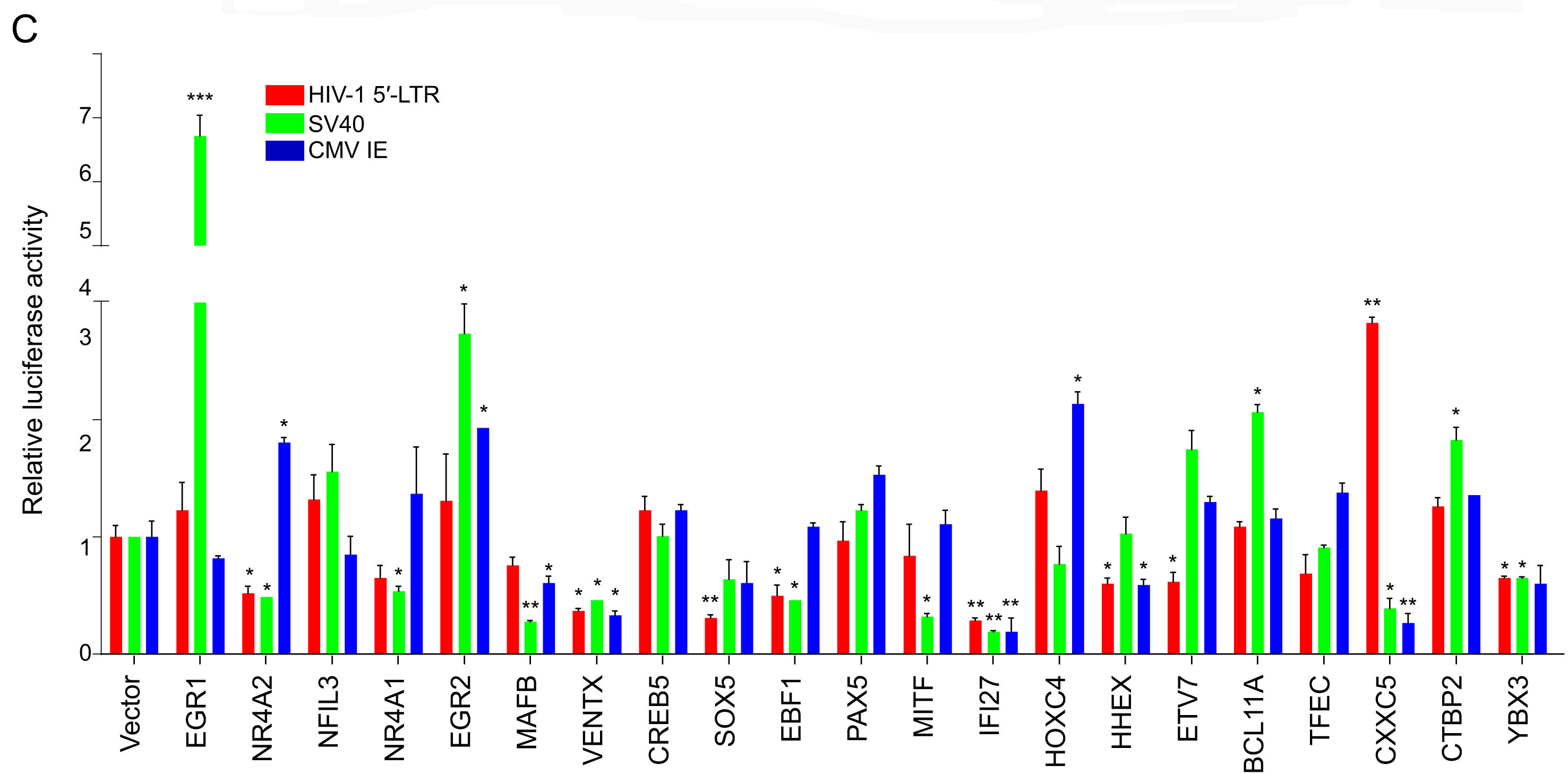
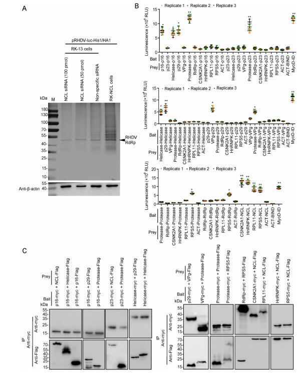
Due to our negligence, the original version of this article, published online on 12 January 2022, contained a mistake in Fig. 4A. The lane of β-actin in Western blotting was misused. The correct Fig. 4 is given below. We apologize for our oversight when preparing the figure and state that this does not change the scientific conclusions of the article in any way.
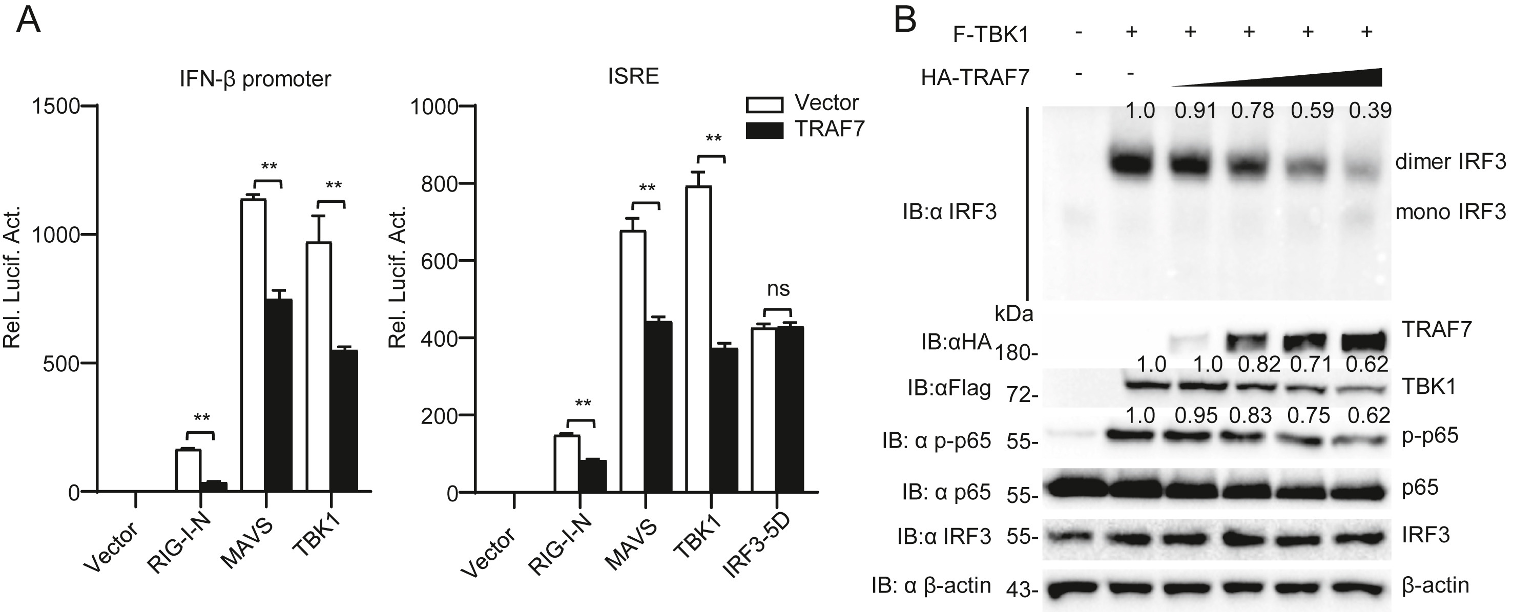
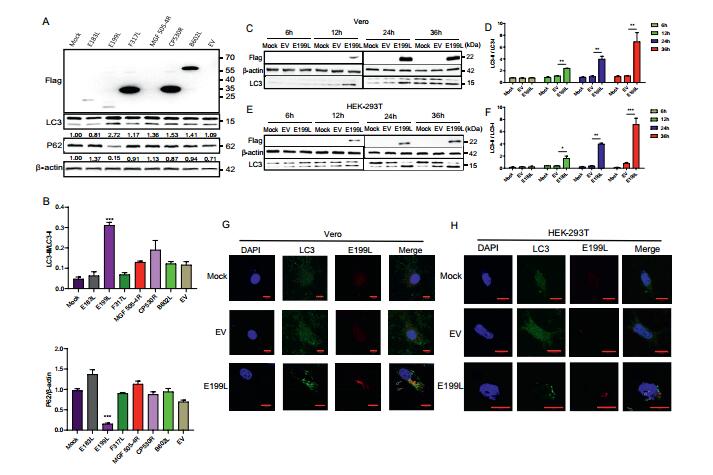
Due to our negligence, the original version of this article, published online on April 08, 2021, contained a mistake in Fig. 1E. The lane of β-actin in Western blotting was misused. The correct Fig. 1 is given below. We apologize for our oversight when preparing the figure and state that this does not change the scientific conclusions of the article in any way.